![]()
![]()
![]()
Use LEFT and RIGHT arrow keys to navigate between flashcards;
Use UP and DOWN arrow keys to flip the card;
H to show hint;
A reads text to speech;
74 Cards in this Set
- Front
- Back
- 3rd side (hint)
|
Leads I II III |
If a pt is connected to a 3 lead ECG monitor, these leads are being recorded |
|
|
|
Increase the paper speed |
Ecg shows ventricular rate of 188, a regular rhythm but pr interval and qrs complex are very hard to assess. What kind of action do you perform to have a better reading |
From Run 25 to Run 50 to stretch it out |
|
|
T wave inversion |
This type of inversion is an indicator of a myocardial ischemia |
|
|
|
Three |
When using a holter test, what is the minimum amount of electrodes to put on a pt |
|
|
|
Myocardial injury |
The ST segment elevation is a characteristic sign of: |
|
|
|
3-5 small boxes |
A normal pr interval should measure 0.12 to 0.20 seconds, which is how many boxes |
|
|
|
Yes, this is a true statement |
An ecg tech is about to place electrodes on the pts chest for holter monitorting test. The pt says "I'm supposed to record any symptoms i experience in the pt diary." Does he understand? |
Holter cannot be taken off or get wet |
|
|
Activate code blue |
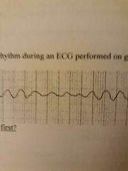
When an ecg shows tracings of ventricular tachycardia on a geriatric pt(pic shown in hint). What is the FIRST thing the tech should do |
|
|
|
The pt has a demand pacemaker |
What does it mean when vertical spikes appear on an ecg tracing sporadically |
It works when needed (when in demand) |
|
|
Continue taking the ecg |
A pt is asymptomatic during an ecg test. What should you do first? |
Asymptomatic= not showing any symptoms |
|
|
Ventricular tachycardia |
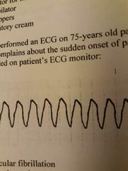
75 yo pt complains of sudden onset of palpitations and lightheadedness. What is this rhythm on the pt ecg |
Remember; we haven't discussed vfib in class |
|
|
Third degree block |
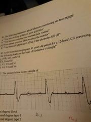
What type of block is this an example of |
Nothing is consistent. P waves and rr intervals are working independantly |
|
|
Hospital's operation manual |
What should you consult when you have questions related to holter monitoring electrodes placement |
Different hospitals have different procedures |
|
|
Sinus arrhythmia |
A pt is experiencing a change of respiratory cycle and variations in vagal tone. What irregularity does this indicate |
Respiratory cycle - respiratory arrhythmia |
|
|
5th intercostal space, junction of left midclavicular line |
Where is the apical pulse located |
V4 |
|
|
A trained athelete |
What kind of individual may normally experience bradycardia |
|
|
|
Brachial artery |
What is the most appropriate site for taking the pulse of an infant |
|
|
|
Corporal interference/somatic |
Ecg tech puts a geriatric pts hands under his butt. What kind of artifact does he expect to observe |
|
|
|
10 |
Standardization mark is how many small boxes |
|
|
|
Ventricles depolarize slowly |
Qrs complex=0.18 sec. What does this mean |
|
|
|
LL |
Theres a broken recording artifact in lead III and avf. Which electrode to check? |
|
|
|
128 BPM |
Stress test on a 60 yo pt at 80% THR. What's the minimum heart rate to acheive? |
220-60=160*0.80 |
|
|
There is a red line on bottom of the paper |
When should the paper be changed in the ecg machine |
|
|
|
Cover the pt with a blanket |
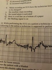
What should a tech do if they see this artifact on a pts ecg |
Somatic |
|
|
aVR |
Which lead is the p wave expected to be inverted |
|
|
|
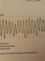
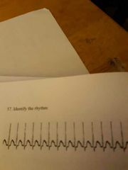
Ventricular tachycardia |
|
|
|
|
39/min |
Rr interval equals 38 small boxes. Whats the hr |
|
|
|
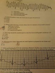
T wave inversion |
Correct indicator of myocardial ischemia |
|
|
|
20 mm |
How high is the standard gain when sensitivity is 2 |
|
|
|
Reattach a new electrode and repeat the recording |
|
|
|
|
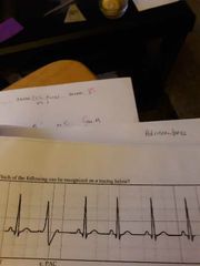
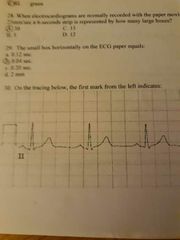
Pr interval |
This represents the time from the beginning of atrial depolarization to the beginning of ventricular depolarization |
|
|
|
1 small box, more than 0.04 sec |
Abnormal q wave measures more than: |
|
|
|
Myocardial injury |
St segment elevation is a sign of: |
|
|
|
20-40bpm |
Ventricles typically produce impulse with a rate of: |
|
|
|
P wave |
This wave is the deflection produced by atrial depolarization |
|
|
|
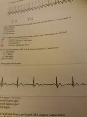
PVC |
|
|
|
|
Bradycardia |
Rr interval equals 7 large boxes, what is the pt experiencing |
|
|
|
RL green |
Grounding electrode during 12 lead ecg test is placed __ and its colored__ |
|
|
|
30 |
25mm/ sec on a 6 second strip represents how many large boxes |
|
|
|
0.04 |
Small box represents how many seconds |
|
|
|
Calibration of the machine |
|
|
|
|
True |
Asystole, isoelectric line, baseline, are synonymous if see no voltage record on ecg. T/f |
|
|
|
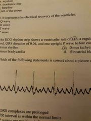
T wave |
This wave represents the electrical recovery of the ventricles |
|
|
|
Sinus tachycardia |
Ventricular rate 146, pr interval 0.14 sec, qrs duration 0.06, one upright p. What's this rhythm |
|
|
|
F waves or sawtooth waves present |
|
|
|
|
Increase the gain to 2 |
Amplitude of waves on ecg is very small. What do you do to improve the quality |
|
|
|
True |
"I can experience mild fatigue and shortness of breath during the test" is patient understanding of stress test. True/false |
|
|
|
Qrs complex |
Ventricular depolarization is represented on ecg strip by: |
|
|
|
Galvonometer |
This part of machine is responsible for changing an electrical activity picked up from the pt skin into a mechanical motion of a stylus |
|
|
|
True |
P wave upright in lead II reflects normal sinus rhythm. T/f |
|
|
|
Hippa |
Informing pts wife about her husband stress test is a violation of: |
|
|
|
Q wave |
First negative reflection after p wave |
|
|
|
True |
Hippa is a national standard. T/f |
|
|
|
Junctional rhythm |
Presence of inverted p wave is what kind of rhythm |
|
|
|
QT interval |
This interval is measured from beginning of qrs to end of t wave |
|
|
|
Av node |
A delay in electrical activity at this level allowing blood to flow from the atria to the ventricles |
|
|
|
Right lower rib cage |
Pt has a whole right lower extremity cast application. Where does the green electrode go? |
|
|
|
Wenckenbach |
When analyzing the strip you notice progressively prolonged pr intervals until a qrs is dropped. What is this |
|
|
|
Orthopnea |
Difficulty breathing when pt is supine |
|
|
|
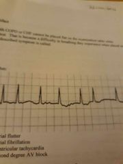
Atrial fibrillation |
|
|
|
|
Lead II |
The lead that is typically recorded to attach to the pts chart |
|
|
|
V7 |
The electrode placed on the 5th intercostal space in a left posterior axillary line |
|
|
|
SVT |
|
|
|
|
25mm/sec |
Ecg normally recorded with paper moving at a speed of |
|
|
|
125 BPM |
Rr interval measures 12 small boxes. Hr? |
|
|
|
Second degree type 1 |
|
|
|
|
PVC |
Wide, bizarre and prolonged qrs complexes |
|
|
|
Inverted |
If seen, the p wave of a rhythm originating in the av junction will appear ___ in lead II |
|
|
|
Lightheaded |
Perspiring Lightheaded Jaundice Flushed Which is a s/s of cardiac compromise and reason to terminate a test |
|
|
|
5 |
How many electrodes do you apply on a pt with a standard telemetry monitor |
|
|
|
V9 |
This electrode is placed on 5th intercostal space in a left paraspinous line |
Spine |
|
|
Inferior infarction |
ST segment elevation in leads II, III, and avf indicates (what surface of heart impairment): |
|
|
|
Atrial fibrillation |
F waves are present. What ectopic rhythm is this |
|
|
|
Myocardial infarction |
While performing a stress test pt experiences heaviness on his chest and diaphoresis. These are symptoms of: |
|