![]()
![]()
![]()
Use LEFT and RIGHT arrow keys to navigate between flashcards;
Use UP and DOWN arrow keys to flip the card;
H to show hint;
A reads text to speech;
56 Cards in this Set
- Front
- Back
|
A-fib is most commonly seen with |
Rheumatic MS |
|
|
What view should you use for a contrast study of an ASD |
Ap 4 |
|
|
What type of r-r interval will have a higher peak velocity |
Longer r-r interval |
|
|
Congenitally stenotic aortic valve can be described as |
Fluttering |
|
|
On M-mode a Flail mitral valve may have the same appearance as |
An infected mv |
|
|
Sympathetic nervous system |
Causes increased heart rate due to flight or fight |
|
|
Parasympathetic nervous system |
Decreases heart rate due to the vagus nerve |
|
|
When Amyl nitrate is administered it |
Increases the systolic anterior motion of the mitral valve |
|
|
ventricular premature beats originate in the |
Ectopic focus |
|
|
The eustachian valve is |
A normal ivc valve seen in subcostal |
|
|
In newborn the right ventricle free wall measures |
2-4mm |
|
|
During valsalva most murmurs |
decrease but ihss increases |
|
|
In M mode The structure used to pinpoint end systole for measurement is |
Maximum anterior motion of the left ventricle posterior wall. The septum is not consistent for measuring due to pressure changes and other factors |
|
|
Trepopnea |
The sensation of dyspnea or palpation, or an uncomfortable feeling that may occur when patients with cardiac disease lie on their left side |
|
|
Lvot obstruction causes the aov to |
Close in mid systole |
|
|
LA dilation is associated with |
Significant mr Increased pulmonary pressure PDA |
|
|
The posterior leaflet of the MV appears smaller because |
It is smaller and the shape is different from the anterior one |
|
|
The e-f slope is reliable In assessment of |
MS and LV function |
|
|
An increase in the size of the A wave of the mitral valve suggests |
AI and LVEDP |
|
|
The criterion that is the most helpful in defining mitral stenosis is |
Left atrial enlargement |
|
|
Myxomatous degeneration used to describe MVP denotes |
Thickening of the MVL |
|
|
M-mode findings on a young patient with congenital AS would show |
Normal leaflets separation |
|
|
The best approach for cw Doppler of AS is |
Suprasternal |
|
|
Overestimation of Doppler peaks in AS occur with coexisting |
AI |
|
|
Reverse doming of the anterior MVL can be observed in |
AR |
|
|
Chagas' disease |
Dcmo posterior and apical thinning, septum usually normal |
|
|
What is the cause of a B notch |
Increased left ventricular end diastolic pressure |
|
|
In elderly patients the a wave is normally |
Equal to or higher than the E wave |
|
|
The best 2-D echo view for Doppler analysis of the tricuspid valve is |
AP4 |
|
|
Tricuspid inflow velocity normally resembles mitral inflow except for |
Tricuspid valve inflow is at a lower velocity |
|
|
Which valve opens first the tricuspid or the mitral |
The TV |
|
|
M-mode recordings of the PV normally show which pulmonary leaflets |
Posterior |
|
|
What is seen with pulmonary hypertension |
Mid systolic notching of the A wave, nonvariation in A wave amplitude and absence of A wave |
|
|
vegetations have been seen at other sites than the valves. These sites are |
Aneurysm of sinus a Valsalva, calcified mitral annulus, infected ventricular septal defect |
|
|
Valve motion in endocarditis is |
Normal |
|
|
Secondary findings to endocarditis are |
Fistula aneurysm and abscess |
|
|
The best way to quantify prosthetic valve motion is |
M mode scanning |
|
|
The best way to quantify prosthetic valve motion is |
M mode scanning |
|
|
How many orifices does a Starr-Edwards ball Prothesis have |
3 |
|
|
The best approach is for obtaining the highest velocities in an aortic prosthesis is |
Apex, super sternal notch and right sternal border |
|
|
What are you can all three struts of an aortic and mitral valve prosthesis be seen |
Parastatal short axis |
|
|
Rounding of the E-point on M-mode with a prosthetic valve indicates |
Some form of obstruction |
|
|
Increased leaflet thickening on a bioprosthetic valve is |
Abnormal |
|
|
Epicardial fat pad is a measurement of visceral fat and has been linked to an increase in |
Coronary artery disease |
|
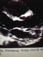
Fluid in the transverse sinus could be |
An abscess or just fluid |
|
|
What is beck's triad |
Signs for cardiac tamponade which include elevated venous pressure hypertension and quiet heart. you can also have jugular vein distention |
|
|
The most sensitive way to diagnose cardiac tamponade is |
Respiratory variation |
|
|
What happens to the hepatic flow during Tamponade |
Reversal of flow during expiration |
|
|
"Breaking" is noted in |
LBBB, Wolf Parkinson White syndrome, and right ventricular pacing |
|
|
What windows should be used to evaluate the interatrial septum |
Apical four, subcostal long and high right parasternal long axis |
|
|
What windows should be used to evaluate the interatrial septum |
Apical four, subcostal long and high right parasternal long axis |
|
|
What view should you use to evaluate the fossa ovalis |
subcostal window |
|
|
Eisenmenger syndrome |
Reversal of a long-standing left to right shunt from pulmonary hypertension shunt is now right to left |
|
|
Eisenmenger syndrome |
Reversal of a long-standing left to right shunt from pulmonary hypertension shunt is now right to left |
|
|
The size of aneurysms during systole |
Increase |
|
|
What type of MI causes papillary muscle rupture |
Inferior MI |