![]()
![]()
![]()
Use LEFT and RIGHT arrow keys to navigate between flashcards;
Use UP and DOWN arrow keys to flip the card;
H to show hint;
A reads text to speech;
56 Cards in this Set
- Front
- Back
|
Pathogenesis of epidermal post-inflammatory hyperpigmentation? |
Increased melanin production and/or transfer to keratinocytes |
|
|
Pathogenesis of dermal post-inflammatory hyperpigmentation? |
Damaged basement membrane allowing "falling" of melanin and subsequent phagocytosis by dermal macrophages |
|
|
Color seen with increased epidermal melanin? |
Tan to dark brown |
|
|
Color seen with increased dermal melanin? |
Gray-blue to gray-brown |
|
|
Which lasers (3) may aid in removal of dermal pigment? |
1. Nd:YAG 2. Q-switched ruby 3. Alexandrite |
|
|
Clinical presentation of erythema dyschromicum perstans? |
Irregularly shaped macules and patches with slate-gray to blue-brown color seen in a symmetric pattern on the trunk with spread to neck, proximal upper extremities, and face |
|
|
Disease course of erythema dyschromicum perstans? |
Slowly progressive; usually does not regress Usually asymptomatic, however can be mildly pruritic |
|
|
Histopathologic findings in erythema dyschromicum perstans? |
1. Vacuolar degeneration of the basal layer 2. Perivascular mononuclear cell infiltrate in upper dermis 3. Increased epidermal melanin and dermal melanophages |
|
|
Two potential drugs that can help treat erythema dyschromicum perstans? |
1. Clofazimine 2. Dapsone |
|
|
Most commonly affected individuals in lichen planus pigmentosus? |
Young to middle-aged adults with skin phototypes III-V |
|
|
Clinical presentation of lichen planus pigmentosus? |
Brown to gray-brown macules and patches in sun-exposed areas or intertriginous zones |
|
|
What can potentially cause lightening of lesions in lichen planus pigmentosus? |
Topical tacrolimus |
|
|
Clinical presentation of melasma? |
Symmetric hyperpigmented patches with irregular outline on the face (also uncommonly on upper chest, forearms) Exacerbated by pregnancy, OCPs, sun exposure |
|
|
Melasma is seen in increased prevalence in what 3 ethnic groups? |
1. Hispanic 2. African 3. Asian/Middle Eastern (i.e. darkly pigmented skin) |
|
|
Pathogenesis of melasma? |
Hyperfunctional melanocytes produce increased amounts of melanin in response to an inducer (hormones, UV radiation) |
|
|
Most common topical regimen to treat melasma? |
Hydroquinone + tretinoin + corticosteroid 2 - 6 month course |
|
|
Clinical presentation of primary (localized) cutaneous amyloidosis? |
Rippled pattern of parallel bands or ridges of hyperpigmentation + pruritus |
|
|
What form of mastocytosis is classically associated with hyperpigmentation? How does it present? |
Urticaria pigmentosa Few to several hundred brown to red-brown papules which urticate after stroking (Darier's sign) |
|
|
What are common medications (4) that cause hyperpigmentation / discoloration? |
1. Chemotherapeutic agents (gleevec, bleomycin) 2. Antimalarials 3. Minocycline 4. Zidovudine i.e. AZT |
|
|
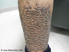
Most common presentation of minocycline-induced hyperpigmentation? |
Gray-blue hyperpigmentation on the b/l shins |
|
|
Where are pigmentary demarcation lines most commonly seen? |
Anterolateral upper arm, posteromedial thigh (demarcation b/w the dorsal and ventral surfaces) Lines are perfectly symmetric and stable over time |
|
|
What percentage of patients taking bleomycin develop flagellate hyperpigmentation? |
10-20% (fairly common side effect) Pigmentation (brown streaks) appears to be dose-dependent |
|
|
Most common location for flagellate hyperpigmentation caused by bleomycin? |
Chest and back |
|
|
What food can cause a flagellate dermatitis that presents as linear streaks of papules or petechiae? |
Shitake mushrooms; raw or partially cooked Pruritus is initially seen; scratching leads to long, flagellate streaks |
|
|
What does hyperpigmentation along the lines of Blaschko indicate? |
Mosaicism (normal variant) |
|
|
Term for hyperpigmentation along the lines of Blaschko? |
Linear and whorled nevoid hypermelanosis (LWNH) |
|
|
What is clinically important about Linear and whorled nevoid hypermelanosis? |
Up to 25% of patients present to pediatrician with extracutaneous findings (neurologic, MSK, cardiac) |
|
|
Inheritance pattern for incontinentia pigmenti? |
X-linked dominant |
|
|
What are the 4 stages of incontinentia pigmenti? |
1. Linear erythema and blisters 2. Verrucous lesions 3. Hyperpigmentation along lines of Blaschko 4. Hypopigmentation along lines of Blaschko |
|
|
What gene is responsible for incontinentia pigmenti? |
Mutations in the NEMO gene |
|
|
Clinical presentation of prurigo pigmentosa? |
Markedly pruritic eruption of erythematous papules and papulovesicles that develop rapidly and involute within a week, leaving macular reticulated hyperpigmentation |
|
|
Markedly pruritic eruption of erythematous papules and papulovesicles that develop rapidly and involute within a week, leaving macular reticulated hyperpigmentation? |
Prurigo pigmentosa |
|
|
Typical patient profile for prurigo pigmentosa? |
Young females in Japan |
|
|
Treatment options for prurigo pigmentosa (3)? |
1. Minocycline 2. Doxycycline 3. Dapsone |
|
|
Clinical presentation of dyskeratosis congenita? |
1. Progressive bone marrow failure 2. Reticulated hyperpigmentation 3. Nail dystrophy 4. Leukoplakia 5. Increased risk of SCC (esp mucosal), AML, and Hodgkin disease |
|
|
Genodermatosis that presents with mucocutaneous triad of reticulated hyperpigmentation + nail dystrophy + leukoplakia, along with progressive bone marrow failure and increased risk of SCC? |
Dyskeratosis congenita |
|
|
Inheritance pattern of dyskeratosis congenita? |
X-linked recessive |
|
|
What enzyme is implicated in dyskeratosis congenita? |
Telomerase mutation is in DKC1 gene which encodes dyskerin, which interacts with telomerase Tissues with high replication rates (bone marrow, skin) are affected the most when telomerase is defective |
|
|
Best method to diagnose dyskeratosis congenita? |
Flow fluorescence in situ hybridization (FISH) of leukocytes detecting short telomere length |
|
|
Inheritance pattern of Naegeli-Franceschetti-Jadassohn syndrome? |
Autosomal dominant |
|
|
What is the mutation responsible for Naegeli-Franceschetti-Jadassohn syndrome? |
Truncating mutations of keratin 14 |
|
|
Clinical features of Naegeli-Franceschetti-Jadassohn syndrome (4)? |
1. Brown or gray-brown reticulated hyperpigmentation of the trunk and periocular/perioral regions 2. Decreased sweat gland function + heat intolerance 3. Palmoplantar keratoderma 4. Dental anomalies |
|
|
Genodermatosis that presents with reticular hyperpigmentation, decreased sweat gland function, palmoplantar keratoderma, and dental anomalies? |
Naegeli-Franceschetti-Jadassohn syndrome |
|
|
What is the mutation responsible for dermatopathia pigmentosa reticularis? |
Heterozygous truncating mutation of keratin 14 gene |
|
|
Clinical features of dermatopathia pigmentosa reticularis? |
1. Reticulated hyperpigmentation 2. Non-scarring alopecia 3. Onychodystrophy |
|
|
Genodermatosis that presents with reticulated hyperpigmentation, non-scarring alopecia, and onychodystrophy? |
Dermatopathia pigmentosa reticularis |
|
|
Genodermatosis with reticulated brown hyperpigmentation that is generalized in male patients and along the lines of Blaschko in female patients? |
X-linked reticulate pigmentary disorder |
|
|
Inheritance pattern in Dowling-Degos disease? |
Autosomal dominant |
|
|
Clinical features in Dowling-Degos disease? |
Reticulated hyperpigmentation in the intertriginous areas (groin, intercluteal, inframammary, neck, inner arms/thighs) |
|
|
Genodermatosis that presents with reticulated hyperpigmentation in the intertriginous areas by the 3rd or 4th decade of life? |
Dowling-Degos disease |
|
|
Genodermatosis that presents as slightly depressed lentigo-like hyperpigmented macules in a reticular pattern on dorsal aspects of hands and feet? |
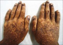
Reticulate acropigmentation of Kitamura |
|
|
Hypermelanosis that presents as both hyper- and hypopigmented macules of varying size on the dorsal surfaces of distal extremities, mostly in Asians? |
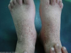
Dyschromatosis symmetrica hereditaria (DSH) |
|
|
Inheritance pattern of dyschromatosis symmetrica hereditaria? |
Autosomal dominant Caused by mutations in ds-RNA specific adenosince deaminase |
|
|
Primary difference between reticulate acropigmentation of Kitamura and dyschromatosis symmetrica hereditaria? |
The former does NOT have hypopigmented macules |
|
|
Hypermelanosis that presents as both hyper- and hypopigmented macules in a generalized distribution, most commonly in Japanese? |
Dyschromatosis universalis hereditaria (DUH) no seasonal change or spontaneous regression with age is seen |
|
|
Hypermelanosis that presents as chronic, asymptomatic gray-brown patches interspersed with hypopigmented macules on the dorsal forearms, seen most commonly in Caucasian women in 5th to 7th decades? |
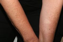
Acquired brachial cutaneous dyschromatosis Likely 2/2 chronic sun exposure |