![]()
![]()
![]()
Use LEFT and RIGHT arrow keys to navigate between flashcards;
Use UP and DOWN arrow keys to flip the card;
H to show hint;
A reads text to speech;
29 Cards in this Set
- Front
- Back
|
What are the four basic tunics of the alimentary canal? |
- Muscosa - Submucosa - Muscularis Externa - Serosa or Adventitia |
|
|
What is the mucosa layer? What are its functions? |

The mucosa is the inner most layer of the alimentary canal lumen. It consists of surface epithelium, a lamina propria, and a muscularis mucosae. It's surface epithelium consists of simple columnar. The major functions of the mucosa are secretion (of enzymes, mucus, hormones, etc), absorption of digested foodstuffs, and protection (against bacterial invasion). |
|
|
What is the submucosa? What are its functions? |

The submucosa is moderately dense connective tissue containing blood and lymphatic vessels, scattered lymphoid follicles, and nerve fibers. It's vessels absorb and transport nutrients, and its abundant elastic fibers help maintain the normal shape of each organ. |
|
|
What is the muscularis externa? What are its functions? |

The muscularis externa is a bilayer of smooth muscles with the inner later running circularly and the outer later running longitudinally. This layer moves the contents of the canal along by segmentation and peristalsis. This tunic is the major regulator of GI motility. |
|
|
What is the serosa? What are its functions? |
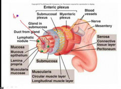
The outermost covering of the intrapertitoneal organs is the serosa, also called the visceral peritoneum. |
|
|
Where can you find adventitia?
|
In the esophagus, which is outside the abdominopelvic cavity, the series is replaced by an adventitia, a layer of coarse fibrous connective tissue that binds the organ to surrounding tissues. The adventitia anchors and protects the surrounded organ. |
|
|
What are the organs of the alimentary canal? |
- Oral cavity or mouth - Pharynx - Esophagus - Stomach - Small intestine - Large intestine |
|
|
The floor of the oral cavity is occupied by the muscular tongue, which is largely supported by the _________. |
Mylohyoid muscle |
|
|
What is the oral vestibule? |
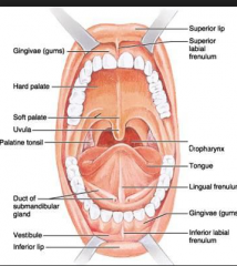
The oral vestibule is the space between the lips and cheeks and the teeth and gums is the oral cavity proper. |
|
|
What is the pharynx? |
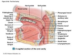
The pharynx is a common passageway for food, fluid, and air. The pharynx is subdivided anatomically into three parts: - Nasopharynx ( pseudo-stratified columnar) - Oropharynx (straified squamous epithelium) - Laryngopharynx (stratified squamous epithelium) |
|
|
What is the esophagus? |
The esophagus, or gullet, extends from the pharynx through the diaphragm to the gastroesophageal sphincter in the superior aspect of the stomach. The esophagus has no digestive or absorptive function. The walls at its superior end contain skeletal muscle, which is replaced by smooth muscle in the area nearing the stomach. |
|
|
What is the gastroesophageal sphincter?
|
The gastroesophageal sphincter is a slight thickening of the smooth muscle layer at the esophagus-stomach junction. |
|
|
What is the stomach? |
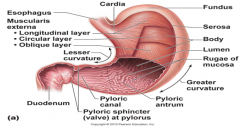
The stomach is on the left side of the abdominal cavity and is hidden by the liver and diaphragm. The stomach is made up of several regions. - The cardial part is the area surround the cardinal orifice through which food enters the stomach. - The funds is a dome-shaped portion of the stomach found superolaterally to the cardia. - The body forms the mid portion of the stomach, which leas to the funnel-shaped pyloric part. - The pyloric part terminates in the pylorus. The pylorus is continuous with the small intestine through the pyloric sphincter or valve. |
|
|
What is the small intestine? |
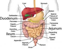
The small intestine is suspended by a double layer of peritoneum. The intestine has three subdivisions: - The duodenum - The jejunum - The ileum Enzymes produced by the pancreas and ducted into the duodenum largely via the main pancreatic duct complete the axiomatic digestion process in the small intestine. Bile, also enters the duodenum via the bile duct in the same area. Nearly all nutrient absorption occurs in the small intestine, where three structural modifications increase the absorption surface of the mucosa: the microvilli, villi, and circular folds. |
|
|
What are the large intestine? |
The large intestine encircles the small intestine on three sides and consists of the following subdivisions: cecum, appendix, colon, rectum, and anal canal. The colon is divided into several distinct regions. The ascending colon travels up the right side of the abdominal cavity and makes a right-angle turn at the right colic flexure to cross the abdominal cavity as the transverse colon. It then turns at the left colic lecture and continues down the left side of the abdominal cavity as the descending colon, where it takes an S-shaped course as the sigmoid colon. The major function of the large intestine is to consolidate and propel the unusable fecal matter toward the anus and eliminate it form the body. While it does that choice, it provides a site where intestinal bacteria manufacture vitamins B and K and reclaims most of the remains water from the undigested food, thus conserving body water. |
|
|
What are the accessory organs of the digestive system? |
- Teeth - Salivary Glands - Liver - Gallbladder - Pancreas |
|
|
What are the classification of the teeth? |
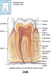
The teeth are classified as - Incisors (2) - Canines (1) - Premolars (2) - Molars (3) |
|
|
Describe the incisors. |
The incisors are chisel shaped and exert a shearing action use in biting. |
|
|
Describe the canines. |
The canines are cone shaped fanglike, the latter description being much more applicable to the canine of animals whose teeth are used for the tearing of food. |
|
|
What are the salivary glands? |
- Parotid glands - Submandibular glands - Sublingual glands |
|
|
What are the parotid glands?
|
The large glands located anterior to the ear and ducting into the mouth. |
|
|
What are the submandibular glands? |
They are located along the medial aspect of the mandibular body in the floor of the mouth, and ducting under the tongue to the base of the lingual frenulum. |
|
|
What are the sublingual glands? |
Small glands located most anteriorly in the floor of the mouth and emptying under the tongue via several small ducts. |
|
|
Saliva consists primarily of viscous glycoprotein called _____ |
Mucin |
|
|
Mucin moistens food and helps to bind it together into a mass called a _____ |
Bolus |
|
|
What is the liver? |
The liver is the largest gland in the body. It's digestive function is to produce bile. |
|
|
What is the gallbladder? |
When digestive activity is not occurring in the digestive tract, bile backs up into the cystic duct and enters the gallbladder. Bile is stored there until needed for digestive process. |
|
|
What is the pancreas? |
The pancreas is a soft triangular gland that extends horizontally across the posterior abdominal wall from the spleen to the duodenum. The pancreas has both an endocrine function producing the hormones insulin and glucagon and an exocrine function. |
|
|
What makes bile? |
Bile is continuously being made by the hepatocytes. |