Reading...
![]()
Play button
![]()
Play button
![]()
Use LEFT and RIGHT arrow keys to navigate between flashcards;
Use UP and DOWN arrow keys to flip the card;
H to show hint;
A reads text to speech;
69 Cards in this Set
- Front
- Back
- 3rd side (hint)
|
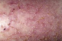
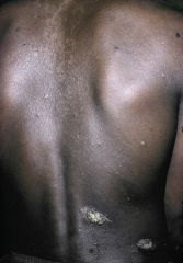
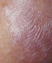
Figure 3-37 Asteatotic eczema (xerosis)
|
|
|
|
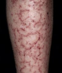
Figure 3-38 Asteatotic eczema (xerosis)
|
|
|
|
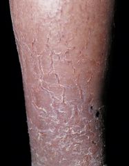
Figure 3-39 Asteatotic eczema (eczema craquel?)
|
|
|
|
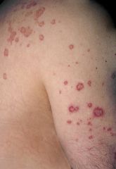
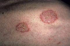
Figure 3-40 Nummular eczema
|
|
|
|
Figure 3-41 Nummular eczema
|
|
|
|
Figure 3-42 Nummular eczema
|
|
|
|
Figure 3-43 Nummular eczema
|
|
|
|
Figure 3-44 Nummular eczema
|
|
|
|
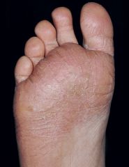
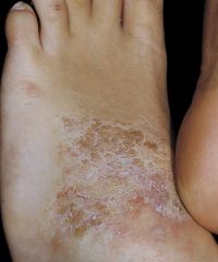
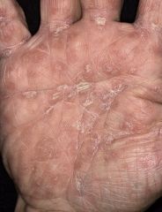
Figure 3-45 Chapped fissured feet
|
|
|
|
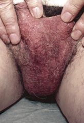
Figure 3-47 Lichen simplex chronicus
|
|
|
|
Figure 3-48 Lichen simplex chronicus
|
|
|
|
Figure 3-49 Lichen simplex chronicus
|
|
|
|
Figure 3-50 Lichen simplex chronicus
|
|
|
|
Figure 3-51 Lichen simplex chronicus
|
|
|
|
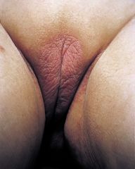
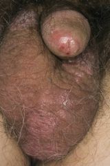
Figure 3-55 Red Scrotum Syndrome
|
|
|
|
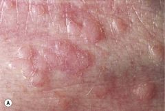
Figure 3-56 Prurigo nodularis
|
|
|
|
Figure 3-57 Prurigo nodularis
|
|
|
|
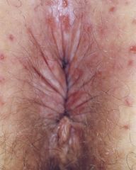
Figure 3-58 Neurotic excoriations
|
|
|
|
Figure 3-59 Neurotic excoriations
|
|
|
|
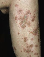
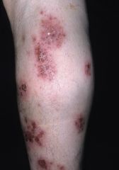
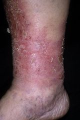
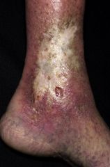
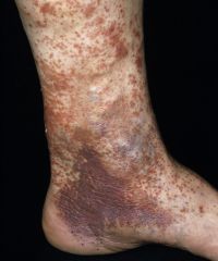
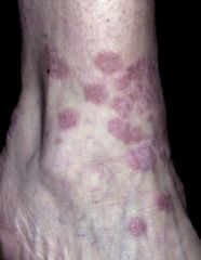
Figure 3-62 Stasis dermatitis
|
|
|
|
Figure 3-64 Stasis dermatitis
|
|
|
|
Figure 3-65 Stasis dermatitis
|
|
|
|
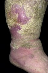
Figure 3-68 Stasis papillomatosis
|
|
|
|
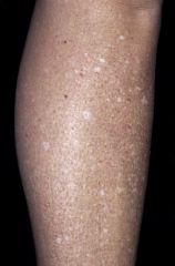
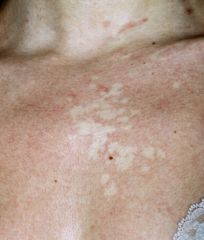
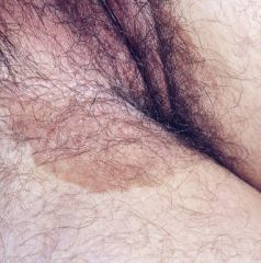
Figure 19-33 Idiopathic guttate hypomelanosis
|
|
|
|
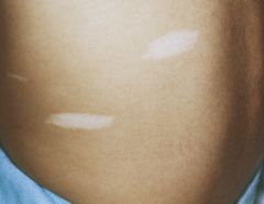
Figure 19-34 Nevus anemicus
|
|
|
|
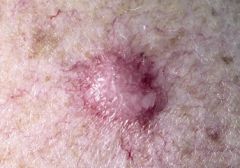
Figure 26-18 Adenoma sebaceum
TUBEROUS SCLEROSIS |
|
|
|
Figure 26-20 Ash-leaf macules (hypomelanotic macules)
TUBEROUS SCLEROSIS |
|
|
|
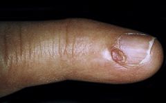
Figure 26-21 Tuberous sclerosis. Periungual fibromas.
|
|
|
|
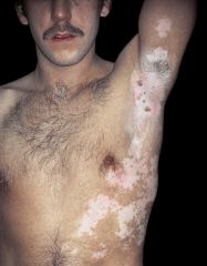
Figure 19-29 Vitilio
|
|
|
|
Figure 22-17 Becker’s nevus
|
|
|
|
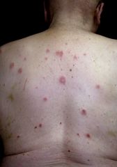
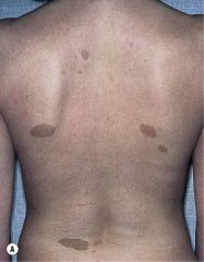
Figure 26-12 von Recklinghausen’s neurofibromatosis. Caf?-au-lait spots
|
|
|
|
Figure 13-19 Erythrasma: a bacterial infection (Corynebacterium minutissimum)
|
|
|
|
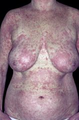
Figure 14-44 Exfoliative erythroderma
|
|
|
|
Figure 22-2 Junction nevus
|
|
|
|
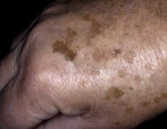
Figure 19-36 Lentigo
|
|
|
|
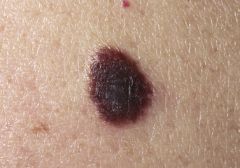
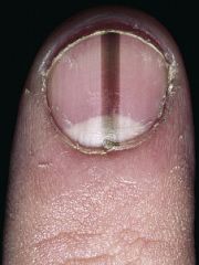
Figure 22-3 Acral Lentiginous Melanoma. The sudden appearance of a pigmented band at the proximal nailfold is suggestive of melanoma
|
|
|
|
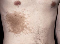
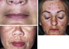
Figure 19-38 Melasma
|
|
|
|
Figure 19-27 Doxycycline-induced phototoxicity
Tetracyclines (doxycycline [Figure 19-27], tetracycline) Fluoroquinolones (ciprofloxacin, ofloxacin, levofloxacin) Sulfonamides |
|
|

|
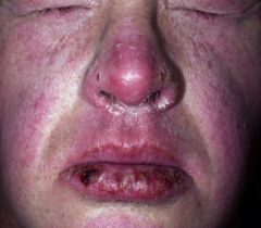
Figure 10-9 Primary syphilis
|
|
|
|
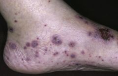
Figure 10-15 Secondary syphilis
|
|
|
|
Figure 10-15 Secondary syphilis
|
|
|
Lesions usually progress from red, painful, and vesicular to “gun-metal grey” as the rash resolves
|
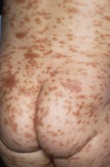
Figure 10-16 Secondary Syphillis
|
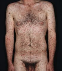
A diffuse eruption consisting of macules, papules, and pustules. Typical early lesions are usually less than 20, round, discrete, nonpruritic, and symmetric macules distributed on the trunk and proximal extremities. Red papular lesions also may appear on the palms, soles, face, and scalp and may become necrotic. Patchy and nonpatchy alopecia may occur. In intertriginous areas, papules may coalesce to form highly infectious lesions called condylomata lata. Lesions usually progress from red, painful, and vesicular to “gun-metal grey” as the rash resolves.
|
|
|
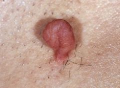
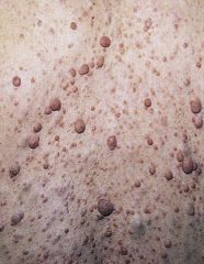
Figure 20-20 SKIN TAGS (ACROCHORDON)
|
|
|
Pigs Skin
|
Figure 26-19 Shagreen patch Tuberous Sclerosis
|
|
|
|
Figure 7-7 Comedones (blackheads)
|
|
|
|
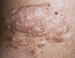
Figure 12-14 Flat warts (verruca plana)
|
|
|
|
Figure 26-2 Granuloma annulare
|
|
|
|
Lichen nitidus
|
|
|
|
Figure 8-64 Lichen sclerosous
|
|
|
|
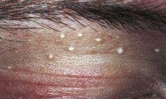
Figure 7-43 Milia
|
|
|
|
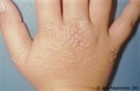
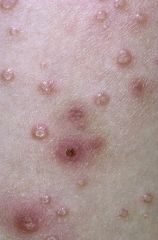
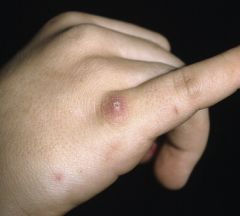
Figure 12-26 Molluscum contagiosum
|
|
|
|
Figure 26-13 von Recklinghausen’s neurofibromatosis
|
|
|
|
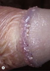
Pearly Penile Papules
|
|
|
|
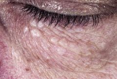
Figure 20-54 Senile sebaceous hyperplasia
|
|
|
|
Figure 20-55 Syringoma
|
|
|
|
Figure 20-25 Dermofibroma
|
|
|
|
Figure 6-31 Cutaneous mastocytosis (urticaria pigmentosa)
|
|
|
|
Disseminated Superficial Actinic Porokerotosis DSAP
|
|
|
|
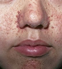
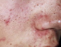
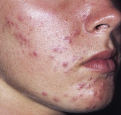
Figure 7-11 Papular and pustular acne
|
|
|
|
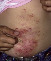
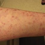
Figure 5-1 Eczematous dermatitis
|
|
|

|
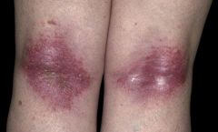
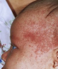
Figure 5-7 Atopic dermatitis
|
|
|
The lesion is painful but does NOT itch. The patient just got a new pet. Can you guess what type of pet?
|
Figure 15-41 Cat-scratch disease
What would you check next? |
Lymph nodes for lymphadenopathy
|
|
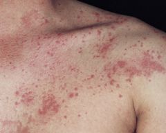
16 year old white male reports getting this rash after jogging
|
Figure 6-16 Cholinergic Urticaria
|
|
|
|
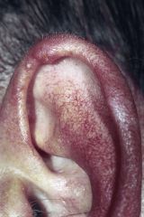
Figure 20-42 Chondrodermatitis nodularis helicis
|
|
|
|
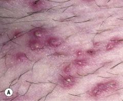
Figure 9-18 Folliculitis
|
|
|
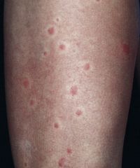
What probably caused these papules?
|
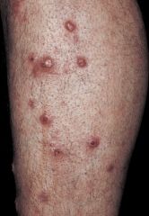
Figure 15-46 Papular urticaria
Bug Bites |
|
|
|
Figure 5-27 Keratosis pilaris
|
|
|
|
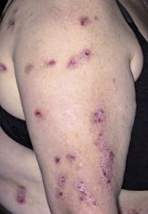
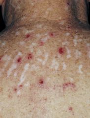
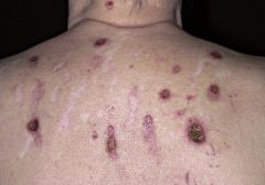
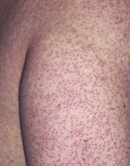
Figure 18-13 Vasculitis
|
|
|
|
Figure 7-65 Miliaria crystallina
|
|