![]()
![]()
![]()
Use LEFT and RIGHT arrow keys to navigate between flashcards;
Use UP and DOWN arrow keys to flip the card;
H to show hint;
A reads text to speech;
77 Cards in this Set
- Front
- Back
|
Nervous System |
The nervous system, along with the endocrine and immune system and the sensory organs, is responsible for receiving various stimuli (Sensory Impulses) and coordinating the reactions of the organism. The nervous system receives stimuli that affect the body surface and/or insides. The stimuli cause impulses that are transmitted, processed and answered in the form of passive or active reactions. In short, the nervous system enables the body to interact, adapt and react to the environment. |
|
|
Division of the nervous system |
1 Central Nervous System (CNS): consisting of brain and spinal cord. 2 Peripheral Nervous System (PNS): consisting of cranial nerves, spinal nerves and their associated ganglia (aggregation of nerve cell bodies). |
|
|
Embryological origin |
Nervous system originate embryologically from the Neural plate of Ectoderm. |
|
|
The Brain |
Encephalon
The brain is the control organ of the body, and is responsible for the regulation, coordination and integration of the rest of the nervous system. |
|
|
Location of Brain |
Formation of cranial cavity: Dorsally: Frontal, Parietal, and Interparietalbones. Ventrally: Basilar part of the Occipital,Sphenoid and Presphenoid bones. Caudally: Occipital bone. Cranially: Ethmoid and Crista gallae. Laterally: Temporal bone. |
|
|
Cranial Cavity Diagram |

|
|
|
Coverings of the Brain |
Meninges
The brain is covered by 3 layers:
1. Dura mater 2. Arachnoid mater 3. Piamater |
|
|
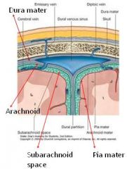
Meninges 3 Layers |
1. Dura mater: made up of dense connective tissue. 2. Arachnoid mater: made up of loose connective tissue with arachnoid villi for the dainage of cerebrospinal fluid (CSF). Space below arachnoid is called subarachnoid space through which CSF is circulated. 3. Piamater: This layer is closely invest the brain and rich of blood supply. |
|
|
Meninges Diagram |
|
|
|
Leptomeninges |
The pia mater and arachnoid together is called leptomeninges because these two membranes are thin in comparison to the Dura mater (Pacchymenix - because dura is thick).
In some places of the brain pia mater entered into the brain and form plexus known as choroid plexus which secretes CSF. |
|
|
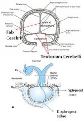
Modification of meninges |
1. Falx cerebri: Fold of dura mater in the longitudinal fissure of cerebrum.
2. Tentorium cerebelli: (means tent of the cerebellum). It is extension of dura mater which separates cerebellum from cerebrum.
3.Diaphragma sellae: It is the circular fold of the dura mater that covers part of the pituitary gland which lies on the sphenoid bone and completing the roof of the sella turcica. |
|
|
Modification of meninges Diagram |
|
|
|
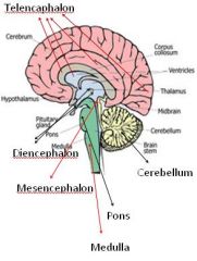
Parts of Brain |
1. Prosencephalon: Telencephalon and Diencephalon (forebrain)
2. Mesencephalon: (mid brain)
3. Rhombencephalon (hind brain): a) Metencephalon: Pons and cerebellum b) Myelencephalon: Medulla oblongata |
|
|
Parts of Brain Diagram |
|
|
|
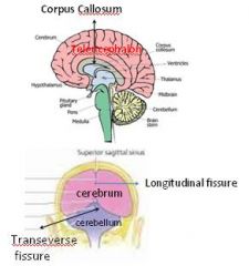
Parts of the Prosencephalon (forebrain) |
Telencephalon or the distant brain (far brain). It consist of: Paired cerebral hemisphere which separated by longitudinal fissure, and connected by corpus callosum (rostrum, genu, body and splenium).
|
|
|
Parts of the Prosencephalon Diagram |
|
|
|
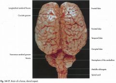
Gyrus and Sulcus |
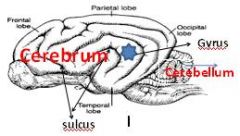
Gyrus and Sulcus: Grey matter (nerve cells) and White matter (fiber) A fold or ridge in the cortex is termed a gyrus (plural gyri) and a groove or fissure is termed a sulcus (plural sulci) |
|
|
Lobes of the brain |
Frontal, Parietal, Occipital, Temporal, Piriform (in ruminant), Olfactory (in dog), and Optic lobe (in bird).
|
|
|
Lobes of the brain Diagram |
|
|
|
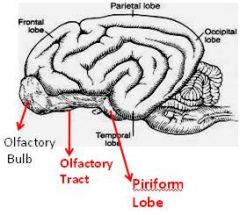
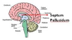
Parts of Forebrain |
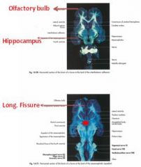
Rhinencephalon begins with the olfactory bulb, olfactory tract and ends into piriform lobe.
Two lateral ventricles : cavity of brain separated by a membrane known as septum pellucidum. |
|
|
Parts of Forebrain Diagram |
|
|
|
Function of Telencephalon |
Olfaction, visual activity, Hearing, intelligence, fear, emotion , hunger, thirst etc.
|
|
|
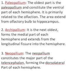
Pallium or Cortex of Cerebral Hemisphere |
|
|
|
Pallium Diagram |
|
|
|
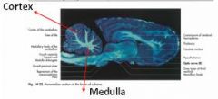
Cerebral Cortex |
The cerebral cortex is the cerebrum's (brain) outer layer of neural tissue in humans and other mammals. It is divided into two cortices, along the sagittal plane: the left and right cerebral hemispheres divided by the medial longitudinal fissure. |
|
|
Cortex and Medulla of Brain |
Cortex of brain located peripherally and consists of nerve cell bodies (grey matter) and medulla located centrally, white in nature consists of nerve fibers (white matter) |
|
|
Internal structure of Cerebral hemisphere (basal ganglion) |
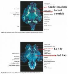
Corpus Striatum: It is an accumulation of grey matter within the white matter.e.g. basal ganglia (nucleus of the brain): a) Caudate, (b) Putamen, (c) Claustrum and (d) Amygdaloid body. |
|
|
Basal Ganglion Diagram |
|
|
|
Internal structure of Cerebral hemisphere (Fibers of brain) |
There are 3 types of gross fibers which connect different parts of brain or separates different structures of brain:
External capsule: It is thin and separates claustrum. Projection fibers: Internal capsule is thick and separates putamen. This fibers projects within the same hemisphere. Commissural fibers: Corpus callosum which connects two cerebral hemisphere. |
|
|
Lymbic System of Cerebral Hemisphere |
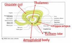
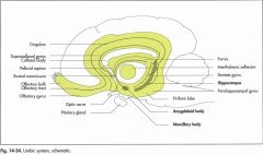
The term lymbic system means some parts of brain structure involved with emotional behavior. It consists of:
- Cingulate gyri, Piriform lobe and hippocampus of cortex of brain. - Thalamus and hypothalamus of diencephalon, and amygdaloid body of basal ganglion. |
|
|
Lymbic System Diagram |
|
|
|
Lymbic System Diagram |
|
|
|
Diencephalon (Parts of Prosencephalon) |
Diencephalon: It is also known as twin brain. It is visible in sagittal section view and ventral view and comprises: - Epithalamus - Thalumus - Subthalumus, and - Hypothalumus
|
|
|
Diencephalon Diagram |
|
|
|
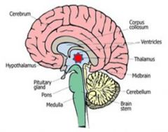
Epithalamus and Thalamus |
1. Epithalamus comprises: Pineal gland and Habenula (nucleus and fibers for olfactory pathway).
2. Thalamus is a large rounded mass composed of large number of nuclei through which input of cerebral cortex in channelled including sensory information from gustatory (taste), optic (vision), vestibulo-cochlear (hearing and balance).
|
|
|
Thalamus Diagram |
|
|
|
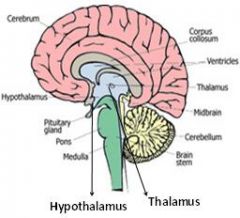
Subthalamus and Hypothalamus |
3. Subthalumus is ventral to the thalumus contains subthalamic nuclei and it is the relay station of extrapyramidal motor pathway. 4. Hypothalumus consist of optic chiasma, mammilary body tuber cinereum through which infundibulum protruded for the suspension of pituitary gland. 5. Third ventricle: Around the thalamus a narrow strip is the 3rd ventricle. |
|
|
Function of hypothalamus |
It regulates sexual activity, role in behaviour including eating and drinking and regulating body temparature. |
|
|
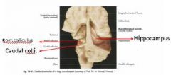
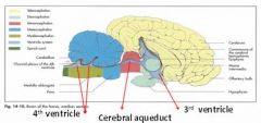
Different parts of Mesencephalon (mid brain) |
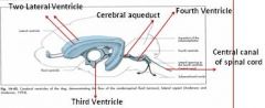
1. Tectum: Roof of mesencephalon and comprises corpora quadrigemina (rostral colliculus and caudal colliculus). 2. Tegmentum: Floor of mesen-cephalon. It contains nucleus of cranial nerve III and IV. 3. Cerebral aqueduct: a channel which connect 3rd ventricle rostrally and 4th ventricle caudally. 4. Cerebral peduncle: Visible on the ventral aspect of the brain just caudal to the optic tract. |
|
|
Mesencephalon Diagram |
|
|
|
Rhombencephalon (hind brain) |
Rhombencephalon consists of:
1. Metencephalon: Pons and Cerebellum
2. Myelencephalon: Medulla oblongata |
|
|
Rhombencephalon Diagram |
|
|
|
Brain - Horse |
|
|
|
Metencephalon |
1.Pons: a bulging structure at the ventral part of the brain and caudaul to the cerebral peduncle. 2.Tegmentum: It is the floor of the metencephalon. 3.Rostrum medullary vellum: It is the roof of the 4th ventricle. 4.Cerebellum: The cerebellum is the second largest part of the metencephalon and is located above the 4th ventricle. |
|
|
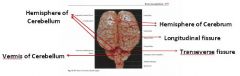
Structure of Cerebellum |
- The cerebellum consists of lobes, lobules and smallest folia. - Outer part is called cortex. - Inner part is called medulla. - Vermis located centrally. - Hemisphere on either side of the vermis. |
|
|
Function of Cerebellum |
Body balance, coordination of skeletal muscle, and control motor function. Control pyramidal and Extrapyramidal system of brain.
Deficit of cerebellar function: Loss of balance and in coordination of muscles.
|
|
|
Cerebellum Diagram |
|
|
|
Myelencephalon - Medulla Oblongata |
Medulla oblongata is continuous with pons cranially and spinal cord caudally.
-It comprises nuclei of the cranial nerves from VI (abducens) to XII (hypoglossal n.) - It also comprises nuclei of the respiratory and circulatory center. |
|
|
Function of Medulla Oblongata |
Control respiration, circulation, food intake, reflex for the protection of eye. |
|
|
Medulla Oblongata Diagram |
|
|
|
Brain Stem |
When cerebellum and cerebral hemisphere is removed the remaining part of the brain is called brain stem.
Brain stem consists of medulla oblongata, pons, and mid brain. |
|
|
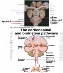
Clinical Neurology - Pyramidal System |
In higher vertebrates damage of the cortex of one side make permanent paralysis of the skeletal muscles of the contralateral side. Why ? Because some of the motor nerves originate from pyramidal cells of one cerebral cortex travel to the spinal cord (cortico-spinal tract) via the pyramid of the medulla to another side. |
|
|
Pyramidal System Diagram |
|
|
|
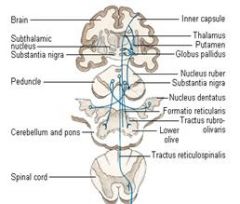
Clinical Neurology - Extrapyramidal System |
In extrapyramidal system the motor nerve originate from basal ganglia, substantia nigra, subthalamic nuclei, red nuclei, and reticular formation and don’t reach their targets by travelling through the pyramid of the medulla. Function and control: - Maintenance of posture - Coordination of muscular activity. |
|
|
Extrapyramidal System Diagram |
|
|
|
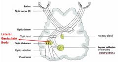
Vision Pathways |
|
|
|
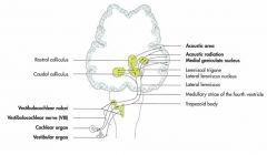
Hearing Pathways |
|
|
|
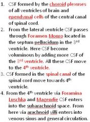
Cerebrospinal Fluid Circulation |
|
|
|
CSF Circulation Diagram |
|
|
|
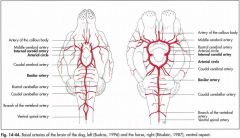
Blood Circulation of Brain |
Brain receives blood from two sources: 1.Ventral spinal artery, and 2.Internal carotid artery. These two arteries forms a circle like pattern at the ventral aspect of the brain known as circle of Willis. Branches: 1.Rostral cerebral artery 2.Middle cerebral artery 3.Caudal cerebral artery 4.Rostral cerebellar artery 5.Caudal cerebellar artery.
|
|
|
Blood Circulation of Brain Diagram |
|
|
|
Dorsal View of Brain of horse |
|
|
|
Dorsal View - Horse |
|
|
|
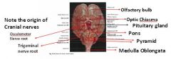
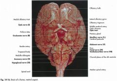
Ventral view of Brain of Horse |
|
|
|
Ventral view - Horse |
|
|
|
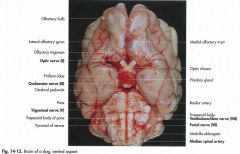
Ventral view - Dog |
|
|
|
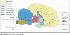
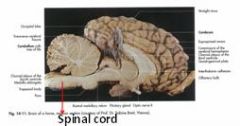
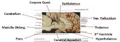
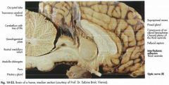
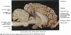
Sagittal view of brain of horse |
|
|
|
Median view - Horse |
|
|
|
Median view - Horse |
|
|
|
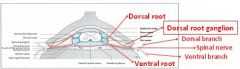
Spinal Cord and its Covering |
Meninges: Similar to the Covering of Brain Denticulate Ligament Thickening of Dura mater due to accumulation of collagen fibers in some areas of the dura.
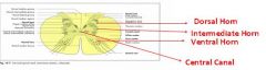
Root of Spinal Nerve: dorsal and ventral root originate from dorsal and ventral horn of the spinal cord.
|
|
|
Spinal roots Diagram |

|
|
|
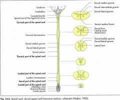
Different Segments of the Spinal Cord |

|
|
|
Segments of the Spinal Cord |
|
|
|
Conus medullaries and Cauda Equina |
•Conus Medullaries: The spinal cord tapers at the mid of the sacrum and looks like a cone shaped. •Cauda Equina: From the conus medullaries several spinal nerves originate to innervate in the muscles, fascia and skin of tail.
|
|
|
Formation of Spinal Nerve |
|
|
|
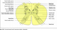
Grey and White mater of the spinal cord |
|
|
|
Mater of the spinal cord Diagram |
|