![]()
![]()
![]()
Use LEFT and RIGHT arrow keys to navigate between flashcards;
Use UP and DOWN arrow keys to flip the card;
H to show hint;
A reads text to speech;
30 Cards in this Set
- Front
- Back
|
When do we image a tumor? |
Deep tumors that cannot be evaluated Tumors involving vital structures Tumors involving bone Tumors adjacent to bone Treatment planning |
|
|
How do we determine tumor type? |
Histology or cytology. |
|
|
How many views should we taken and what technique of imagine would we use for checking metastasis in the lungs? |
3 views right and left lateral and dorsoventral using radiography. |
|
|
What would we use ultrasound and endoscopy for? |
Examining internal organs and things |
|
|
What is schintigraphy? |
Administering radio-pharmaceuticals which localise to the area of inflammation or tumor using technetium-99M |
|
|
When would we use CT scanning? |
CT imaging is used for planning treatment and also useful for lesions in the skull in combination with a contrast agent. |
|
|
When would we use MRI? |
CNS problems. Soft tissues not very helpful with cortical bone etc. |
|
|
What must we do to all tumors? |
Biopsy them!!! |
|
|
What type of biopsy should not lead to increased incidence of metastasis? |
Properly planned and preformed |
|
|
Things we must take from a biopsy? |
Is it something we can ignore Does it warrant further diagnosis Epithelial or mesenchymal Mast cell, lymphoma, soft tissue sarcoma |
|
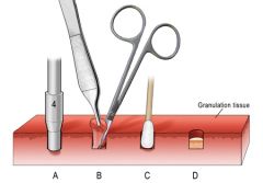
What type of technique is this? |
Dermal punch biopsy |
|
|
What is an excisional biopsy? |
Removal of whole tumor |
|
|
Benefits of excisional biopsy? |
May be curative, tissue margins and orientation evaluated. |
|
|
When would we do an excisional biopsy? |
If tumor is easily removed, area allows wide margins |
|
|
Downfall of excisional biopsy? |
Carries the same risk as removal under anesthetic |
|
|
What is an incisional biopsy? |
Removal of a small piece if hard to get the whole tumor |
|
|
Downfall of incisional biopsies? |
1) Not curative 2) Doesn't represent the entire tissue 3) Complicated further surgery 4)Seeding areas such as the bladder 5)Hemorrhage risk or viscous rupture |
|
|
How do we properly manage tissue? |
Avoid cautery and lasers, crush, dessication Label correctly and fix Margins important Impression smears Sample without formalin |
|
|
How can we mark out margins of a tumor? |
Inks and dyes, seperate slice in a container |
|
|
Reasons you might come to the wrong diagnosis with a biopsy? |
Poorly fixed/prepared Poor sampling Failure to recognize signs Species inexperience Lack of orientation Incomplete requisition |
|
|
What factors must we consider before taking a biopsy? |
Amount Position Type (Fluid) Anatomic location |
|
|
Benefits of cytology of a tumor? |
Easily recovered, little disruption to the tumor and surrounding tissues. Multiple sites can be sample during a single procedure Rapid mounting and staining |
|
|
When would we do a needle biopsy? |
Remove small core of tumor tissue from solid lesion without surgery. |
|
|
Advantages of needle biopsy? |
Recovers more tissue than needle aspirates. Tissue retains architecture Good for inaccessible sites without surgery Multiple samples removed at single approact. |
|
|
What are disadvantages of needle biopsies? |
More complications post biopsy than fine needle aspirates. Hemorrhage, oedema etc. |
|
|
When would we use a skin punch biopsy? |
Skin and superficial soft tissue tumors |
|
|
Benefits of skin punch biopsy? |
Recovers lots of tissue compared to FNA Tissue architecture preserved. Multiple samples through single approach. |
|
|
When would we do a bone marrow biopsy? |
To diagnose myeloid or lymphoid problems. |
|
|
Which areas can be used for a bone marrow biopsy and how should animal be place? |
Femur, humerus or iliac crest Femur and humerus lateral recumbency Sternal for iliac crest. |
|
|
How do we perform a bone marrow biopsy? |
1) Anaesthetise animal 2) Scrub and clip area to be done 3) Stab incision to site 4) Bone needle advanced into site and corkscrewed into bone 5) Stylet removed and 10 ml syringe attached 6) Work quick as bone marrow clots fast 7) Smear like a blood |