Reading...
![]()
Play button
![]()
Play button
![]()
Use LEFT and RIGHT arrow keys to navigate between flashcards;
Use UP and DOWN arrow keys to flip the card;
H to show hint;
A reads text to speech;
138 Cards in this Set
- Front
- Back
- 3rd side (hint)
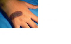
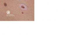
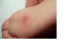
What type of primary lesion is this?
|
macule
Small (< 1cm), Flat, No Scale, Not palpable Ex: Freckle |
Small (< 1cm), Flat, No Scale, Not palpable
Ex: Freckle |
|
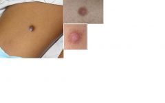
What type of primary lesion is this?
|
Patch
Large (> 1cm), Flat, No Scale, Not palpable Ex: Vitiligo |
Large (> 1cm), Flat, No Scale, Not palpable
Ex: Vitiligo |
|
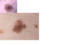
What type of primary lesion is this?
|
Papule
Small (<1cm), Solid lesion, Palpable Ex: mole, pimple, wart |
Small (<1cm), Solid lesion, Palpable
Ex: mole, pimple, wart |
|
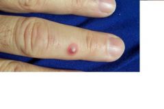
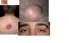
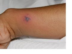
What type of primary lesion is this?
|
Nodule
Large (>1cm), Solid lesion, Palpable Ex: Large cysts, lymphoma, some melanoma, BCC |
Solid lesion, Palpable
|
|

What type of primary lesion is this?
|
Vesicle
Small (< 1 cm), Circumscribed, Elevated, Clear Fluid Filled Ex: Chicken Pox, herpes simplex |
Small (< 1 cm), Circumscribed, Elevated, Clear Fluid Filled
Ex: Chicken Pox, herpes simplex |
|

What type of primary lesion is this? (2nd)
|
Bulla
Large (> 1cm), Circumscribed, Elevated, Clear Fluid Filled Ex: severe sun burn, poison ivy, bulla disorders, some drug eruptions (EM, TEN) |
Large (> 1cm), Circumscribed, Elevated, Clear Fluid Filled
Ex: severe sun burn, poison ivy, bulla disorders, some drug eruptions (EM, TEN) |
|
What type of primary lesion is this?
|
Pustule
Circumscribed, Superficial, Contains a purulent exudate that may be white, yellow or green Ex: folliculitis, acne pimple, rosacea, insect bite reaction |
Circumscribed, Superficial, Contains a purulent exudate that may be white, yellow or green
Ex: folliculitis, acne pimple, rosacea, insect bite reaction |
|
What type of primary lesion is this?
|
Plaque Large (> 1cm), Usually well defined , Often formed by confluence of papules, Plateau like lesion
Ex: Psoriasis, Congenital Nevus (much larger than a centimeter), Ring worm (tinea corporis), Eczema |
Large (> 1cm), Usually well defined , Often formed by confluence of papules, Plateau like lesion
Ex: Psoriasis, Congenital Nevus (much larger than a centimeter), Ring worm (tinea corporis), Eczema |
|
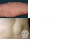
What type of primary lesion is this?
|
Wheal
Round and/or flat-topped, Edematous – resulting from infiltration, Pale red papule or plaque, Disappears within hours (will develop new spots, but individual spots are transient) Ex: Allergic reaction, hives |
Round and/or flat-topped, Edematous – resulting from infiltration, Pale red papule or plaque, Disappears within hours (will develop new spots, but individual spots are transient) Ex: Allergic reaction, hives |
|
What type of secondary lesion is this?
|
Keloid
- Firm, nodular scar, Extends beyond area of injury – history of keloid is indication for recurrance, any trauma to skin – removal could cause further keloid |
- Firm, nodular scar, Extends beyond area of injury – history of keloid is indication for recurrance, any trauma to skin
|
|
What type of secondary lesion is this
|
Thickened epidermis induced by scratching (itching), Skin lines are accentuated. Ex: Lichen Simplex chronicus, Chronic Eczema
|
Thickened epidermis induced by scratching (itching), Skin lines are accentuated. Ex: Lichen Simplex chronicus, Chronic Eczema
|
|
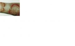
What type of secondary lesion is this?
|
Crust
– Dried residue of serum, pus or blood. Ex: impetigo, scab |
Dried residue of serum, pus or blood. Ex: impetigo, scab
|
|
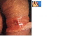
What type of secondary lesion is this?
|
Ulcer
Deeper loss of tissue surface (epidermis and dermis), Always heals with scar formation. Ex: bed sore, stasis dermatitis |
Deeper loss of tissue surface (epidermis and dermis), Always heals with scar formation. Ex: bed sore, stasis dermatitis
|
|
|
What is this??
Loss of skin surface (epidermis), Surface is moist, but does not bleed, Does not leave scar – skinning your knee |
Erosion
|
|
|
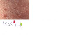
What type of secondary lesion is this?
|
Scale
– Thin flake of exfoliated dermis, Excess dead epidermal cells Ex: Dandruff, dry skin, tinea (fungal), actinic keratosis |
Thin flake of exfoliated dermis, Excess dead epidermal cells
Ex: Dandruff, dry skin, tinea (fungal), actinic keratosis |
|
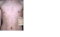
Identify.
|
ACNE
Comedones – Open – blackheads – Closed – whiteheads – Can extract with extractor but will reoccur without treatment Papules – pink acne bump Papulopustule – bump comes to head Cystic – larger, scar and pores are clogged deeper Nodular – same as above Caring for acne – Cleansing – no more than 2-3 times a day, use only hands, wash cloths increase oil – soap plays a small role and unless skin is extremely oily a mild soap should always be used to limit irritation – Sunlight – can help temporarily, will dry up oil glands and bacteria. But exfoliation after tan will clog pores and increase acne – Cosmetics – doesn’t matter as long as oil free – Don’t pick!! – leads to scarring and inflammation |
|
|
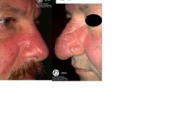
Identify.
|
Rosacea.
|
|
|

Identify
|
SEBORRHEIC DERMATITIS
|
|
|
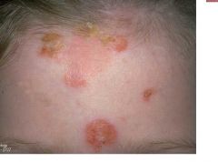
Identify
|
Seborrheic Dermatitis
|
|
|

Identify.
|
Seborrheic Dermatitis
|
|
|

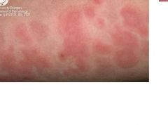
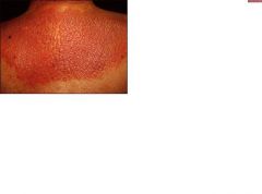
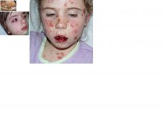
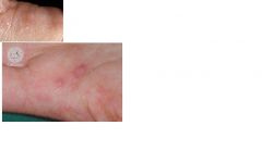
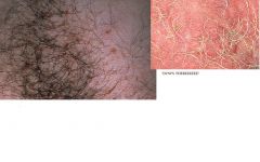
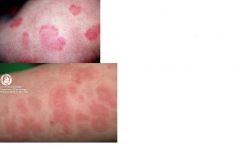
Identify
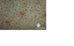
|
Guttate Psoriasis
Guttate: Appears as small red scaly paps or plaques on the skin |
Appears as small red scaly paps or plaques on the skin
type of psoriasis |
|
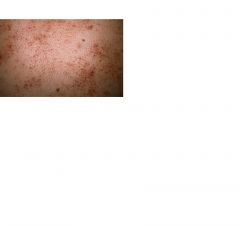
Identify
|
Guttate Psoriasis
Guttate: Appears as small red scaly paps or plaques on the skin |
type of psoriasis
|
|

Identify.
|
Psoriasis
|
|
|
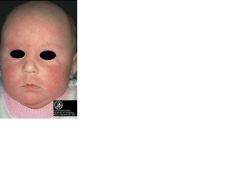
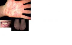
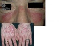
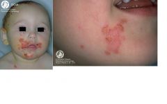
Identify.
|
atopic dermatitis
– severe itching!!! In infants often seen as an oozing, red, crusting condition on face and scalp because they keep scratching After infancy, skin tends to be less red. Is dry, pink, with scale or thickening. May see bleeding or infection. |
– severe itching!!!
In infants often seen as an oozing, red, crusting condition on face and scalp because they keep scratching After infancy, skin tends to be less red. Is dry, pink, with scale or thickening. May see bleeding or infection. |
|
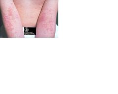
Identify.
|
atopic dermatitis
In teens and adults, most commonly occurs on hands and feet. Other common areas; ACF (infants), back of knees (infants), ankles, wrists, face, neck, and upper chest. Hypo/hyper pigmentation – even after clears will see hypopigmentation, hyper because of itching and exfoliating Symptoms of atopic dermatitis VERY ITHCY Itch, scratch, rash cycle. “the itch that rashes” Burning and stinging from constant itching. Diagnosis criteria for atopic dermatitis = pruritus, morphology, distrubution and must consider: (1) a personal or family history of atopic disease (asthma, allergic rhinitis, atopic dermatitis), (2) xerosis-ichthyosis, (3) facial pallor with infraorbital darkening, (4) elevated serum IgE, (5) fissures under the ear lobes, (6) a tendency toward nonspecific hand dermatitis, (7) a tendency toward repeated skin infections, and (8) nipple eczema. |
VERY ITHCY
Itch, scratch, rash cycle. “the itch that rashes” Burning and stinging from constant itching. |
|

Identify.
|
VERY ITCHY
Itch, scratch, rash cycle. “the itch that rashes” Burning and stinging from constant itching. In teens and adults, most commonly occurs on hands and feet. Other common areas; ACF (infants), back of knees (infants), ankles, wrists, face, neck, and upper chest. Hypo/hyper pigmentation – even after clears will see hypopigmentation, hyper because of itching and exfoliating |
VERY ITCHY
Itch, scratch, rash cycle. “the itch that rashes” Burning and stinging from constant itching. |
|

Identify
|
VERY ITCHY
Itch, scratch, rash cycle. “the itch that rashes” Burning and stinging from constant itching. In teens and adults, most commonly occurs on hands and feet. Other common areas; ACF (infants), back of knees (infants), ankles, wrists, face, neck, and upper chest. Hypo/hyper pigmentation – even after clears will see hypopigmentation, hyper because of itching and exfoliating |
VERY ITCHY
Itch, scratch, rash cycle. “the itch that rashes” Burning and stinging from constant itching. |
|

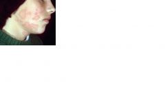
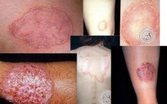
Identify
|
lichen simplex chronicus
|
Very itchy.
Itch, scratch, rash cycle. “the itch that rashes” Burning and stinging from constant itching. |
|

Identify
|
LICHEN SIMPLEX CHRONICUS (circumscribed neurodermatitis)
Is a localized form of lichenification usually occurring in circumscribed scaly plaques. Seen in people with chronic excema (atopic dermatitis) Seen on nape of neck, wrists, external surfaces of forearms, lower legs, scrotum and vulva Results from chronic itching, rubbing and scratching |
Is a localized form of lichenification usually occurring in circumscribed scaly plaques.
Seen in people with chronic excema (atopic dermatitis) Seen on nape of neck, wrists, external surfaces of forearms, lower legs, scrotum and vulva Results from chronic itching, rubbing and scratching |
|
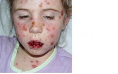
Identify.
|
Pityriasis Rosea
Mild, acute inflammatory disease – Very common condition Has a characteristic course. First lesion appears, resembles ringworm = fawn colored, scaly oval eruption. Two weeks later, numerous smaller “spots” appear. Etiology and epidemiology of Pityriasis Rosea Cause is unknown. A virus may cause the rash. Most common age 10-35 y/o Usually occurs in spring and fall Rash can last from several weeks to several months. - will fade away on its own with no treatment - typically won’t reoccur |
Mild, acute inflammatory disease – Very common condition
Has a characteristic course. First lesion appears, resembles ringworm = fawn colored, scaly oval eruption. Two weeks later, numerous smaller “spots” appear. Etiology Cause is unknown. A virus may cause the rash. Most common age 10-35 y/o Usually occurs in spring and fall Rash can last from several weeks to several months. - will fade away on its own with no treatment - typically won’t reoccur |
|
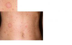
Identify
|
Pityriasis Rosea
|
CHRISTMAS TREE PATTERN
|
|

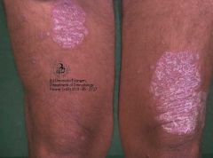
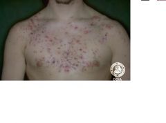
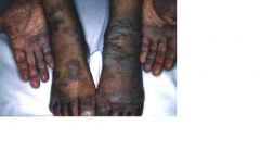
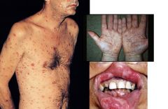
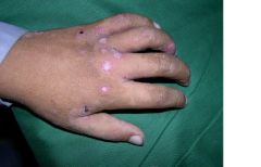
what are all of these examples of....
|
psoriasis
Signs and symptoms Usually starts as little red bumps, gradually grows larger and thick white scale forms (scales shed) Can have mild itching Can be very disfiguring – knees and elbows most common Arthritis Psychosocial impact can be major factor |
types:
Plaque: Most common form of the disease Guttate: Appears as small red scaly paps or plaques on the skin Inverse: Occurs in armpits, groin and skin folds – need to make sure not yeast infection because will get worse with steroid treatment Pustular: White blisters surrounded by red skin – tobacco use indicated as risk (hand) Erythrodermic: Intense redness over large areas (back), emergency because whole skin is inflamed, can be adverse drug infection and |
|
Identify
|
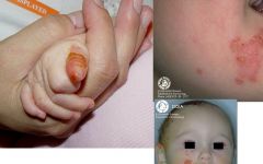
Impetigo
Superficial infection (involving the epidermis) caused by Staphylococcus Aureus or Group A Strep. Infection may arise as primary infections in minor superficial breaks in the skin or secondary infections of preexisting dermatoses |
Superficial infection (involving the epidermis) caused by Staphylococcus Aureus or Group A Strep.
Infection may arise as primary infections in minor superficial breaks in the skin or secondary infections of preexisting dermatoses |
|
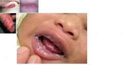
Identify.
|
Impetigo
Superficial infection (involving the epidermis) caused by Staphylococcus Aureus or Group A Strep. Infection may arise as primary infections in minor superficial breaks in the skin or secondary infections of preexisting dermatoses |
Superficial infection (involving the epidermis) caused by Staphylococcus Aureus or Group A Strep.
Infection may arise as primary infections in minor superficial breaks in the skin or secondary infections of preexisting dermatoses |
|
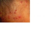
Identify
|
Folliculitis
Usually non-tender May be pruritic Red Papule or pustule confined to the hair follicle. May find erythema around hair follicle Sycosis = deep seated, chronic and recalcitrant on head and neck |
Usually non-tender
May be pruritic Red Papule or pustule confined to the hair follicle. May find erythema around hair follicle Sycosis = deep seated, chronic and recalcitrant on head and neck |
|
Identify.
|
Folliculitis
|
|
|
Identify.
|
pityrosporum folliculitis
|
|
|
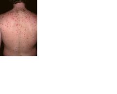
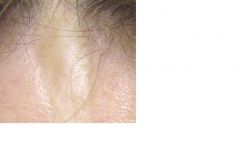
What are all of these examples of..?
|
Folliculitis
|
|
|
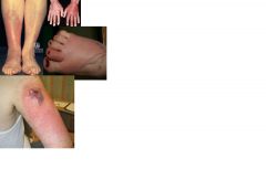
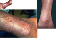
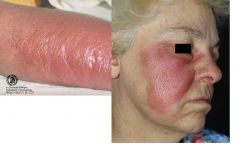
what are these examples of?
|
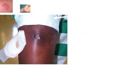
cellulitis
|
|
|
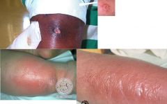
the top and the bottom are different things theres 2 answers on this card
|
Erysipelas on bottom
mrsa on top |
|
|
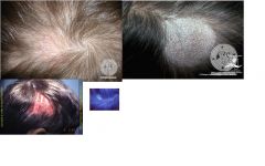
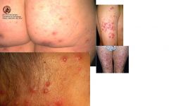
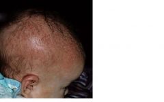
Identify.
|
Tinea Capitus
|
|
|
|
Candidiasis
|
|
|
|
common warts
|
|
|
|
flat warts
|
|
|
|
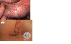
genital warts
|
|
|
|
herpes simplex
|
|
|
|
herpes zoster
|
|
|

|
Molluscum Contagiosum
|
|
|
|
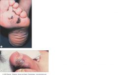
plantar warts
|
|
|
|
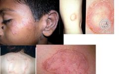
Tinea Corporis
|
|
|

|
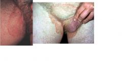
Tinea Cruris
|
|
|
|
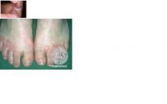
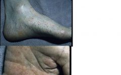
Tinea Pedis
|
|
|
|
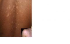
Tinea Versicolor
|
|
|
|
Tinea Cruris
|
|
|
|
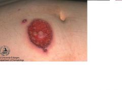
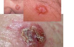
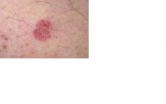
basal cell carcinoma
|
|
|
|
basal cell carcinoma
|
|
|
|
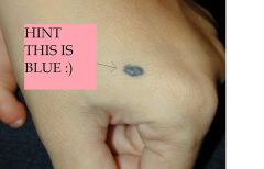
blue nevi
|
|
|
|
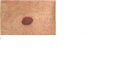
common nevi
|
|
|
|
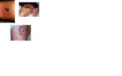
congenital nevi
|
|
|
|
dermatofibroma
|
|
|
|
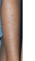
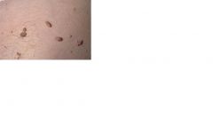
dysplastic nevi
|
|
|
|
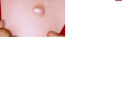
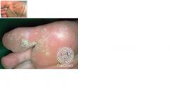
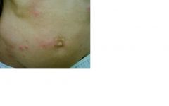
epidermal inclusion cysts
|
|
|
|
halo nevi
|
|
|
|
lipoma
|
|
|
|
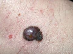
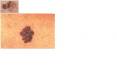
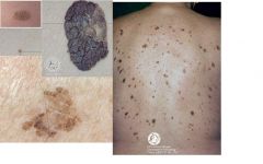
malignant melanoma
|
|
|
|
malignant melanoma
|
|
|
|
malignant melanoma
|
|
|

|
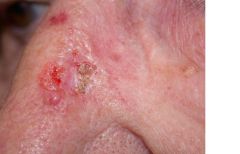
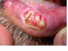
squamous cell carcinoma
|
|
|
|
squamous cell carcinoma
|
|
|
|
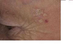
Seborrheic Keratosis
|
|
|
these are...
|
skin tags
|
|
|
|
squamous cell carcinoma
|
|
|
|
look at acanthosis nigricans, its not in here but i dont feel like finding the picture :)
|
damn thing says i need an answer to save this card.. here it is:
ALLIE (im the answer to everything) |
|
|
|
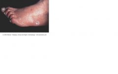
arterial leg ulcers
|
|
|
|
bed bugs
|
|
|

who did this?
|
black widow spider
|
|
|
what was crawling in this persons bed that night?
|
brown recluse spider
|
|
|
creeping eruptions?? yeh im giving you the answer because its not in the ppt,or that i could find, just in last years lecture notes
|
ALLIE
|
|
|

|
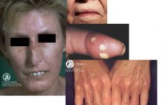
Dermatomyositis
|
|
|
|
Dermatomyositis
|
|
|
|
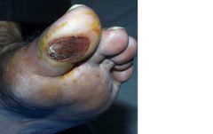
diabetic ulcer
|
|
|
|
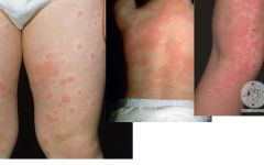
drug induced acute urticaria
|
|
|
|
erythema migrans rash of lyme
|
|
|
|
erythema multiforme major and minor
dont know which one is which lol |
|
|
|
exanthematous drug eruption
|
|
|
who did this?
|
fire ants
|
|
|

|
fleas.
|
|
|
|
gonococcemia
|
|
|
|
gonococcemia
|
|
|

|
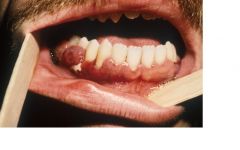
kaposi sarcoma
|
|
|
|
kaposi sarcoma
|
|
|
|
kaposi sarcoma
|
|
|
|
scleroderma
|
|
|

|
lyme
|
|
|
|
Meningococcemia
|
|
|
|
Meningococcemia
|
|
|

|
lice found on the body
|
|
|
|
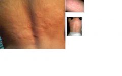
urticaria
|
|
|
|
urticaria
|
|
|
|
tertiary syph
|
|
|
|
systemic lupus
|
|
|
|
SJS TEN .. listed under both
TEN when >30% BSA covered SJS when <10% BSA covered |
|
|
|
stasis ulcers
|
|
|
|
SJS TEN .. listed under both
TEN when >330% BSA covered SJS when <10% BSA covered |
|
|
|
secondary syph
|
|
|
|
scleroderma
|
|
|

|
scabies
|
|
|
|
scabies
|
|
|
|
scabies
|
|
|
|
rocky mtn spotted fever
|
|
|
|
primary syph
|
|
|

|
physical urticaria
|
|
|
|
pediculosis pubis
pubic lice |
|
|
|
head lice- pediculosis capitis
|
|
|
|
pediculosis capitis
head lice |
|
|

|
bacterial endocarditis
|
|
|

|
bacterial endocarditis
|
|
|
|
dermatomyositis
|
|
|
|
localized scleroderma
|
|
|

|
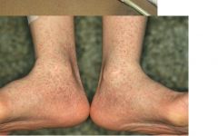
diaper dermatitis
|
|
|
|
erysipelas
|
|
|
|
folliculitis
|
|
|
|
herpes simplex
|
|
|
|
herpes zoster
|
|
|
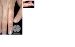
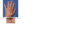
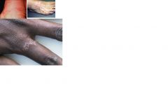
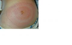
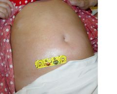
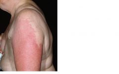
what type of lesion is this
|
wheal
|
|
|

|
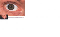
vitiligo
|
|
|
|
tinea corporis
|
|
|
|
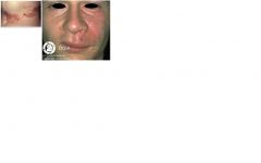
sebhorrheic dermatitis
|
|
|
|
|
s aureus cellulitis
|
|
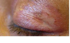
|
|
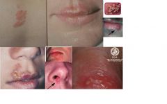
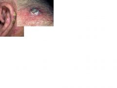
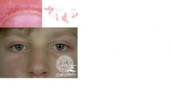
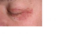
periorbital dermatitis
|
|
|
|
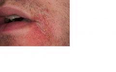
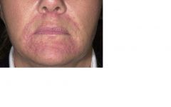
perioral dermatitis
|
|
|
|
s aureus cellulitis
|
|
|
|
nummular dermatitis
|
|
|
|
mrsa
|
|
|

|
lichen simplex chronicus
|
|
|
|
impetigo
|
|
|
|
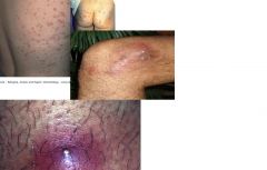
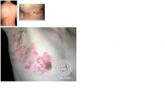
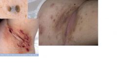
Hidradenitis suppurativa
|
|
|
|
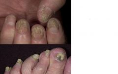
Onychomycosis (tinea unguium)
|
|