![]()
![]()
![]()
Use LEFT and RIGHT arrow keys to navigate between flashcards;
Use UP and DOWN arrow keys to flip the card;
H to show hint;
A reads text to speech;
78 Cards in this Set
- Front
- Back
|
What are X-rays? |
Ionized electromagnetic radiation that has shorter wavelengths than visible light |
|
|
What is the difference between SOFT X-RAYS and HARD X-RAYS? |
Soft x-rays are closer to UV light wavelength Hard x-rays are closer to gamma wavelength |
|
|
How are x-rays measured? |
Roentgen, rad, rem |
|
|
What do curie's measure? |
Nuclear decay per second in a sample of radionuclide |
|
|
What is a radiologist? |
physiciansthat train and subspecialize in different types of imaging |
|
|
What are radiographers? |
technicianstrained to perform specific types of imaging |
|
|
What is radiology? |
amedical specialty that uses of imaging to both diagnose and treat diseasevisualized within the human body |
|
|
What imaging technologies do Radiologists use to diagnose or treat diseases? |
X-ray Radiography Ultrasound CT - computed tomography Nuclear medicine PET - positron emission tomography MRI - magnetic resonance imaging |
|
|
What is interventional radiology? |
a subspecialty of radiology in whichdisease is diagnosed and treated nonoperatively. |
|
|
Who takes the medical images? |
The radiographer or radiologic thechnologist |
|
|
What role do clinicians play in the imaging system? |
1. interpretation of the image 2. Correlate clinical findings with imaging information |
|
|
What is the clinician's responsibility? |
Always recognize that if the result of any imaging study does not fit the physical findings, further clinical evaluation and diagnostic investigations are needed |
|
|
What is the purpose of radiology? |
noninvasivetest used to identify and screen for lung or heart disease, fractures,dislocations,bone growth,foreign objects |
|
|
How is radiology described? |
x-rayphotons pass through the body and are captured on plain film or digitally@9?Bo |
|
|
What is a radiograph |
a recorded image of an anatomic part acquired by the passage of x-rays through the body, used to assist with the diagnosis of musculoskeletal problems |
|
|
What do radiographs produce? |
2 dimensional planar images, therefore at least 2 films are required to localize the lesion |
|
|
Is a film an xray? |
a film is NOT an x-ray. X-rays are invisible things that float aroundin the air. The thing you view on theview box is called a Radiographic Film Interpretation or simply a Radiograph>D |
|
|
What is a conventional radiograph? |
aradiograph made without contrast enhancement or other equipment modification |
|
|
What are the basic components of x-ray technology? |
x-ray tube and the image receptor |
|
|
What are the categories of receptors used to capture the x-ray image? |
1. Film/screen 2. Fluoroscopy 3. Digital |
|
What is this category of receptor? |
Digital |
|
What category of receptor is this? |
Film/screen |
|
What category of receptor is this? |
Fluoroscopy |
|
|
What 3 things do you need to produce an x-ray? |
1. A source of electrons 2. A force to move them 3. Something to stop them |
|
|
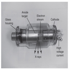
What is in an x-ray tube? |
A cathode and an anode in a vacuum glass tube |
|
|
How does the X-ray Tube work? |
1. The cathode has a heated thoriated tungsten filament that makes the ELECTRONS when kilovoltage is applied 2. The electrons strike the anode and decelerate creating the x-rays due to energy conversion |
|
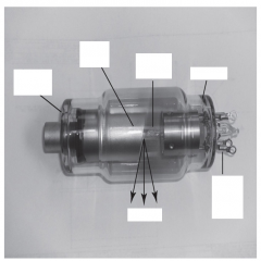
Label the xray tube |
|
|
|
How is a radiograph produced? |
•Thex-rays passes through a patient after being attenuated •Theremnant beam has an aerial image of the patient •Theremnant beam interacts with film (in the image receptor) •Thefilm is called a “latent image” because it must be developed •Thevisual image is a Radiograph |
|
|
What is a film/screen? |
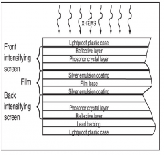
The combination of film and crystal coated intensifying screens |
|
|
Where is the film located? |
in the cassette |
|
|
What is a cassette? |
a lightproof plastic cases that holds the film between layers |
|
|
What does fluroscopy images require in order to create an image? |
High doses of radiation |
|
|
How is radiodensity determined? |
a structure's material composition and its thickness |
|
|
What does it mean when you say, a structure is more radiodense? |
The object will absorb more x-rays and leave the film white (EX: Bones) |
|
|
What does it mean when you say, a structure is less radiodense? |
The object will not absorb as much X-radiation allowing penetration to the film turning it black (EX: air) |
|
|
What does Radiolucent mean? |
Anything that lets x-rays pass, these images are made visible with contrast medium. Used for patients with osteoporotic bone, tumors, and infections |
|
|
What is Radiopaque? |
Anything that blocks x-rays. Usually metal implants, shields, and dental work. Not used for human tissue except for things like calcified gallstones |
|
|
What is radiodense? |
Substances that will not allow x-rays or radiation to pass |
|
|
How will air, fat, water, and bone appear on a radiograph? |
Air = black Fat = darker grey Water = Lighter Grey Bone = white |
|
Identify each tissue. |
1. Air or gas 2. Fat 3. Water, muscle, and other soft tissue 4. Bone |
|
|
What is radiographic density on film? |
the amount of darkness on the radiograph |
|
|
What affects the radiographic density? |
The amount of time of exposure measured in milliamperage seconds |
|
|
How is the quality of a radiograph evaluated? |
1. Density (mAs, time of exposure) 2. Contrast (kVp, the force pushing the x-rays through the body) 3. Detail (how clear or sharp the object appears) 4. Distortion (no movement) |
|
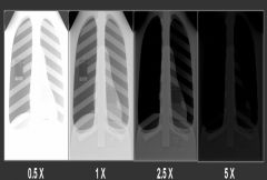
Which of these films are properly exposed? |
1X |
|
|
True or False: the higher the kVp = the more mAs needed |
False. mAs and kVp are inverse. Higher kVp = less mAs needed |
|
|
What is the best method to get a good radiograph? |
Highest kVp (force) and lowest mAs (time exposed to the radiation) |
|
What view is this? |
Swimmer's View |
|
|
What is recorded detail? |
sharpness, resolution, definition of the shape |
|
|
What affects detail in the radiograph? |
Movement, distance from the tube, and beam size |
|
|
What is radiographic distortion? |
difference between the patient object being filmed and the recorded image |
|
|
What is the Collimator? |
a device for limiting the size and shape of a radiation beam, used to reduce scatter radiation. Located on the x-ray tube |
|
|
What is required to view all three dimensions of a structure? |
at least 2 images as close to 90 degrees to each other as possible |
|
|
What is an invalid image? |
ONE VIEW IS NO VIEW!!!! |
|
|
What are the 3 positions and projections to provide greatest visualization with minimal radiation exposure? |
1. AP 2. Lateral 3. Oblique |
|
what is the view on the left and the view on the right? |
Left = P to A view Right = Lateral view |
|
|
Position refers to what 2 things? |
1. The patients general position: standing, seated, supine, prone, erect, recumbent, trendelenburg 2. Which body part is closest to the image receptor (bucky) |
|
|
What does it mean if the image was taken "upright"? |
Patient was seated or standing, Usually done to see "what happens in weight bearing/functional view" |
|
|
What does it mean if the image was taken in "decubitus"? |
specifically used to describe pt in a horizontal position AND a horizontal x-ray beam Used to view fluid/air levels in the chest and abdomen |
|
|
what is projection? |
the path the xray beam takes as it travels from the tube, through the patient, and to the bucky |
|
|
What are the 4 most common projections? |
1. AP anteroposterior 2. PA posteroanterior 3. Lateral 4. Oblique (taken in the spine, wrist, hand, ankle, and foot) |
|
|
True or False. The right and left tell you what side is closest to the X-ray. |
False. The right and left tell you what side is closes to the Bucky |
|
|
The bony structure that is at a (blank) degree angle to the x-ray tube will appear the most clearly defined and least distorted. |
at a 90 degree angle to the x-ray tube
|
|
|
True or False. The farther the structure is to the film plate/receptor, the less distortion and greater definition will be perceived. |
False. The CLOSER the structure is to the film plate the less distortion and greater definition will be perceived. |
|
|
What are the standardizing distances radiographers strive to keep consistent to ensure good quality radiographs? |
40 inches for most radiographers Laterals of the C-spine and chest and P-A chest it is 72 inches |
|
|
When viewing film, the person is oriented on the monitor in anatomic position except for what two structures? |
The hand and foot. They are normally place with the digits and toes directed upward in a P-A view. |
|
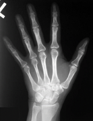
This a radiograph of which hand? |
LEFT DUH!!! |
|
|
The radiograph should be placed on the view box as if the practitioner is looking at the patient face to (blank). |
Face lol. |
|
|
Hands and feet are placed with the digits pointing up and from what projection? |
P to A projection |
|
|
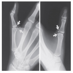
What is placed near the patient's point of pain on the radiograph as a better reference for the physician? |
Sometimes a BB is taped to the place near the patient's point of pain |
|
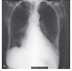
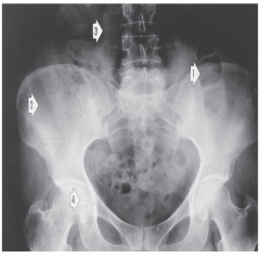
What caused the heart to appear enlarged? |
the patient was angled incorrectly to the central ray, causing a distortion of the internal organs |
|
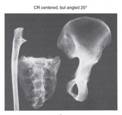
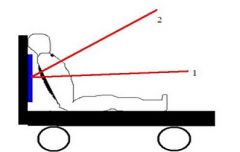
an example of the central ray angled at 25 degree |
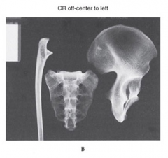
An example of the central ray being placed too far to the left and off center to the objects |
|
|
What are the advantages of radiology? |
quick, easy, portable, and relatively inexpensive |
|
|
What the disadvantages of radiology? |
ionizing radiation to the body, poor at visualizing soft tissues, and small fractures |
|
|
Why do we as PT's study radiology? |
- More comprehensive patient evaluation - Professional communication with other health providers to improve patient care - Research - Patient Education |
|
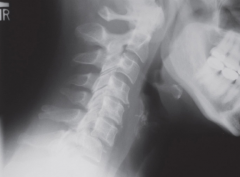
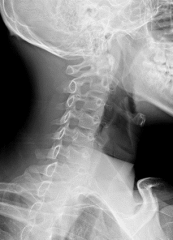
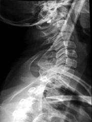
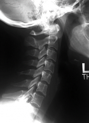
What view was this radiograph taken in and what is happening in the image? |
Lateral cervical spine Anterolisthesis of C6 on C7 |
|
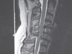
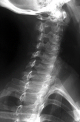
Is this an X-ray? What is the image demonstrating? |
This is a Sagittal MRI demonstrating C6 anterolisthesis, spinal canal encroachment, and moderate spinal cord compression. |
|
Label all structures you see...lol. |
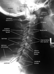
Stupidest slide:) |
|
|
One drunk man says to the other, my wife drove me to drinking... |
The other man says, "you're lucky! my wife makes me walk" |