Reading...
![]()
Play button
![]()
Play button
![]()
Use LEFT and RIGHT arrow keys to navigate between flashcards;
Use UP and DOWN arrow keys to flip the card;
H to show hint;
A reads text to speech;
28 Cards in this Set
- Front
- Back
|
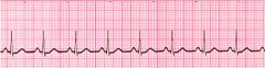
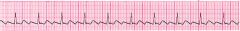
Normal Sinus Rhythm
"Rhythm - Regular Rate - (60-100 bpm) QRS Duration - Normal P Wave - Visible before each QRS complex P-R Interval - Normal (<5 small Squares. Anything above and this would be 1st degree block) Indicates that the electrical signal is generated by the sinus node and travelling in a normal fashion in the heart. Treatment: PT Stable - no Cardiac treatment PT unstable - Possible PEA - Fix underlining causes" |
|
|
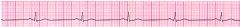
Sinus Bradycardia
"A heart rate less than 60 beats per minute (BPM). Rhythm - Regular Rate - less than 60 beats per minute QRS Duration - Normal P Wave - Visible before each QRS complex P-R Interval - Normal TREATMENT Usually benign and often caused by patients on beta blockers PT Stable - (Could be normal for runners / physically fit person) PT unstable - Possible cause of Brain injury (contra indication of D50 due to increased intracranial Pressure - Possible Over dose on Beta Blockers - Glucagon Dose - 1 to 2 mg - Possible low glucose -consider D50 25 mg (or 12.5 half dose) - Possible Pacing required - Speed up rate with Atropine .5 mg Rapid push" |
|
|
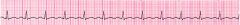
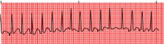
Sinus Tachycardia
"An excessive heart rate above 100 beats per minute (BPM) which originates from the SA node. Causes include stress, fright, illness and exercise. Not usually a surprise if it is triggered in response to regulatory changes e.g. shock. But if their is no apparent trigger then medications may be required to suppress the rhythm - Regular Rate - More than 100 beats per minute QRS Duration - Normal P Wave - Visible before each QRS complex P-R Interval - Normal The impulse generating the heart beats are normal, but they are occurring at a faster pace than normal. Seen during exercise TREATMENT PT Stable - Could be normal - if caused from a known stress, freight, illness or exercise - no treatment PT unstable - possible cardio vert with " |
|

|
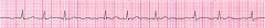
Supraventricular Tachycardia (SVT) Abnormal
"A narrow complex tachycardia or atrial tachycardia which originates in the 'atria' but is not under direct control from the SA node. SVT can occur in all age groups Rhythm - Regular Rate - 140-220 beats per minute QRS Duration - Usually normal P Wave - Often buried in preceding T wave P-R Interval - Depends on site of supraventricular pacemaker Impulses stimulating the heart are not being generated by the sinus node, but instead are coming from a collection of tissue around and involving the atrioventricular (AV) node Treatment: PT Stable - PT unstable - |
|
|
Atrial Fibrillation
"Many sites within the atria are generating their own electrical impulses, leading to irregular conduction of impulses to the ventricles that generate the heartbeat. This irregular rhythm can be felt when palpating a pulse Rhythm - Irregularly irregular Rate - usually 100-160 beats per minute but slower if on medication QRS Duration - Usually normal P Wave - Not distinguishable as the atria are firing off all over P-R Interval - Not measurable The atria fire electrical impulses in an irregular fashion causing irregular heart rhythm Treatment: PT Stable - PT unstable - " |
|
|
Atrial Flutter
"Rhythm - Regular Rate - Around 110 beats per minute QRS Duration - Usually normal P Wave - Replaced with multiple F (flutter) waves, usually at a ratio of 2:1 (2F - 1QRS) but sometimes 3:1 P Wave rate - 300 beats per minuteP-R Interval - Not measurable As with SVT the abnormal tissue generating the rapid heart rate is also in the atria, however, the atrioventricular node is not involved in this case. Treatment: PT Stable - PT unstable - High Flow O2, IV, Cardisem (deltisem) 15 to 20 mg, 20 to 25mg " |
|

|
1st Degree AV Block
"1st Degree AV block is caused by a conduction delay through the AV node but all electrical signals reach the ventricles. This rarely causes any problems by itself and often trained athletes can be seen to have it. The normal P-R interval is between 0.12s to 0.20s in length, or 3-5 small squares on the ECG. Rhythm - Regular Rate - 60 to 100 QRS Duration - Normal P Wave - Ratio 1:1 P Wave rate - Normal P-R Interval - Prolonged (>5 small squares) Treatment: PT Stable - PT unstable - |
|

|
2nd Degree Block Type 1 (Wenckebach) (Mobitz I)
"Another condition whereby a conduction block of some, but not all atrial beats getting through to the ventricles. There is progressive lengthening of the PR interval and then failure of conduction of an atrial beat, this is seen by a dropped QRS complex. Rhythm - Regularly irregular Rate - Normal or Slow QRS Duration - Normal P Wave - Ratio 1:1 for 2,3 or 4 cycles then 1:0. P Wave rate - Normal but faster than QRS rate P-R Interval - Progressive lengthening of P-R interval until a QRS complex is dropped Treatment: PT Stable - PT unstable - " |
|

|
2nd Degree Block Type 2 (Mobitz II)
"When electrical excitation sometimes fails to pass through the A-V node or bundle of His, this intermittent occurrence is said to be called second degree heart block. Electrical conduction usually has a constant P-R interval, in the case of type 2 block atrial contractions are not regularly followed by ventricular contraction Rhythm - Regular Rate - Normal or Slow QRS Duration - Prolonged P Wave - Ratio 2:1, 3:1 P Wave rate - Normal but faster than QRS rate P-R Interval - Normal or prolonged but constant Treatment: PT Stable - PT unstable - " |
|

|
3rd Degree Block
"3rd degree block or complete heart block occurs when atrial contractions are 'normal' but no electrical conduction is conveyed to the ventricles. The ventricles then generate their own signal through an 'escape mechanism' from a focus somewhere within the ventricle. The ventricular escape beats are usually 'slow' Rhythm - Regular Rate - Slow QRS Duration - Prolonged P Wave - Unrelated P Wave rate - Normal but faster than QRS rate P-R Interval - Variation Complete AV block. No atrial impulses pass through the atrioventricular node and the ventricles generate their own rhythm Treatment: PT Stable - PT unstable - " |
|

|
Bundle Branch Block – (Extra)
"Bundle Branch Block – (Extra) Abnormal conduction through the bundle branches will cause a depolarization delay through the ventricular muscle, this delay shows as a widening of the QRS complex. Right Bundle Branch Block (RBBB) indicates problems in the right side of the heart. Whereas Left Bundle Branch Block (LBBB) is an indication of heart disease. If LBBB is present then further interpretation of the ECG cannot be carried out. Rhythm - Regular Rate - Normal QRS Duration - Prolonged P Wave - Ratio 1:1P Wave rate - Normal and same as QRS rate P-R Interval - Normal Treatment: PT Stable - PT unstable - " |
|

|
Premature Ventricular Complexes (PVC)
"Due to a part of the heart depolarizing earlier than it should Rhythm - Regular Rate - Normal QRS Duration - Normal P Wave - Ratio 1:1 P Wave rate - Normal and same as QRS rate Treatment: PT Stable - PT unstable - |
|

|
Junctional Rhythms
" Rhythm - Regular Rate - 40-60 Beats per minute QRS Duration - Normal P Wave - Ratio 1:1 if visible. Inverted in lead IIP Wave rate - Same as QRS rate P-R Interval - Variable Treatment: PT Stable - PT unstable - " |
|

|
Ventricular Tachycardia (VT) Abnormal
"Rhythm - Regular Rate - 180-190 Beats per minute QRS Duration - Prolonged P Wave - Not seen Results from abnormal tissues in the ventricles generating a rapid and irregular heart rhythm. Poor cardiac output is usually associated with this rhythm thus causing the pt to go into cardiac arrest. Shock this rhythm if the patient is unconscious and without a pulse Treatment: PT Stable - PT unstable - " |
|

|
Ventricular Fibrillation (VF) Abnormal
"Disorganized electrical signals cause the ventricles to quiver instead of contract in a rhythmic fashion. A patient will be unconscious as blood is not pumped to the brain. Immediate treatment by defibrillation is indicated. This condition may occur during or after a myocardial infarct. Rhythm - Irregular Rate - 300+, disorganized QRS Duration - Not recognizable P Wave - Not seen This patient needs to be defibrillated!! QUICKLY Treatment: PT Stable - PT unstable - CPR, Pads on and Shock ASAP, CPR, Bag valve Mask + High Flow O2, Shock every 2 min if necessary, IV, EPI, 1mg 1:10,000, amiodarone 300 mg, EPI 1mg 1:10,000, amiodarone 150 mg, find and fix any H's and T's, maybe some D50, fluid boils, Narcan. then maybe call Medical Control and possibly call the code. " |
|

|
Asystole - Abnormal
"Rhythm - Flat Rate - 0 Beats per minute QRS Duration - None P Wave - None Carry out CPR!! Treatment: PT Stable - PT unstable - |
|

|
Myocardial Infarct (MI)
"Rhythm - Regular Rate - 80 Beats per minute QRS Duration - Normal P Wave - Normal S-T Element does not go isoelectric which indicates infarction. However this is not diagnostic unless associated with a 12 lead ECG Treatment: PT Stable - PT unstable - " |
|

|
Paced Rhythm
"Rhythm - Irregular Rate - 60 Beats per minute QRS Duration - Normal P Wave - Normal S-T Element does not go isoelectric which indicates infarction. However this is not diagnostic unless associated with a 12 lead ECG Treatment: PT Stable - PT unstable - |
|

|
NSR with unifocal PVC
"Rhythm - irregular Rate – (60-100 bpm) QRS Duration – normal when present P Wave – visible before each QRS complex PVC in one direction Treatment: PT Stable - High Flow O2, SAMPLE History, IV, Fluid PT unstable - (Chest Pain) - High Flow O2, SAMPLE History, IV, Fluid boils, Aspirin, .4 nitro up to three times, Morphine 2mg" |
|

|
Normal Sinus with multifocal Couplet PVC
"Rhythm - irregular Rate – (60-100 bpm) QRS Duration – normal when present P Wave – visible before each QRS complex PVC in both directions Treatment: PT Stable - High Flow O2, SAMPLE History, IV, Fluid PT unstable - Chest Pain - High Flow O2, SAMPLE History, IV, Fluid boils, Aspirin, .4 nitro up to three times, Morphine 2mg" |
|

|
Junctional Brady
" Rhythm: Regular Rate: 40-60bpm P waves: Inverted or absent PR interval: Short or Absent QRS width: Normal Interventions: If hemodynamically stable = No tx-If not stable, tx like sinus Brady = Atropine Treatment: PT Stable - PT unstable - Atropine .5 mg rapid push" |
|

|
Slow V-Tach
"Wide complex regular rhythm with rate 40-120 Runs may last few minutes Treatment:*** Observe only! rhythm is self-terminating, NOT destabilizing If hypertensive, consider atrial pacing*** Danger: if you treat this with lidocaine or amiodarone or procainamide, rhythm will go to asystole, patient will die. Treatment: PT Stable - PT unstable - " |
|

|
Fine Ventricular Fibrillation
"Rhythm - chaotic Rate – 0 bpm QRS Duration – none present P Wave –absent or chaotic varying in size and shape. Treatment: PT Stable - PT unstable - |
|

|
Torsades de pointes
"Rhythm – irregular Rate – 60-100 bpm or 150-300 depending on where pace is coming from. Torsades de pointes is a polymorphic ventricular tachycardia that differs from general ventricular tachycardia in both appearance and cause. The phrase “torsades de pointes” means “twisting on a point” which explains the action of the QRS complex and how it varies from beat to beat. Each QRS complex is wide and changes in size with each beat. The QT interval is increased to 600 milliseconds or more. Torsades can occur as both a perfusing or non-perfusing rhythm. It is a shockable rhythm. Treatment: PT Stable - PT unstable - " |
|

|
Pulseless Electrical Activity (PEA)
"Any rhythm shown on monitor when the patient does NOT have a pulse. Interventions: CPR, intubation, ACLS drugs -Tx cause Treatment: PT Stable - PT unstable - " |
|

|
Agonal rhythm
"Rhythm: Slightly irregular to totally irregular Rate:20-40 bpm pattern of slowing in later stages P wave: usually not present AV conduction. PR interval is not present and not measured Ventricular conduction ARS complexes are extremely wide and may exceed .60 seconds. Treatment: PT Stable - PT unstable - " |
|

|
Idioventricular rhythm
"Rhythm: regular Rate: 30-45 bpm QRS duration: complexes are wide (> 0.12 sec, often > 0.16 sec)ventricular signal is transmitted by cell-to-cell conduction between cardiomyocytes and not by the conduction system. Treatment: PT Stable - PT unstable - " |
|
|
Multifocal Atrial Tachycardia
"Rhythm: Irregular Rate: 100-250 bpm P wave: two or more ectopic P waves with different morphologies QRS duration: Normal Treatment: PT Stable - High Flow O2, IV, Monitor, Fluid Bolus PT unstable - " |