Reading...
![]()
Play button
![]()
Play button
![]()
Use LEFT and RIGHT arrow keys to navigate between flashcards;
Use UP and DOWN arrow keys to flip the card;
H to show hint;
A reads text to speech;
31 Cards in this Set
- Front
- Back
|
What neural structures make up gray matter? White Matter? |
Gray matter: Processing and modification of information
- Nerve cell bodies - Axons - Dendrites White matter: Transmission of information over long distances - Axons |
|
|
How do gray matter and white matter appear on a myelin stain?
|
Grey matter: grey
White matter: black |
|
|
What is the usefulness of a Nissl stain on neuronal sections?
|
Cell bodies stain dark purple
|
|
|
|
|
|
What are the key roles of the spinal cord and what kinds of structures orchestrate these roles?
|
Motor:
- Motor neuron cell bodies - Descending motor tracts Sensory: Somatosensory information from body; some processing - Ascending somatosensory tracts Autonomic: Contributes to homeostasis - Preganglionic sympathetic neurons (all) - Preganglionic parasympathetic neurons (S2 - S4) - Descending tracts to regulate outflow from spinal cord |
|
|
|
|
|
What are the four major components that make up the brain stem?
What are their functions? |
Cranial nerves and nuclei
Long tracts: - Carry motor and sensory information between spinal cord and higher CNS areas Cerebellar circuitry: - Assists cerebellar motor control Reticular formation - Regulates vital functions (HR, respiration, consciousness) |
|
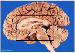
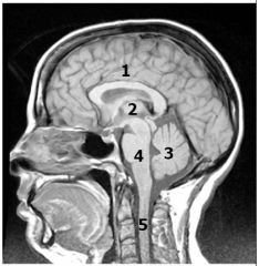
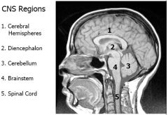
What region of the CNS is this?
|
Diencephalon
|
|
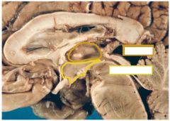
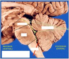
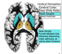
What are these structures?
Are they paired or unpaired? These structures form the walls of what structure? |
Paired
3rd ventricle |
|
What are the functions of these structures?
|

|
|

|
|
|
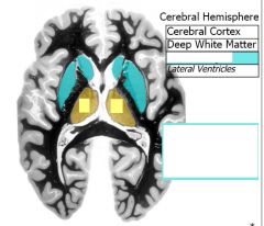
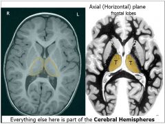
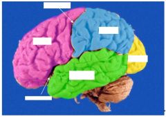
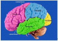
What are these structures?
What are their functions? |
Cerebral cortex:
- All the grey matter on the outside of the cerebral hemispheres Deep white matter - Connects regions of cortex and subcortical structures Basal ganglia - Buried grey matter structures in cerebral hemisphere |
|
|
Arrows pointing to precentral gyrus and postcentral gyrus
|
|
|
|
|
|
|
|
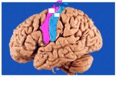
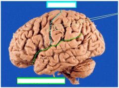
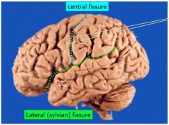
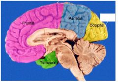
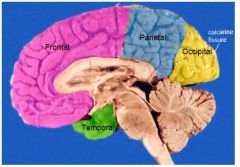
What are these two regions of the cerebral cortex?
What do they do? |
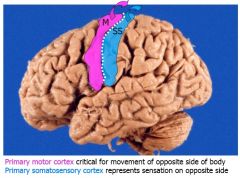
Precentral gyrus = primary motor cortex
Postcentral gyrus = primary somatosensory cortex |
|
|
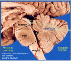
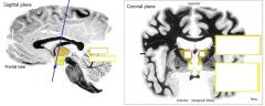
Describe the path of CSF through the ventricular system and approximately where in the CNS each of these regions is found
|
1. Lateral ventricles (2) (Curves through cerebral hemispheres
↓ Foramen of Monroe (2) 2. Third ventricle (midline cavity of diencephalon ↓ Cerebral aqueduct (Midbrain) 3. Fourth ventricle (Midbrain, Pons, Medulla) ↓ Medial and lateral foramina Subarachnoid space |
|
|
What is the septum pellucidum?
|
Membrane that separates the two lobes of the lateral ventricles
|
|
|
What structures produce CSF?
Where are they located? |
Choroid plexus:
- Lateral ventricles - Roof of third ventricle - Posterior of 4th ventricle |
|
|
What are arachnoid granulations?
What is their function? Where are they located? |
Large groups of villi that project through arachnoid space to the dural venous sinuses
Reabsorb CSF Seen mainly in superior saggital sinus |
|
|
What are the meningeal layers and where are they found in reference to the CNS?
|
Dura (outermost)
Arachnoid Pia (innermost) |
|
|
What are the functions of the meninges?
|
Cover the brain and spinal cord
Stabilize the shape and movement |
|
|
What arteries supply the dura with blood?
|
Meningeal arteries
|
|
|
What causes an epidural hematoma?
Where does the blood pool? |
Rupture of a meningeal artery
Blood creates a space between the dura and the skull |
|
|
What are the falx cerebri and tentorium cerebelli?
What are their functions? |
Deep dural folds into cranial cavity
Provide stability when the head is moving |
|
|
Where are the major venous sinuses located?
|
Along the edges of the dural folds
|
|
|
What causes a subdural hematoma?
Where does blood pool in this hematoma? |
Rupture of a bridging vein (traveling from the arachnoid to the dura)
Blood pools in the potential subdural space |
|
|
Where do major blood vessels that supply the brain travel?
|
Subarachnoid space
|
|
|
How is the pia attached to the brain and arachnoid mater?
|
Hugs gyri and dips into sulci
Connected to arachnoid by long strands of connective tissue |
|
|
What are the two main blood supplies to the brain?
What regions do each of these supply? |
Internal carotid branches ("anterior circulation")
- Retina - Hemispheres - Lateral cortex - Most basal ganglia - Medial cortex (anterior) Vertebral-basilar branches ("posterior circulation") - Medial cortex (posterior) - Most diencephalon - Brainstem - Cerebellum - Upper cervical spinal cord |
|
|
What supplies the spinal cord with blood?
|
Aortic branches
|