![]()
![]()
![]()
Use LEFT and RIGHT arrow keys to navigate between flashcards;
Use UP and DOWN arrow keys to flip the card;
H to show hint;
A reads text to speech;
241 Cards in this Set
- Front
- Back
|
which teeth are more mineralized between primary and permanent |
permanent |
|
|
what is the difference in arch space/length from the primary to permanent dentition |
2-4mm |
|
|
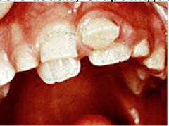
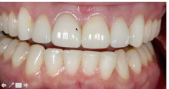
what does the presence of mamelons after the age of 10 indicate |
open bite |
|
|
at what age does the calcification of the primary roots is completed |
3-4 y.o. |
|
|
what is the typical tooth eruption sequence for primary teeth
|
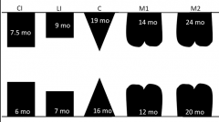
central lateral 1st molar canine 2molar |
|
|
what are 2 general pattern for primary teeth eruption |
-front to back except for canines -lowers before uppers except for lateral |
|
|
describe the primary eruption sequence Rule of 4 |
-eruption of 4 teeth every 4 months -start with 4 teeth at 7 months |
|
|
where does the primate space develop in the maxillary |

between lateral and canine primary dentition |
|
|
where does the primate space develop in the mandibular |
between the canine and the 1st premolar primary dentition |
|
|
what causes the primary spacing for anterior teeth |
most frequently caused by growth of the dental arches |
|
|
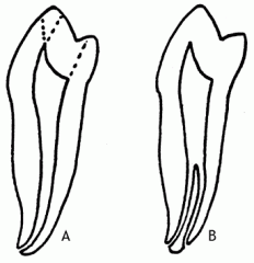
describe the direction of enamel rods of primary teeth |
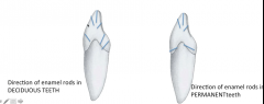
-the direction of the rod in the cervical third grows in an occlusal direction unlike the permanent dentition where the rods grown in a more cervical (away from occlusal surface) |
|
|
between the primary or permanent molars, which have roots that are more divergent |
primary |
|
|
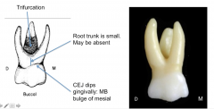
describe the root trunk of primary molars |
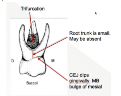
lacks an identifiable root trunk -> root trunk may be small or absent |
|
|
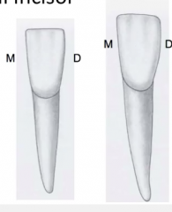
which primary tooth has the smallest F-L crown dimension |
mandibular central incisors |
|
|
which teeth are most bilaterally symmetrical tooth |
primary AND permanent mandibular central incisors |
|
|
in delayed resorption of primary incisors, where does the permanent incisors usually erupt |
lingually (shark teeth) to the primary incisors |
|
|
where does the primary maxillary central incisor exhibit a prominent cervical ridge |
both on the facial and lingual surfaces |
|
|
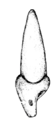
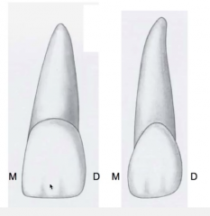
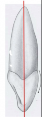
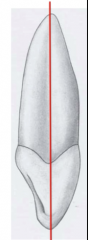
from the facial view, does the crown of a primary canine has a longer slope on the mesial or distal |

mesio-incisal ridge is longer than distal-incisal |
|
|
the cusp tip of a primary canine is generally off set to which direction |
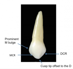
distally, therefore making the medial slope longer in length |
|
|
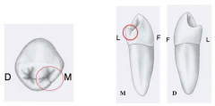
which teeth does the crown of a maxillary 1st primary molar resembles |
permanent 1st premolar |
|
|
which teeth does the roots of the maxillary 1st primary molars resembles
|
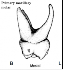
a typical permanent maxillary molar |
|
|
where is the cervical ridge most prominent for the primary maxillary 1st molar |
on the MF surface |
|
|
which primary tooth generally has an oblique ridge |

Maxillary 2nd molar |
|
|
which primary teeth is the only one to have an oblique ridge & transverse ridge & DL groove |
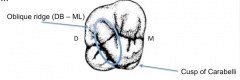
Maxillary 2nd molar |
|
|
which primary teeth exhibits a cusp of Carabelli |

Maxillary 2nd molar |
|
|
which primary tooth exhibits more cusps, maxillary 1st or maxillary 2nd molar |
maxillary 2nd molar |
|
|
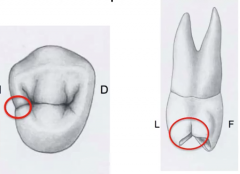
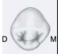
which primary tooth has the MOST distinct prominent facial cervical ridge |

mandibular 1st molar (mesial side) |
|
|
On the facial view where is the CEJ most apically positioned |
medial side on the cervical third |
|
|
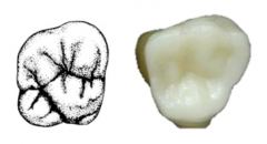
what triangular fossa does the primary mandibular 1st molar usually exhibit |

a distal triangular fossa |
|
|
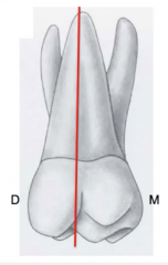
where is the central fossa usually displaced in the primary mandibular 1st molar |
distally |
|
|
which primary tooth has the most distinctive transverse ridge |
mandibular 1st molar |
|
|
which permanent tooth does the primary mandibular molar look like |
none. the primary mandibular 1st molar differs most from any permanent teeth |
|
|
which cusp is the highest and sharpest on the primary mandibular 1st molar |
mesiolingual |
|
|
which teeth has primarily biting functions |
maxillary incisors |
|
|
which teeth has eh greatest facio-lingual axial inclination |
maxillary central incisors |
|
|
which teeth has the greatest cervical curvature on the medial side of any other tooth |
maxillary central incisors |
|
|
In general, what are the usual patterns of how the CEJ dips on each tooth |
-Anterior teeth > posterior teeth -Maxillary teeth > Mandibular teeth -Mesial side > distal side |
|
|
which teeth are the only ones where the distance is wider mesio-distally than facio-lingually |
maxillary central incisors |
|
|
which anterior teeth has the greatest mesio-distal crown dimension |
maxillary central incisors |
|
|
which teeth has nearly identical measurements for mesio-distal and inciso-cervical |
maxillary central incisors |
|
|
why is the lingual embrasures larger than the facial embrasures for maxillary incisors |

the contact between a maxillary central and lateral incisor makes the lingual embrace larger than the facial |
|
|
which install embrasure is the smallest for the maxillary incisors |
embrasure between the maxillary centrals is smaller than the embrasure between the central and lateral incisors |
|
|
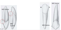
which tooth has the most crown shape variation |
maxillary lateral incisors -except for the third molars, the maxillary lateral incisors exhibit the most deviation in crown morphology |
|
|
maxillary lateral incisors most often are in abnormal relation and contact with adjacent teeth |
true |
|
|
except for the third molars, which tooth is most often congenitally missing |
maxillary lateral incisors |
|
|
which non-molar tooth most frequently has a mesial and distal pulp horn |
maxillary central incisor |
|
|
which non-molar tooth that is LEAST likely to have a bifurcated root |
maxillary central incisors |
|
|
which anterior teeth would most likely have lingual pit caries |

maxillary lateral incisors |
|
|
what anatomical features of a maxillary lateral incisor that may complicate root planning |
the distal lingual groove |
|
|
which maxillary anterior teeth has the greatest convexity in the disto-incisal angle |
maxillary lateral incisors |
|
|
which anterior teeth generally have the most prominent marginal ridges |
maxillary lateral incisors |
|
|
which anterior teeth has the most distinct and deepest lingual fossa |
maxillary lateral incisors |
|
|
which maxillary tooth has the smallest mesio-distal crown width |
maxillary lateral incisors |
|
|
which anterior teeth has nearly identical facio-lingual & mesio-distal measurements |

maxillary lateral incisors |
|
|
which maxillary incisors have a more narrow mesio-distal width |
maxillary lateral incisors |
|
|
described the contact location of maxillary anterior teeth |

I Just Jacked Michael Jackson's Moped |
|
|
which maxillary teeth has the farthest cervical (away from gingiva) contact |
maxillary lateral incisors |
|
|
describe the distal contact of a maxillary lateral incisor |
distal contact is centered both inciso-cervially and facio-lingually |
|
|
describe the maxillary lateral incisor root length |
is usually equal to or longer than the maxillary central incisor root |
|
|
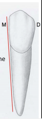
describe the cervical curvature of mandibular central incisors |
mesial cervical curvature is greater than the distal |
|
|
describe a common feature about mandibular incisors in terms of what you see in the Mesial and Distal root surfaces |

surfaces tend to be concave |
|
|
which teeth has the smallest crown dimensions |

mandibular central incisors |
|
|
which teeth has the most symmetrical crown |
mandibular central incisors -difficult to tell between the left & right mandibular central incisors |
|
|
which teeth has the sharpest incisal angles |

mandibular central incisors (mesial and distal) |
|
|
describe the contact points for the mandibular central incisors |
-proximal contact at the same levels mesial and distal (incisal third) -contact points at the same inciso-cervical level |
|
|
which teeth generally only occlude with one opposing tooth |

mandibular central incisors AND maxillary third molars |
|
|
which succedaneous tooth to erupt first in the mouth |
permanent mandibular central incisors *remember permanent 1st molars are NOT succedaneous |
|
|
describe the buccal and lingual embrasures of mandibular central incisors |
they are the same size |
|
|
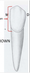
in relation to its long axis, where does the crown of a mandibular lateral incisor tilts |
distally -can see the distal marginal ridge if looking straight from the mesial side |
|
|
compare the mesio-distal width of the mandibular central versus lateral |
the lateral is larger |
|
|
which anterior teeth has the greatest cervical prominence |
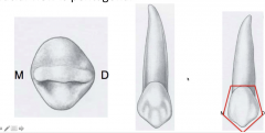
maxillary canines |
|
|
describe the vertical axis of a maxillary canine from a proximal view |
vertical line is straight along its axis (mesial or distal) |
|
|
which tooth has the greatest total tooth length |

maxillary canines |
|
|
which tooth has the longest root of ANY teeth |
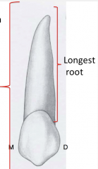
maxillary canines |
|
|
which anterior tooth has the greatest facio-lingual crown dimension |
maxillary canines |
|
|
describe the canine distal contact |
it is centered (middle third) |
|
|
which teeth has the potential of contacting both anterior and posterior teeth |
maxillary canines |
|
|
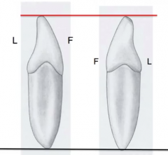
describe the cusp tip location of the maxillary canine |
located facially to the long axis -cusp is centered or slightly facial -> so if you are looking at the tooth from the incisal view, the lingual side is more visible located at the middle facial lobe (opposite to mandibular canine) |
|
|
describe the crown of a maxillary canine |
-has a distal bulge (mesial and distal are not symmetrical) -has pentagonal shape from facial view |
|
|
which tooth has the straightest mesial alignment of crown to root |
mandibular canines -mesial surface is almost parallel to the long axis |
|
|
which teeth has the longest crown dimension |
mandibular canines |
|
|
compare the canines in terms of -cingulum size -distance mesio-distally |
-maxillary canine has larger cingulum
-maxillary canine has larger width mesio-distally |
|
|
which anterior teeth most frequently exhibit a bifurcated root |
mandibular canine -when bifurcation is present it creates a facial and lingual root |
|
|
which mandibular tooth has the longest root length |
mandibular canine *remember that maxillary canines has the longest root of ANY tooth |
|
|
describe how the cross section of the root of a mandibular canine would look |
-at the CEJ = ovoid (wider mesio-disally at the labial) -at cervical = flatten in a mesio-distal direction |
|
|
how does the mandibular canine contacts differs from the maxillary canines |

contact is located more incisally |
|
|
describe the shape of a mandibular canine when viewed from the inter proximal side -from incisal to apical end -from crown tip to root apex |
-has a continuous convex facial surface
-makes a C shape |
|
|
describe the incisal edge of a mandibular canine |
is lingual to long axis when viewed at the incisal side (opposite of maxillary canine) -therefore you see more of the facial aspect of the tooth when looking at the occlusal view |
|
|
which maxillary teeth has the most pronounced developmental marginal groove |

maxillary 1st premolar (on the mesial side) |
|
|
why is it difficult to adapt a matrix band to the maxillary 1st premolar |
it has a mesial concavity |
|
|
describe the shape of a cross section of a maxillary 1st premolar at the CEJ -root -pulp chamber |
-kidney shape root -kidney shape pulp chamber floor |
|
|
which non-molar tooth most frequently exhibits 3 roots |
maxillary 1st premolar -this tooth normally has 2 roots |
|
|
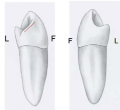
describe the facial cusp of a maxillary 1st premolar |
it is offset to the distal -> has longer mesio-facial cusp ridge than disto-facial *note: is also true for PRIMARY maxillary canines |
|
|
which premolars have the steepest cusp inclines |
maxillary 1st premolar |
|
|
describe the lingual cusp of a maxillary premolars |
it is offset to the mesial (opposite of the buccal cusp which is offset to the distal) |
|
|
which posterior teeth has the greatest cervico-occlusal crown height |
maxillary 1st premolar |
|
|
which non-molar teeth have the sharpest demarcation between the pulp chamber and canal |
maxillary 1st premolar |
|
|
compare the cusp position between the maxillary 1st versus 2nd premolar |
-the size and position of the cusps on the 2nd premolar are more identical -the height of the cusps on the 2nd premolar are equal in height unlike the 1st premolar |
|
|
which posterior tooth is the most symmetrical |
maxillary 2nd premolars |
|
|
describe the grooves of a maxillary 2nd premolar |
has a short central groove with lots of supplemental grooves that that make it look "wrinkly" |
|
|
which teeth has the fossa that are closest in size |

maxillary 2nd premolar |
|
|
describe the triangular ridge of a mandibular 1st premolar |
-has uniquely prominent triangular ridge which makes the tooth looks like it has "snake eyes" -has no central grooves -has a separate mesial and distal pit due to the triangular ridge |
|
|
describe the developmental groove on the mandibular 1st premolar |
-mesio-lingual developmental groove originates from the occlusal pit -mesio-lingual developmental groove extends onto the proximal surface |
|
|
what is a identifying characteristic of a mandibular 1st premolar |
the only tooth with a mesio-lingual groove
|
|
|
why does the mandibular 1st premolar have a mesial marginal ridge that runs at a 45 degrees angle and how does this affect the occlusal view looking from the distal versus the mesial side |
-due to the presence of the mesio-lingual developmental groove -the mesial marginal ridge is located more cervically than the distal marginal ridge (more occlusally) -> therefore more of the occlusal surface can be seen when looking from the mesial side |
|
|
what is the occlusal shape of a mandibular 1st premolar |
diamond |
|
|
in the rare event of a second canal for a mandibular 1st premolar, where is it most likely located |

to the lingual |
|
|
which premolar is the only premolar to frequently have only one pulp horn |
mandibular 1sr premolar |
|
|
compare the height between the lingual and facial cusps of a mandibular 1st premolar |

the lingual cusp height is ~2/3 of the height of the facial cusp *note: the lingual cusp is similar in development to cingulum of a canine |
|
|
which teeth does the lingual cusp of the mandibular 1st premolar occlude during normal occlusion |
none |
|
|
which posterior teeth has the most variation in cusp height between the facial versus lingual cusps |
mandibular 1st premolar |
|
|
where is the facial attached gingival narrowest |
on the facial aspect of mandibular premolars |
|
|
which posterior tooth has the smallest facio-lingual measurements |
mandibular 1st premolar |
|
|
which mandibular teeth has dimensions in the facio-lingual and mesio-distal that are closest in diameter |

mandibular 1st premolar *note the maxillary lateral incisors have closest dimensions for ANTERIOR teeth |
|
|
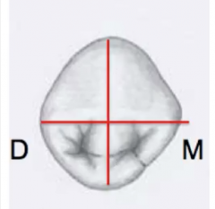
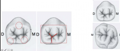
what is the basic coronal outline shape of mandibular 2nd molar viewed in the occlusal side |
pentagon *note: occlusal table shape is RECTANGULAR |
|
|
which premolar is most likely to have a crescent-shaped central developmental groove |

mandibular 2nd premolar -Y, U or H shape (Y most common) |
|
|
where is the shortest interdental papilla located |

between mandibular 2nd premolar and 1st molar |
|
|
describe the cusps and pits on a mandibular 2nd molar with Y type grooves |
-cusp: has 1 facial and 2 lingual cusp -pits: 3 (same as maxillary 1st molar) -> mesial, central & lateral |
|
|
which premolar are the only ones that have multiple lingual cusps |

mandibular 2nd premolars |
|
|
which premolar are the only ones what have a lingual groove |

mandibular 2nd premolars (located more distally unlike mandibular 1st premolar which is located mesio-lingual) |
|
|
which premolars are the only ones to have a central fossa |
mandibular 2nd premolars |
|
|
which premolar most frequently has a single central pit |
mandibular 2nd premolar |
|
|
which premolar is the most congenitally missing premolar |
mandibular 2nd premolar |
|
|
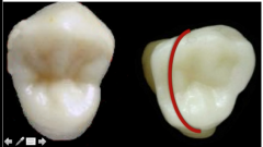
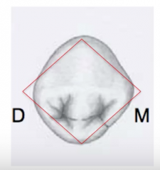
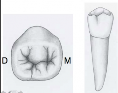
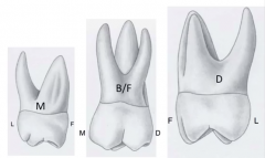
what shape is the occlusal outline form of a maxillary first molar |
rhomboidal -the mesio-facial and disto-lingual angles tend to form an acute angle while the mesio-lingual and disto-facial angles tend to form an obtuse angle |
|
|
which angles on the maxillary 1st molar makes up its oblique ridge |
mesio-lingual and disto-facial |
|
|
describe how the maxillary 1st molar is tapered |
towrds the facial and therefore the buccal embrasure is larger than the lingual |
|
|
what are the most prone part of the facial and lingual surfaces of molars |
the lingual of maxillary and the facial of mandibular |
|
|
which root is the largest in a maxillary 1st molar |
the palatal (lingual side) |
|
|
what is the smallest root in a maxillary 1st molar |
the disto-buccal root -mesio-buccal root needs room for MB2 so it will be bigger than the disto-buccal root |
|
|
from the facial view, where is the apex of the lingual root alined |
lingual root is in line with re facial groove of the tooth |
|
|
from the lingual view, where is the apex of the lingual root alined |
lingual root is lined with the midpoint of the mesio-distal diameter |
|
|
when a 4th pulp canal is present in a maxillary 1st molar, where is it located |
in the mesio-buccal canal |
|
|
for a maxillary 1st molar, list distance from the cervical line of the 3 furcations in ascending order |
mesial < buccal < distal
|
|
|
for a maxillary 1st molar, what forms the distal boundary of the central fossa |
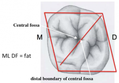
the oblique ridge
|
|
|
for a maxillary 1st molar, the center of the oblique ridge is at the same level with what |
height of the oblique ridge is at the same level/height with the marginal ridge |
|
|
where does the mesio-lingual cusp of a maxillary first molar occlude |
in the central fossa of the mandibular molars |
|
|
which teeth has the greatest facio-lingual crown diameter of ALL teeth |
maxillary 1st molar |
|
|
which maxillary posterior teeth has the closest facio-lingual and mesio-disto measurements with each other |
maxillary 1st molar |
|
|
compare the mesio-disto dimension of a maxillary 1st molar on the facial versus the lingual side |
the facial side is wider than the lingual side |
|
|
for a maxillary 1st molar, which cusp is not part of the molar's cusp triangle
|
the disto-lingual cusp |
|
|
for a maxillary 1st molar, which cusp is the largest and longest |
the mesio-lingual |
|
|
which tooth is most likely forced into the maxillary sinus during an extraction |
maxillary 1st molar |
|
|
how does the maxillary 1st molar create a problem for matrix placement |
the distal concavity makes it challenging to place the matrix band around the tooth |
|
|
compare the disto-lingual groove of a maxillary 1st versus a maxillary 2nd molar |
the disto-lingual groove of the 1st molar is shorter than the 2nd molar |
|
|
for a maxillary 2nd molar, which cusp is not part of the molar's cusp triangle |
the disto-lingual cusp |
|
|
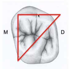
what is the shape of a maxillary 2nd molar if the disto-lingual cusp is missing |
3 cusp type heart shape -as you move progressively towards the posterior the cusps gets smaller and therefore the disto-lingual cusp of a maxillary 2nd molar is sometimes missing |
|
|
compare the inclination and deviation of the roots between a maxillary 1st and 2nd molar |

the roots of a maxillary 2nd molar tend to be LESS divergent and greater inclinations compare with the maxillary 1st molar |
|
|
for a maxillary 2nd molar, what is the shape of the cross sectional outline at the cervical |
roughly triangular |
|
|
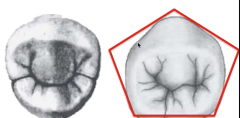
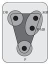
for the mandibular 1st molar, what pattern describes the grooves |
Y or Dryopethicus pattern |
|
|
what is the occlusal outline of the mandibular 1st molar |
pentagon |
|
|
for the mandibular 1st molar, list the cusp size in descending order |
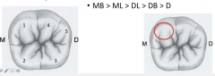
MB > ML > DL > DB > D |
|
|
which teeth has the largest mesio-distal crown dimension of ANY tooth |
mandibular 1st molar, -think of how this tooth has 3 cusps on the buccal |
|
|
which mandibular teeth are the only one that has a wider mesio-distal measurement over the facio-lingual |

mandibular 1st molar |
|
|
which posterior teeth are the only one that has a wider mesio-distal measurement over the facio-lingual |

mandibular 1st molar -has the greatest mesio-disto diameter of all the molars |
|
|
which mandibular teeth has the largest facio-lingual dimension |
mandibular 1st molar *note that the maxillary 1st molar has the largest facio-lingual length of ALL teeth |
|
|
which teeth has the largest occluso-cervical crown dimension of any mandibular molars |
mandibular 1st molar |
|
|
describe the facial surface of a mandibular 1st AND 2nd molar in respect to its position with the mandible ramus |
facial surfaces of mandibular molars are located medial to the border of the ascending rams |
|
|
how can you differentiate between mandibular 1st versus 2nd molars according developmental grooves |
mandibular 2nd molar has 1 buccal groove versus the mandibular 1st molar has 2 grooves (buccal and disto-facial) |
|
|
what is the name of the developmental groove between the disto-buccal and distal cusp of a mandibular 1st molar |
disto-facial groove |
|
|
describe the usual number of roots and canals for a mandibular 1st molar |
-2 roots -mesial is bigger -3 canals -mesial has 2 canals |
|
|
which teeth have the longest root of any molars |

mandibular 1st molar *note that eh maxillary canine has the longest root of ANY tooth |
|
|
which teeth have the greatest root separation of ANY other teeth |

mandibular 1st molar |
|
|
which tooth has the greatest facio-lingual dimension of ANY other root |
mandibular 1st molar |
|
|
where does the disto-buccal cusp of the mandibular occlude |
in the central fossa of the maxillary molar |
|
|
what is the ideal position and height of the lingual cusp of a mandibular 1st molar |
one that accommodates working movements |
|
|
where is the shortest interdental papilla located |
between the mandibular 2nd premolar and 1st molar |
|
|
what is the groove pattern for a mandibular 2nd molar |
cross (+) pattern or cruiform occlusal pattern |
|
|
how can you distinguish between the mesial and distal side of a mandibular 2nd molar |
the largest facio-lingual diameter is in the mesial 1/3 side |
|
|
describe general pattern of mandibular molars in terms of their measurements mesio-distal and facio-lingual |
-mandibular molars are the only posterior teeth that are wider mesio-distally over facio-lingually -mandibular molars are the only mandibular teeth that are wider mesio-distally over facio-lingually |
|
|
how does the crown of a mandibular 2nd molar incline |
inclines to the mesial and lingual *think of how the larger mesio-lingual cusps weighs down the rest of the tooth |
|
|
describe the position of a mandibular 2nd molar in terms of: -long axis of the root -long axis of the crown |
-long axis of the root apices is more facially located -long axis of the crown is more lingually located |
|
|
which molars are the only tooth to contact only 1 opposing tooth in occlusion |
maxillary 3rd molars *note: mandibular central incisors alos occlude with only 1 tooth |
|
|
which molars most frequently has only 3 cusps |
maxillary 3rd molars |
|
|
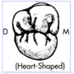
what is the shape of a maxillary 3rd molar in occlusal view |
heart shape -usually missing the disto-lingual cusp |
|
|
which tooth has the most variation in crown morphology of ANY tooth -general morphological variation as well |
mandibular 3rd molar |
|
|
which tooth has the greatest distal root inclination of any other tooth |
mandibular 3rd molar |
|
|
which mandibular tooth has the shortest root |
mandibular 3rd molar |
|
|
describe the general root pattern and canals of both maxillary and mandibular teeth (not including 3rd molars) |
|
|
|
how does the condyles move when the mandible move from centric occlusion -> edge to edge (central incisors touching each other) |
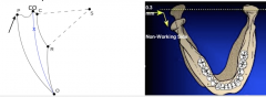
the condyles move forward and downwards |
|
|
how does a non-working condyle move |

downward, forward, and medially *note: working side is the side where the mandible move towards |
|
|
which guidance plays the greatest role in discludingthe posterior teeth in latero-protrusive |
anterior guidance -when your jaw move out of centric occlusion using canine guidance for example, your posterior teeth do not touch each other in occlusion anymore |
|
|
how are teeth positioned during NON masticatory swallowing |
teeth are in contact in intercuspal position |
|
|
what almost exclusively determines intercuspal position (or centric occlusion) |
tooth contact |
|
|
compare and contrast how centric relation and centric occlusion are guided into position |
-centric relation is a ligament guided position -centric occlusion is a tooth guided position (most comfortable position when your teeth comes together) |
|
|
in Posselt's envelop of motion, where is the maximum intercuspal position located |
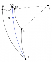
at the most superior point *on the diagram it is at "CO" |
|
|
what is centric occlusion synonymous to what |

intercuspal position |
|
|
define Bennet movement |
the side swipe of the mandible -the bodily shift of the mandible towards the woking condyle -occurs at eh earliest stage of lateral movement |
|
|
define Postral position |

physiological resting position -usually 2-4mm below ICP/CO |
|
|
what determines the mandibular postural position |
almost exclusively by the behavior of the mandibular musculature -is a muscle guided position |
|
|
what muscles will you use when moving from the postural position to centric occlusion |

anterior fibers of temporalis muscle (elevates the mandible therefore closing the mouth) |
|
|
define the Curve of Spee |
anterior-posterior curvature of the occlusal surfaces as seen in a buccal view *think of weeeee as you slide down |
|
|
define Curve of Wilson |
right to left curvature of the occlusal plane of posterior teeth (perpendicular to Curve of Spee) |
|
|
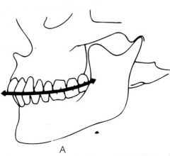
contrast overjet versus overbite |
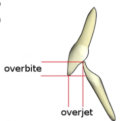
overjet = horizontal overlap (front and back) -usually 2-4mm overbit = vertical overlap (up and down) |
|
|
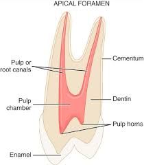
what is the hardest dental tissue and what is it made of |
enamel -made of inorganic matter (NOT collagen) |
|
|
define Perikymata |
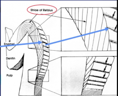
depressions outside the tooth as a result of normal enamel apposition |
|
|
define Retzius |

development disturbances inside the tooth that run obliquely from the the DEJ to enamel surfaces |
|
|
where does dentin continue to form on a tooth that has multiple roots |
dentin continues to form MOST rapidly at the floor and roof of the pulp chamber |
|
|
what type of dentin does caries stimulate to form |
tertiary dentin |
|
|
when does 2nd dentin form |
formed after root formation is complete, normally after the tooth has erupted and is functional -> maintains its incremental aspect of growth |
|
|
what percentage of dentin is organic |
20-30% |
|
|
what is the primary function of the dental pulp |
is to form dentin *it is NOT to nourish the tooth or provide sensation (these are functions but NOT the main function) |
|
|
how is dentin formed
|
by the dental papilla and the pulp |
|
|
which dentin is more mineralized |
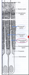
intratubular or peritubular dentin -these wrap around the odontoblast extensions |
|
|
where is the dentinoenamel junction (DEJ) located |

at the junction between the dental papilla and the inner enamel epithelium |
|
|
what are the dental papilla cells that are in contact with the inner enamel epithelium called |
odontoblasts |
|
|
what is dental papilla synonymous with |

dental pulp |
|
|
what is the softest dental tissue |
cementum |
|
|
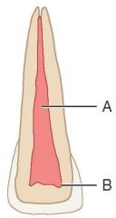
describe the two types of cementum |
-Acellular: located at the coronal 2/3 -Cellular: located at the apical 1/3 (where all the nerves and blood supply are) |
|
|
what shape do the arrangements of teeth make when they are view from the occlusal side |
parabolic (a parabola) -> U shape |
|
|
describe the arrangement of mandibular posterior teeth in the arch from an occlusal view |
the 4 posterior teeth in the mandibular arch are alined in a straight line |
|
|
generally how does the number of lobes relate to the number of cusps for posterior teeth |
the number of lobes that form the posterior teeth coincides with the number of cusp it has |
|
|
name 4 exception to the general rule that all teeth develop from 4 lobes |
-mandibular 1st molar (5 lobes b/c 5 total cusp) -maxillary 1st molar (5 lobes b/c of cusp of Carabeli) -maxillary 2nd molar 3 cusp type -3rd molars may develop from 4 or 5 lobes |
|
|
what do developmental groove separates |
separates cusp ridges from marginal ridges *ie mandibular 1st molar has 2 buccal grooves that separates the different lobes |
|
|
how are transverse ridges form |
from the union of the facial and lingual triangular ridges *image is of max 1st molar |
|
|
which teeth normally has a cingulum |
all 12 anterior teeth |
|
|
what percentage of the total facio-lingo dimension do the occlusal table of a posterior teeth make up |
55-66% |
|
|
what does the periodontium comprise of |
gingiva, PDL, cementum & alveolar bone *the epithelial attachment (junctional epithelium) is often considered part of a tooth's periodontium |
|
|
how does thickness of PDL change with age |
is usually 0.2mm wide -> decrease to 0.1mm with old age due to deposition of cementum and bone |
|
|
what does PDL fibers attach to |
PDL fibers attach tooth (cementum) to dental alveolar bone |
|
|
what does gingival fibers attach to |
gingival fibers attach tooth (cementum) to gingiva |
|
|
what is the predominant tissue for PDL fibers |
Type 1 collagen |
|
|
name the 3 types of PDL fibers and where they are located |
-transseptal fibers: tooth to adjacent tooth *these are actually gingival fibers, NOT PDL -oblique fibers: tooth (cementum) to alveolar bone -inter-ridicular fibers: root to root |
|
|
which PDL fibers provide the major support for a tooth during function |
oblique fibers -reduces the likelihood of forceful impaction into the alveolus |
|
|
which PDL fibers are most likely to be found in the middle third of a root |
oblique fibers *they are the most prevalent fibers of the three |
|
|
about what percentage of root formation is completed at the time of tooth eruption for permanent teeth |
50%
|
|
|
when is the apex of a tooth fully formed after it erupts in the mouth |
2-3 years |
|
|
where is the facial height of contour found for most teeth |
at the cervical third EXCEPT for mandibular molars |
|
|
where is the lingual height of contour found for most teeth |
-Anterior teeth = cervical third -posterior teeth = middle third *EXCEPT man 2nd PM is in occlusal third |
|
|
describe the 4 general rule about the position of a cervical line on a tooth |
CEJ dips deeper (towards the crown) on -maxillary over mandibular -Anterior over posterior teeth -mesial side over the distal side -greatest on the mesial of the maxillary central incisor |
|
|
define hypercementosis |
excess of calcified tissue (cementum) formation at the root apex |
|
|
where are supernumerary teeth usually found |
on maxilla, between centrals or sometimes rarely as 4th molars |
|
|
define concresecence |
when cementum of two teeth join together |
|
|
define oligodontia |
a developmental abnormality characterized by the presence of fewer teeth than normal |
|
|
define anodontia |
condition of missing all teeth |
|
|
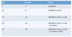
describe the eruption pattern of permanent dentition |
|
|
|
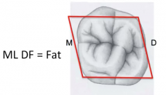
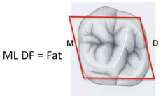
what are the 3 cardinal eruption rule for permanent teeth |
1. girls teeth erupt before boys 2. mandibular erupts before maxillary 3. teeth of skinny kids erupt before fat kids |
|
|
when do permanent teeth begin to form |
at 4 moths in utero (mandibular 1st molar) |
|
|
what is the 1st succedaneous tooth to erupt |
mandibular central incisors *note: mandibular 1st molar is NOT a succedaneous tooth |
|
|
describe the general calcification of 1st molars and central incisors |
-1st molars: at birth -central incisors: 3-4 moths after birth |
|
|
describe 2 general rule for root formation |
at eruption: 50% root -active eruption occurs after 1/2 of root is form 2-3 years after eruption: 100% root -apex of root fully develop by 2-3 years after eruption |
|
|
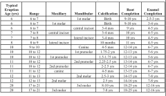
describe general eruption sequence, calcification, and root completion for permanent teeth (this is a reference slide) |
|