![]()
![]()
![]()
Use LEFT and RIGHT arrow keys to navigate between flashcards;
Use UP and DOWN arrow keys to flip the card;
H to show hint;
A reads text to speech;
62 Cards in this Set
- Front
- Back
|
Hydrophobic Amino Acids Def |
-water fearing, non polar -found burried in the interior of proteins -prefer contact with one another over h20 |
|
|
Name Hydrophobic Alipathic Amino Acids & (ABBR) |
|
|
|
Name Hydrophobic Aromatic Amino Acids (ABBR) |
|
|
|
3 Subdivisions of Hydrophillic Amino Acids and their features |
1) Polar-Neutral 2) Polar- Acidic- Negative 3) Polar - Basic -Positive -attracted to polar h20 -on surface of proteins -R-Groups have h20 affinity |
|
|
Name the Polar Acidic Amino Acids & (ABBR) |
|
|
|
Name the Polar Neutral Amino Acids (ABBR) |
|
|
|
Name the Polar Basic Amino Acids (ABBR) |
|
|
|
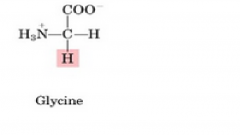
Glycine (G) Side Chain Structure |
|
|
|
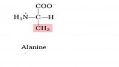
Alanine (a) Side Chain Structure |
|
|
|
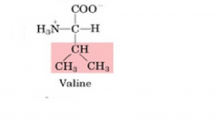
Valine (v) Side Chain |
|
|
|
Leucine (L) Side Chain Structure |

|
|
|
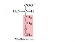
Methionine (M) Side Chain Structure |
|
|
|
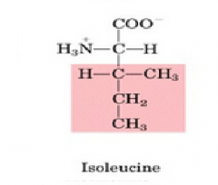
Isoleucine (I) Side ChainStructure |
|
|
|
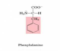
Phenylalanine (F) Side ChainStructure |
|
|
|
Tyrosine (Y) Side Chain & Pka |
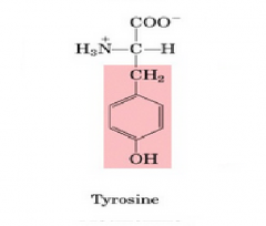
Pka=10.07 |
|
|
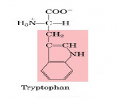
Tryptophan (w) Side Chain Structure |
|
|
|
Serine (s) Side ChainStructure |

|
|
|
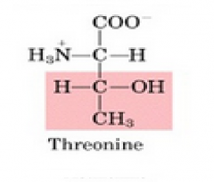
Threonine (T) Side ChainStructure |
|
|
|
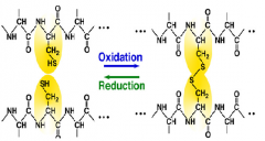
Cysteine (c) Side Chain & Pka |
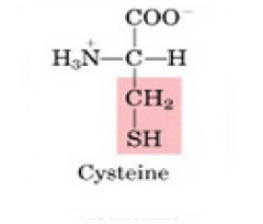
forms disulfide bonds Pka= 8.0 |
|
|
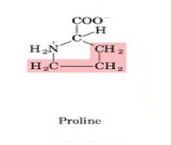
Proline (p) Side Chain Structure |
|
|
|
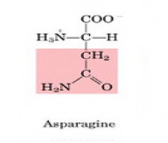
Aspararagine (N) Side Chain Structure |
|
|
|
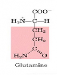
Glutamine (Q) Side Chain Structure |
|
|
|
Lysine (K) Side Chain & Pka |
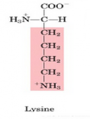
Pka= 10.53 |
|
|
Arginine (R) Side Chain & Pka |
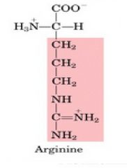
Pka= 12.48 |
|
|
Histidine (H) Side Chain & Pka |
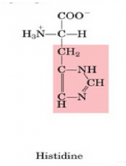
Pka= 6.10 |
|
|
Aspartate- Aspartic Acid-(D) Side Chain & Pka |
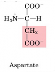
Pka= 3.86 |
|
|
Glutamate - Glutamic Acid-(E) Side Chain & Pka |
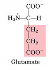
Pka= 4.07 |
|
|
Peptide Bond (Amide Bond) Formation [Def & Drawing] |
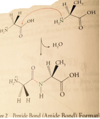
Amide attacks carboxyl carbon releasing 1 H20 |
|
|
Describe the N to C formation of polypeptides |
The N terminal starts a peptide chain, is connected to the left of the alpha carbon, the carboxyl group is connected to the right c-terminal end of the chain Thus Polypetides Move in the N to C Direction |
|
|
Describe the Primary Structure of Proteins and Primary R Groups |
Primary structure is the amino acid sequence of protein chain primary r groups determine folding of protein and final 3-D structure |
|
|
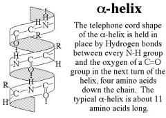
Describe the formation of a Secondary Structure A-Helix |
initial folding of the protein where hydrogen bonding between the amide hydrogens and carbonyl oxygens of the peptide bonds occur |
|
|
Describe a type of Fibrous protein and how they are formed |
a-keratins are a type of secondary structure that form a hair cell 1-an a-helix bundles with 2 other helices to form a protofibril 2-many protofibrils combine to form a microfibril (insoluble) 3- tons of microfibrils make a macrofibril making a hair cell |
|
|
Name the two types of secondary structures |
|
|
|
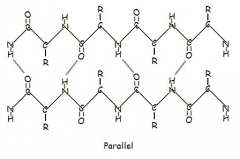
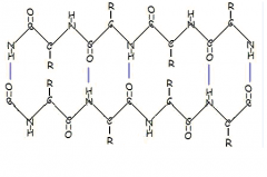
Describe a B pleated sheet and the two types of formations |
all of the carbonyl oxygens and amide hydrogens are involved in boding
|
|
|
Describe the formation of Tertiary Structure and their shape? |
Globular Proteins further folding of secondary structure 1 chain |
|
|
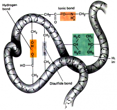
Name the Tertiary structure side chain forces and describe them |
1) Van der walls: hold r groups of nonpolar amino acids that are hydrophobic 2) Hydrogen bonds: hydrogen bonds between polar (- to +) H+ to -OR groups 3) Disulfide Bridges: Oxidation of 2 cysteine SH groups to form s-s bond 4) Salt Bridges: ionic bonds between oppositely charged Carboxyl O- and +NH3 Amino groups 5) Covalent bonds of thiol amino acids (aromatics with circles) |
|
|
Describe the formation of Quateranary Structure and the forces that it has |
Multiple polypetide side chains interact to form an activated protein or enzyme uses the same side chain forces as tertiary structure to hold multiple chains together |
|
|
Describe Carbohydrates and their functions |
simple sugars Function: provide an energy source for cellular metabolism because they are easily broken down to co2 via oxidation |
|
|
Name two Complex Carbs |
starches and cellulose |
|
|
Describe Monnosaccharides and name examples |
single sugar unit (monomer) 6 Carbon Monosaccharides
5 Carbon Monosaccharide
|
|
|
Naming Monosaccharides Ketoses and Aldoses |
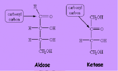
Ketose: has a ketone functional group Aldose: aldehyde functional group # OF CARBONS
|
|
|
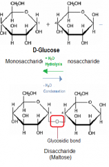
Name D-Glucose Isomers and describe their structures |
A-Glucose has C-1 hydroxyl above ring B-Glucose has C-1 hydroxyl below ring |
|
|
Describe how Disaccharides are connected and name examples and the monomers that make them |
two monosaccharides connected via a glycosidic linkage, a Glucose attached to something else
|
|
|
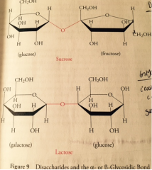
Glycosidic Bonds [Alpha vs Beta] Describe their structures |
A 1-4 linkages occur when hydroxyl groups on 1 and 4 are both in the beta position (down) makes a v shaped link B 1-4 linkages occur when hydroxyl groups on 1 and 4 are opposite creating a straight bond |
|
|
Describe the linkages of Maltose |
made of 2 a-d-glucose molecules with an a 1-4 linkage |
|
|
Describe the linkages of lactose |
made of one molecule of B-D-Galactose and one of either a or b glucose. b1-4 linkage |
|
|
Describe the linkages of Sucrose |
water soluble sugar of plants product of a-d-fructose and b-d-fructose via an a1-->b2 anomeric linkage |
|
|
Describe the different Polysaccharides and their function |
Homopoly: made up of a single monosaccharide hetero: made up of two or more monosaccharides 1) Glycogen- animal glucose storage 2) Starch- corn plant glucose storage 3) Cellulose- plant structure |
|
|
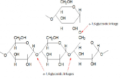
Glycogen Description and formation |
storage molecule for glucose, found in the liver and skeletal muscles made of many glucose molecules connected via a 1-4 and many short glucose side branches connected via a 1-6 linkages |
|
|
Describe the breakdown of Amylose to form Glucose |
amylose is a starch that is broken down by (A Amylase and B Amylase hich cleave maltose off then is converted to glucose via maltase |
|
|
Function of hydrolyzing glycosidic linkages and how they are named |
the hydrolysis of polysaccharides such as maltose into two units of glucose allows glucose to enter metabolic pathways as an energy source the enzymes that hydrolyze glycosidic bonds are named after the sugar they work on and are highly specific |
|
|
Describe how Polysaccharides regulate the break down of glycogen |
Because the hydrolyzation of polysacharides requires a high energy of activation it allows us to use enzymes as gate keepers: when we need energy from glucose we open the gate of glycogen hydrolysis |
|
|
Alpha and Beta 1-4 connections disaccahrides Image |
|
|
|
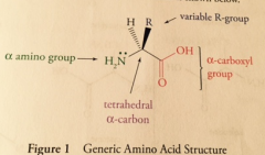
General Amino Acid Structure Image |
|
|
|
Proline Unique Features |
unique amino acid, its amino group is covalently bonded to its own side chain creating a secondary alpha amino group and a ring like structure |
|
|
|
|
|
|
Glycogen Bonding Image |
|
|
|
Hydrolysis of Disaccharide Maltose Bond Image |
|
|
|
Alpha Helix Bonding Image |
|
|
|
Parallel B pleated Sheet Image |
|
|
|
Anti Parallel B-Pleated Sheet Image |
|
|
|
Tertiary Structure Image |
|