Reading...
![]()
Play button
![]()
Play button
![]()
Use LEFT and RIGHT arrow keys to navigate between flashcards;
Use UP and DOWN arrow keys to flip the card;
H to show hint;
A reads text to speech;
175 Cards in this Set
- Front
- Back
|
Division of the Skeleton
Bones of the Skull, Vertebral Column, and Thoracic Cavity (Sternum, Ribs) |
Axial Skeleton
|
|
|
Bones of the upper and lower extremities, and the associated bones that connect the limbs to the trunk at the shoulders and pelvis.
|
Appendicular Skeleton
|
|
|
Serves as articulations or muscle and ligament attachments
|
Projections or Elevations
|
|
|
Any roughened protrusion or bump*
*For ligament or tendon (skeletal muscle) attachment |
Process
|
|
|
Any pointed process or slender ridge*
*For ligament or tendon (skeletal muscle) attachment |
Spine
|
|
|
Roughened, curved ridge*
*For ligament or tendon (skeletal muscle) attachment |
Crest
|
|
|
Small, rounded nodule*
*For ligament or tendon (skeletal muscle) attachment |
Tubercle
|
|
|
Large, rough process*
*For ligament or tendon (skeletal muscle) attachment |
Tuberosity
|
|
|
Very large, blunt projection*
*For ligament or tendon (skeletal muscle) attachment |
Trochanter
|
|
|
Smooth, rounded knob forming a joint (articulation)
|
Condyle
|
|
|
Prominence near a condyle*
*For ligament or tendon (skeletal muscle) attachment |
Epicondyle
|
|
|
Shallow, blind pocket, often forming a joint (Articulation)
|
Fossa
|
|
|
Small pit, for ligament attachment
|
Fovea
|
|
|
Groove, often for blood vessels
|
Sulcus
|
|
|
Flat, smooth pad, forming a joint (Articulation)
|
Facet
|
|
|
Communicating hole for blood vessels/nerves
|
Foramen
|
|
|
Narrow slit or gap for passage of vessels/nerves
|
Fissure
|
|
|
Deep socket for the eyeball
|
Orbit
|
|
|
Entrance into a tubular canal within a bone
|
Meatus
|
|
|
Concealed, hollow cavity for air. (Makes bone lighter)
|
Sinus
|
|
|
What are the functions of the Skeletal System?
|
Support, Leverage/Locomotion, Storage of Minerals, Blood Cell Production, Other Special Functions
|
|
|
Function of the Skeletal System
Support |
The skeletal system provides structural support for the entire body. Individual bones or groups of bones provide a framework for the attachment of soft tissues and organs.
|
|
|
Function of the Skeletal System
Storage of Minerals |
The calcium salts of bone represent a valuable mineral reserve that maintains normal concentrations of calcium and phosphate ions in body fluids. Calcium is the most abundant mineral in the human body, 98% of Calcium is deposited in the bones of the skeleton.
|
|
|
Function of the Skeletal System
Blood Cell Production |
Red blood cells, white blood cells, and platelets are produced in the red marrow, which fills the internal cavities of many bones. Red Bone Marrow (Reticular Connective Tissue), found in the spongy bone.
|
|
|
Function of the Skeletal System
Protection |
Delicate tissues and organs are often surrounded by skeletal elements. The ribs protect the heart and lungs, the skull encloses the brain, the vertebrae shield the spinal cord, and the pelvis cradles delicate digestive and reproductive organs.
|
|
|
Function of the Skeletal System
Leverage/Locomotion |
Many bones of the skeleton function as levers. They can change the magnitude and direction of the forces generated by skeletal muscles. Bones are moved by muscles. The movements produced range from the delicate motion of a fingertip to powerful changes in the position of the entire body.
|
|
|
Function of the Skeletal System
Other Special Functions |
Wide female pelvis, heavier male skeleton, foot arches, patella (knee cap), ear ossicles, sinus cavities, fused sacrum
|
|
|
What are the several bone disorders that were discussed in lecture?
|
Rickets, Osteomalacia, Osteoporosis, Osteomyelitis
|
|
|
Bone Disorder
Bones become very brittle/break, due to a lack of Calcium in the bone matrix, due to a vitamin D deficiency (diet, sunlight) |
Rickets
|
|
|
Bone Disorder
Bones become “bendy”/flexible due to a lack of Calcium in bones, bones, can occur during pregnancy. |
Osteomalacia
|
|
|
Bone Disorder
Brittle, very porous bone, due to less active osteoblasts that form bone matrix (less bone matrix). Occurs with low hormones, as in menopause. |
Osteoporosis
|
|
|
Bone Disorder
Destruction of bone by bacterial infection |
Osteomyelitis
|
|
|
True or False: Bone Tissue is Vascularized (blood vessels) and Innervated (nerves)
|
True
|
|
|
Dense, concentric layers of parallel osteons or Haversian systems
|
Compact (Cortical) Bone
|
|
|
Composed of a network of bony struts (“Trabeculae,” spicules)
|
Spongy Bone (Cancellous)
|
|
|
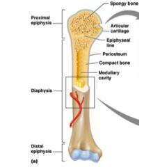
Shaft of the long bone
|
Diaphysis
|
|
|
The ends of a long bone
|
Epiphysis
|
|
|
The space within a bone that contains the yellow bone marrow
|
Medullary Cavity
|
|
|
Holes in compact bone for larger blood vessels
|
Nutrient Foramen
|
|
|
Dense irregular connective tissue, layer that surrounds a bone, consisting of an outer fibrous and inner cellular region. (except at the ends of the bone)
|
Periosteum
|
|
|
Thin, internal, cellular membrane that contains osteoblasts and osteoclasts
|
Endosteum
|
|
|
Hyaline Cartilage located on the ends of the epiphysis, provides a protective layer that prevents bone on bone contact.
|
Articular Cartilage
|
|
|
What are the 9 Gross Anatomy items of the Long Bone discussed in lecture?
|
Compact bone, Spongy bone, Diaphysis, Epiphysis, Medullary Cavity, Nutrient Foramen, Periosteum, Endosteum, Articular Cartilage
|
|
|
What is the Matrix (Ground Substance and Fiber) of Bone?
|
Ground Substance: Hard Solid. Osteoid (all organic molecules secreted by the cells that live in the bone that form bone matrix.
Fiber: Collagen |
|
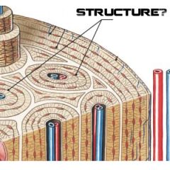
The basic histological unit of compact bone, consisting of osteocytes organized around a central canal and separated by concentric lamellae.
|
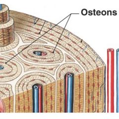
Haversian or Osteon System
|
|
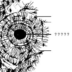
Longitudinal canal in the center of an osteon that contains blood vessels and nerves. Within an osteon the osteocytes are arranged in concentric layers around the central canal. Runs parallel to the surface of the bone.
|
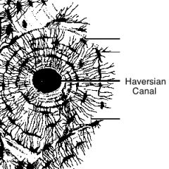
Haversian (Central) Canal
|
|
|
Concentric layers of bone within an osteon. It is the bone matrix: Osteoid, Collagen Fibers, and Mineral Salts.
|
Lamellae
|
|
Small chambers sandwiched between concentric layers of calcified matrix (lamellae). Osteocytes are found inside.
|

Lacunae
|
|
|
Microscopic passageways between cells; permits the diffusion of nutrients and wastes to and from osteocytes.
|
Canaliculi
|
|
|
Mature bone cells responsible for the maintenance and turnover of the mineral content of the surrounding bone. Located within small chambers called Lacunae, that are sandwiched between concentric layers of calcified matrix (Lamellae).
|
Osteocytes
|
|
|
Immature bone cells that secrete the organic components of the bone matrix (osteoid) within connective tissue (intramembranous ossification) or cartilage (endochondral ossification). Responsible for the production of new bone, process called osteogenesis. Found in the cellular layer of the periosteum and endosteum.
|
Osteoblasts
|
|
|
Found on the innermost layer of the periosteum and in the endosteum lining the medullary cavity. Stem cell that gives rise to osteoblasts. The ability to produce additional osteoblasts becomes extremely important after a bone is cracked or broken.
|
Osteoprogenitor Cells
|
|
|
Large, multi-nucleated cells that secretes acids, through the exocytosis of lysosomes, that dissolve the bony matrix and release amino acids and the stored calcium and phosphate. Located in the periosteum and endosteum.
|
Osteoclasts
|
|
|
The process of breaking down the mineral matrix of bone
|
Osteolysis
|
|
|
What are the two methods of ossification?
|
Intramembranous Ossification
Endochondral Ossification |
|
|
Bone Ossification
Forms in a membrane. The formation of bone within a connective tissue without the prior development of a cartilaginous model. Forms the flat bones of skull, some facial bones, clavicle formation is in a mesenchyme membrane. |
Intramembranous Ossification
|
|
|
Forms all other bones a skeleton precursor made of hyaline cartilage is replaced by bone tissue.
|
Endochondral Ossification
|
|
|
Review the Steps for Intramembranous Ossification
|
No Really... Go Read About It
|
|
|
Review the Steps for Endochondral Ossification
|
No Really... Go Read About It
|
|
|
Reticular Connective Tissue, forms all types of blood cells. Found in spaces of spongy bone.
|
Red Bone Marrow
|
|
|
Adipose Connective Tissue, fills the medullary cavity, nutrient storage.
|
Yellow Marrow (Adipocytes)
|
|
|
Functions of Joints
|
Articulation, Locomotion, and Support
|
|
|
What are the two ways to categorize joints?
|
Function (Movement) and Structure (Type of Material)
|
|
|
Joints categorized by function (movement)?
|
Synarthrosis, Amphiarthrosis, Diarthrosis
|
|
|
No Movement, Example: Suture of the Skull
|
Synarthrosis
|
|
|
Little Movement, Example: Pubic Symphysis of Pelvis
|
Amphiarthrosis
|
|
|
Free Movement, Example: Shoulder, Hip Joints
|
Diarthrosis
|
|
|
What are the joint structure categories?
|
Fibrous Joints, Cartilaginous Joints, Synovial Joints
|
|
|
Sutures, Syndesmoses, and Gomphoses are classified as what type of joint?
|
Fibrous Joints
|
|
|
Joint Category
Fibrous Connective Tissue (Ligaments) Connect Bone to Bone. Dense Regular Connective Tissue |
Fibrous Joints
|
|
|
Join flat bones of the skull. These joints become totally calcified with age to become synostoses. No movement
|
Sutures
|
|
|
Joins parallel bones like radius and ulna, tibia and fibula. Amphiarthrosis
|
Syndesmoses
|
|
|
Binds each tooth to the surrounding bony socket.
|
Gomphoses
|
|
|
Joint Category
Cartilage that holds bones together |
Cartilaginous Joints
|
|
|
What are the two types of Cartilaginous Joints?
|
Synchondroses and Symphysis
|
|
|
Cartilaginous synarthrosis, such as the articulation between the epiphysis and diaphysis of a growing bone. Sternum to Ribs. Hyaline Cartilage.
|
Synchondroses
|
|
|
Fibrous Amphiarthrosis, such as those between adjacent vertebrae or between the pubic bones of the coxae. Fibrocartilage.
|
Symphysis
|
|
|
Only joint with a cavity, All diarthrosis joints (freely moveable); specialized for movement and permits a wide range of motion.
|
Synovial Joints
|
|
|
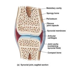
What are the structures of a synovial joint?
|
Articular Cartilage, Fibrous Joint Capsule, Synovial Membrane, Joint Cavity, Synovial Fluid, Accessory Structures, Sensory Nerves and Blood Vessels
|
|
|
Cartilage pad that covers the surface of a bone inside a joint cavity
|
Articular Cartilage
|
|
|
Surrounds the synovial joint, composed of this layer of dense connective tissue.
|
Fibrous Joint Capsule
|
|
|
Lines the joint cavity but stops at the edges of the articular cartilages. Produces synovial fluid. An incomplete layer of fibroblasts confronting the synovial cavity plus the underlying loose connective tissue.
|
Synovial Membrane
|
|
|
The space within the synovial membrane where the synovial fluid is located.
|
Joint Cavity
|
|
|
Provides lubrication and reduces friction, nourishes the chondrocytes, acts as a shock absorber.
|
Synovial Fluid
|
|
|
What are the Accessory Structures of a synovial joint?
|
Pads of Cartilage or Fat, Ligaments, Tendons, Bursae
|
|
|
A fibrous cartilage pad between opposing surfaces in a joint.
|
Menisci (Meniscus)
|
|
|
Often found around the periphery of the joint, lightly covered by a layer of synovial membrane. Provides protection for the articular cartilages and serve as packing material for the joint as a whole.
|
Fat Pads
|
|
|
Dense band of connective tissues fibers that attach on bone to another
|
Ligaments
|
|
|
A collagenous band that connects a skeletal muscle to an elements of skeleton.
|
Tendons
|
|
|
A small sac filed with synovial fluid that cushions adjacent structures and reduces friction.
|
Bursae
|
|
|
Joints
Stability vs. Movability |
The greater the range of motion at a joint, the weaker it becomes.
|
|
|
What are the different types of synovial joints?
|
Ball and Socket, Hinge, Pivot, Plane, Condyloid, Saddle
|
|
|
Triaxial Joint, A round head of one bone rests within a cup shaped depression in another. All combinations of movements, including rotation, can be performed.
|
Ball and Socket
|
|
|
All combinations of movements, including rotation, can be performed
|
Triaxial Joint
|
|
|
Permits angular movement in a single plane
|
Monaxial Joint
|
|
|
Monaxial Joint, Permits angular movement in a single plane, like the opening and closing of a door.
|
Hinge
|
|
|
Give an example of a ball and socket joint
|
Shoulders and Hips
|
|
|
Give an example of a hinge joint
|
Elbows and Knees
|
|
|
Monaxial Joint, They permit only rotation
|
Pivot
|
|
|
Give an example of a pivot joint
|
a pivot joint between the atlas and axis allows you to rotate your head to either side
|
|
|
Permit only small sliding movements
|
Nonaxial
|
|
|
They permit sliding in any direction
|
Multiaxial
|
|
|
Nonaxial and Multiaxial Joints, Flattened or slightly curved faces. The relatively flat articular surfaces slide across one another, but the amount of movement is very slight. Ligaments usually prevent or restrict rotation.
|
Plane Joint
|
|
|
Biaxial Joint, An oval articular face nestles with a depression on the opposing surface. With such an arrangement, angular motion occurs in planes, along or across the length of the oval.
|
Condyloid Joint
|
|
|
Angular motion occurs in planes
|
Biaxial Joint
|
|
|
Give an example of a condyloid joint
|
Connects the fingers to the metacarpal bone, and the toes to the metatarsal bones.
|
|
|
Give an example of a plane joint
|
Plane joints are found at the ends of the clavicles, between the carpal bones, between the tarsal bones, and between the articular facets of adjacent vertebrae.
|
|
|
Biaxial Joint, Have complex articular faces. Each one resembles a _______ because it is concave on one axis and convex on another. Are extremely mobile allowing extensive angular motion without rotation.
|
Saddle Joint
|
|
|
Give an example of a saddle joint
|
Base of the Thumb
|
|
|
What are the functions of synovial fluid?
|
Provides lubrication, reduces friction, reduces heat buildup, nourishes the chondrocytes, acts as a shock absorber.
|
|
|
Bone to bone straps of dense, regular connective tissue.
|
Collateral Ligaments
|
|
|
What are the three types of Arthritis?
|
Degenerative, Rheumatoid, and Gouty
|
|
|
Are ligaments vascularized?
|
No
|
|
|
Inflammation (swelling) of joints
|
Arthritis
|
|
|
List the joint disorders discussed in lecture
|
Arthritis, Degenerative Arthritis (Osteoarthritis), Rheumatoid Arthritis, Gouty Arthritis, Bursitis, Sprains
|
|
|
Brought on by old age, joint abuse or obesity progressive erosion of articular cartilage, leading to joint ossification and immobility.
|
Degenerative Arthritis (Osteoarthritis)
|
|
|
Autoimmune Disease, Causing thickening of synovial membrane and breakdown of cartilage and bone, leading to stiffens, swelling, and pain.
|
Rheumatoid Arthritis
|
|
|
Metabolic disorder in males, high levels of uric acid in blood, leading to crystallization in the synovial sac.
|
Gouty Arthritis
|
|
|
Inflammation (swelling) of bursae, Promoted by prolonged stress or pressure at joints. Joint capsule not directly involved tendon, may calcify leading to stiffness.
|
Bursitis
|
|
|
Stretched or torn ligaments may lead to instability of the joint
|
Sprains
|
|
|
What are the four shared basic properties of muscle tissues?
|
Excitability, Contractility, Extensibility, Elasticity
|
|
|
All muscles are _________ active and provides ________
|
Electrically; Movement
|
|
|
What are the three types of Muscle?
|
Skeletal, Cardiac, Smooth
|
|
|
What are the functions of Skeletal Muscle?
|
Locomotion, Posture/Support, Support Soft Tissue, Regulate Entering and Exiting of Material, and Maintains Body Temperature
|
|
|
What are the three rules of locomotion?
|
1. Muscles shorten when they contract. They can therefore only pull on bones to move them. They DO NOT push bones.
2. Muscles always cross at least one joint and attach two or more bones. They move bones at the joint. 3. One attachment is stationary (origin), the other attachment is on the moving bone (insertion) |
|
|
Function of Skeletal Muscle
Muscle contractions pull on tendons and move the bones of the skeleton. |
Locomotion
|
|
|
Moves the bones of the skeleton
|
Skeletal Muscles
|
|
|
Function of Skeletal Muscle
Constant muscular contraction to keep the body upright without collapsing or stand without toppling over. |
Maintain Posture/Support
|
|
|
The abdominal wall and the floor of the pelvic cavity consist of layers of skeletal muscle. These muscles support the weight of visceral organs and protect internal tissues from injury.
|
Support Soft Tissue
|
|
|
Function of Skeletal Muscle
Openings, or orifices, of the digestive and urinary tracts are encircled by skeletal muscles. These muscles provide voluntary control over swallowing, defecation, and urination. |
Regulate entering and exiting of material
|
|
|
Function of Skeletal Muscle
Muscle contraction require energy, and whenever energy is used in the body, some of it is converted to heat. The heat lost by contracting muscles keeps our body temperature in the range required for normal functioning. |
Maintain body temperature
|
|
|
Voluntary Muscle
|
Skeletal Muscle
|
|
|
Voluntary, Multi-nucleated (outer edges of the fibers), Striated, Unidirectional cell orientation, Bound together by a sacrolemma (cell membrane) and supplied with a branch of one motor nerve, connected by Areolar Connective Tissue
|
Skeletal Muscle
|
|
|
Location of Skeletal Muscle
|
Attached to the Skeleton
|
|
|
Moves blood
|
Cardiac Muscle
|
|
|
Moves Organ Contents
|
Smooth Muscle
|
|
|
Short, Branched, Mono-Nucleated (Centrally located), Involuntary, Auto-rhythmic, Hormones and Nervous System regulate rate and strength of contractions.
|
Cardiac Muscle
|
|
|
Where is Cardiac Muscle located?
|
The Heart
|
|
|
Cell junction that allows electrical signals to spread through tissues.
|
Intercalated Discs
|
|
|
Forms the walls of visceral organs
|
Smooth Muscle
|
|
|
Short, Mono-Nucleated, Non-Striated
Myofibrils: Are less organized, less myosin Properties: Involuntary, Contractions are slow and prolonged, Auto-Rhythmic Contraction, Contraction is regulated by hormones and nervous system |
Smooth Muscle
|
|
|
Tendon Attachments
|
Dense Regular Connective Tissue, Attaches Muscle to Bone
|
|
|
Epimysium, Perimysium, Endomysium
|
Connective Tissue Wrappings
|
|
|
Wraps the muscle organ (dense irregular connective tissue)
|
Epimysium
|
|
|
Wraps Fascicle (bundle of skeletal muscle fibers) (Loose/Reticular CT)
|
Perimysium
|
|
|
Wraps the muscle fiber (areolar connective tissue)
|
Endomysium
|
|
|
Skeletal Muscle Function
Areolar and Dense Irregular Connective Tissue, keeps the muscle fibers aligned |
Connective tissue wrappings
|
|
|
Skeletal Muscle Function
Supply glucose and oxygen to the muscle fibers |
Blood Vessels
|
|
|
Tendon Attachments, Connective Tissue Wrappings, Blood Vessels and Nerves
|
Skeletal Muscle Functions
|
|
|
Skeletal Muscle Functions
Requires innervation to contract, motor nerve, commands the muscle cell to contract. |
Nerves
|
|
|
Fiber Arrangements, and Functional Significance of the Arrangements
|
Affects the muscles Force of contraction and Range of motion
- Maximum Range, Minimal Force - Maximum Force, Minimal Range |
|
|
Maximum Range, Minimal ______
|
Force
|
|
|
Maximal Force, Minimal ______
|
Range
|
|
|
Skeletal Plasma Membrane
|
Sarcolemma
|
|
|
Each muscle cell is innervated by a
|
Motor Unit Nerve
|
|
|
What are the two myofilament proteins?
|
Myosin (Thick) and Actin (Thin)
|
|
|
Myosin is _____ Filament
|
Thick
|
|
|
Actin is _____ Filament
|
Thin
|
|
|
2 types of contractile protein filaments
|
Myosin and Actin
|
|
|
Each sarcomere shortens when myofilaments sliding past each other.
Myosin filament heads bind to actin thin filaments and pull them. More overlap causes more shortening. I band will decrease. |
Sliding Filament Theory of Contraction
|
|
|
Sarcomere structure and appearance in contracted vs. relaxed muscle
|
During a contraction, the A band stays the same width, but the Z lines move closer together and the I Band and H band are reduced in width
|
|
|
The main muscle accomplishing the action
|
Prime Mover
|
|
|
Muscles that contract to help the prime mover
|
Synergists
|
|
|
Muscles that produce the reverse action as a prime mover and its synergists
|
Antagonists
|
|
|
The sarcoplasm of a skeletal muscle fiber contains hundreds to thousands of
|
Myofibrils
|
|
|
Each ______ is a cylindrical structures, responsible for skeletal muscle fiber contraction.
|
Myofibril
|
|
|
Muscles contract in an _________ fashion
|
All-or-None
|
|
|
Cylinders inside cells made of bundles of myofilament proteins
|
Myofibrils
|
|
|
Review The Possible Diagrams
|
Review The Possible Diagrams
|
|

Sarcomere
|
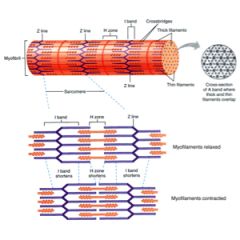
Sarcomere
|
|
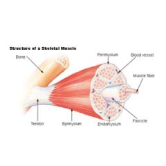
Skeletal Muscle
|

Skeletal Muscle
|
|

Knee Joint
|
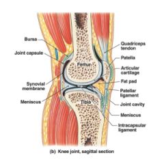
Knee Joint
|
|

Synovial Joint
|
Synovial Joint
|
|

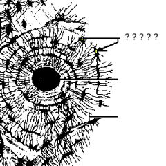
Micro-Anatomy of a Long Bone
|

Micro-Anatomy of a Long Bone
|
|
Long Bone
|

Long Bone
|