Reading...
![]()
Play button
![]()
Play button
![]()
Use LEFT and RIGHT arrow keys to navigate between flashcards;
Use UP and DOWN arrow keys to flip the card;
H to show hint;
A reads text to speech;
93 Cards in this Set
- Front
- Back
|
Connective Tissue.
|
What type of tissue is this?
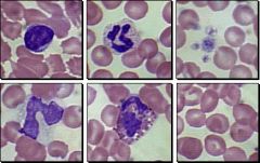
Idnetify the following: Erythrocyte Platelet Leukocyte Neutrophil Lymphocyte Monocyte Eosinophil Basophil |
|
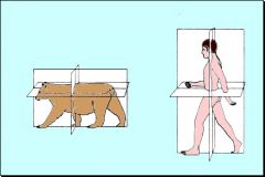
Identify following terms: Sagittal plane
Transverse plane Dorsal plane Cranial Caudal Ventral Dorsal Inferior Superior Anterior Posterior |

hb
|
|
|
Thoracic Limb:
proximal-humerus, radius and ulna Extends to carpus.cranial and caudal surfaces. distal-carpus, metacarpus and phalanges. Dorsal and palmar. Pelvic Limb: proximal-femur, tibia and fibula. Extends to tarsus (hock, ankle). Cranial/caudal surfaces. Distal-tarsus, metatarsus and phalanges. dorsal and plantar. |
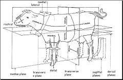
Define Thoracic Limb, Pelvic Limb.
|
|
|
Box 1-Epithelial Tissue-
anchor cells. Protection, absorption, secretion. Box 2-Nervous Tissue- Communication. React to physical/chemical stimuli. conduct to other location. Box 3-Connective Tissue- Connects, holds, supports body tissues. Components: Cells, extracellular fibers(strength), extracellular ground substance(nutrient diffuse from blood vessels) Box 4-Muscle Tissue- ellongated cells (fasciculus=sm bundle) |
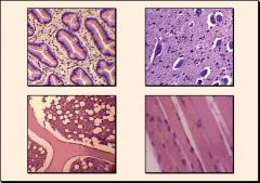
Identify each Box.
|
|
|
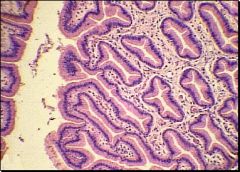
Organ Tissue
Epithelial tissue- anchor cells Connective tissue- connects/holds & supports Muscle tissue- elongated cells (fibers) Nervous tissue- conductive Lumen of ileum-(cavity) Stroma-organ connective tissue |
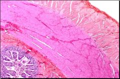
What is shown in this picture?
Identify the following structures: Epithelial tissue Connective tissue Muscle tissue Nervous tissue Lumen of ileum Stroma |
|
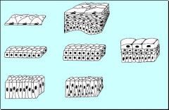
Identify the following epithelia types:
Simple Squamous Simple Cuboidal Simple Columnar Stratified Squamous Stratified Cuboidal Pseudostratified Columnar Transitional |

Simple-one cell layer
Stratified-multiple cell layers Transitional-number of layers and cell shape varies |
|
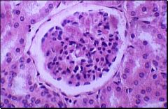
What type of tissue is this?
Identify the following: Simple squamous epithelial cells Bowman's space Simple cuboidal epithelial cells |

Simple Squamous Epithelial Tissue
|
|
|
Where are squamosal cells found?
|
Lining body cavities
(mesothelium) Cardiovascular Lymphatic systems (endothelium) Gas and liquid exchange in kidneys |
|
|
What about its shape is beneficial to a squamousal cell?
|
FLAT & LRG SURFACE AREA:
diffusion and filtration. Line bowman's space in cortex of the kidney. |
|
|
What type of cells line the Bowman's space in the cortex of the kidney?
|
Simple Squamousal epithelial cells
|
|
|
Where are Simple cuboidal epithelial cells found?
|
Lining ducts & tubules
|
|
|
What is the finction of simple cuboidal cells?
|
secretion & absorbtion
(kidney, canine) |
|
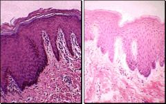
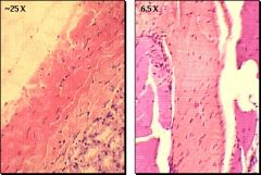
What type of tissue is this?
Identify the following structures: connective tissue basal cell layer dry epithelium keratinized layer wet epithelium |

Stratified Squamous Epithelial Tissue
|
|
|
How is epithelial tissue nourished?
|
diffusion from blood vessels in connective tissue
|
|
|
What is unique about basal cells?
|
Continually dividing
New cells pushed to surface where they are worn off |
|
|
Describe the function of the dry epithelium?
|
(SKIN)
contain keratin (strong protein) resist trauma, bacterial & fungus infections, watertight |
|
|
What is the keratinized layer of the dry epithelium?
|
superficial layer of dead
squamous cells |
|
|
What is different about the wet epithelium?
|
Line mouth, esophagus and vagina
Stratified cell- no cornified surface |
|
What type of tissue is this?
Identify the following structures: Simple cuboidal cell Cell nuclei Kidney tubule lumen |

Simple Cuboidal Epithelial tissue
|
|
|
What are the locations and function of simple cuboidal cells?
|
Lining ducts and tubules
Secretion & absorbtion stratified cuboidal v. rare (kidney, canine) |
|
|
What is the general(simple) finction of the cell nuclei?
|
contains genetic material
controls synthetic & meta bolc activities of cytoplasmic organelles |
|
|
What is necessary for a cell nuclei to be found?
|
Cell must not be dividing
|
|
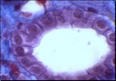
What type of tissue is this?
Identify the following structures: Simple columnar cells Cell nuclei Basal surface Luminal surface |

Simple Columnar Epithelial Tissue
|
|
|
Where are simple columnar cells located and what is their function?
|
Lining stomach, itestines, lrg.
ducts. Secretion & Absorbtion (cilia & microvilli-increase surface area) |
|
|
Where is the cell nuclei found in a columnar cell?
|
Basal portion & form a single
line in cross section |
|
|
What is the basal surface of an epithelial cell?
|
side anchored to basement
membrane. Attaches cell to rest of body. |
|
|
What is the luminal side of an epithelial cell?
|
Side that communicates with lumen of gut or respiratory tract
|
|

What type of tissue is this?
Identify the following: Pseudostratified columnar cells Cell nuclei Basal surface Luminal surface |
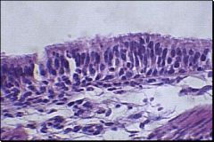
Pseudostratified Columnar Epithelial Tissue
|
|
|
What is the pseudostratified columnar epithelium?
|
Simple columnar epithelium that
appears to be multiple cell layers. Not all columns extend equally to luminal surface |
|
|
How do cell nuclei appear in pseudocolumnar epithelium?
|
nuclei do not line up
|
|
|
Where is pseudocolumnar epithelium located?
|
Upper respiratory tract
|
|
What type of tissue is this?
Identify the following structures: Bladder cavity Epithelial layer Connective tissue |

Trasitional Epithelial Tissue
|
|
|
Why is the bladder cavity lined with transitional epithelium?
|
Allows distention of bladder
lining w/o rupturing or separating cells Protects body from noxious waste |
|
|
What happens when transitional epithelium distends?
|
Number of layers decrease
Top layer becomes squamous |
|
|
What are the three major components of connective tissue?
|
Cells
Extracellular fibers Extracellular ground substance |
|
|
What is the function of the ground substance in connective tissue?
|
nutrients diffuse from blood vessels to cells
|
|
|
What is the function of extracellular fibers in connective tissues?
|
strength
|
|

What type of tissue is this and what is depicted in the picture?
Identify the following: Duct secretory cells |
Epithelial Connective Tissue
Downgrowths-glands-function in secretion & excretion |
|
|
What are the two main types of glands?
|
Exocrine-ducts convey mucus &
enzymes to epithelial surface. Endocrine-no ducts-not connected to epithelium. Release hormone directly into blood for distribution |
|
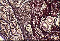
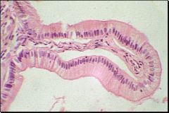
What type of tissue is this?
Identify the following: Reticular fibers Collagen fibers Gut |

Reticular Connective Tissue
|
|
|
What are reticular fibers?
|
Thin collagen fibers
Surrounding blood vessels & nerves |
|
|
What are collagen fibers?
|
Composed of protein collagen
Great strength Inelastic |
|
|
What are elastic fibers?
|
Composed of protein elastin
Stretch easily & return to original length |
|
What type of tissue is this?
Identify the following: Fibroblasts Elastic fibers Collagen fibers |

Elastic Connective Tissue
|
|
|
What is the function of fibroblasts?
|
formation of collagen, elastin & ground substance
|
|
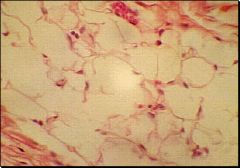
What type of tissue is this?
Identify the following: Adipose cell Adipose cell nuclei Collagen Arteriole |

Loose Connective Tissue
|
|
|
Where is loose connective tissue found?
|
Between muscles-packing material (soft and pliable)
|
|
|
What type of cells make up loose connective tissue?
|
Adipose cells
|
|
|
Where are adipose cell nuclei found?
|
In the corner
|
|
|
What is the arteriole?
|
sm. blood vessel, supported by loose connective tissue
|
|
|
How is dense connective tissue different from loose?
|
More fibers, fewer cells, lessm ground substance
|
|

What type of tissue is this? Identify the following:
Regular dense connective tissue Fibroblast nuclei Irregular dense connective tissue |
o
|
|
|
What characterizes dense connective tissue?
|
tightly packed fibers
few fibroblasts v. strong no elasticity or flexibility |
|
|
What is dense connective tissue?
|
thick wave of collagen fibers
few cells flexibility > strength (dermis, nerve & muscle sheaths) |
|
|
What is plasma composed of?
|
Water, Protein, Solutes
|
|
|
What are erythrocytes & how do they function?
|
Red Blood Cells
transport of oxygen (mammals-lack nuclei) |
|
|
How do platelets function?
|
Clot formation
|
|
|
What are leukocytes and how do they function?
|
White Blood Cells
defense against foriegn invaders |
|
|
What are the structures of leukocytes?
|
Neutrophil
Lymphocyte Monocyte Eosinophil Basophil |
|
|
What is the function of neutrophils?
|
1st line of defense
engulfing & digesting invaders |
|
|
What is the function of lymphocytes?
|
synthesis of antibodies
|
|
|
What is the function of monocytes?
|
leave blood to devour invaders in connective tissues
|
|
|
What is the function of Basophils?
|
release chemicals mediating itch and tissue swelling
|
|
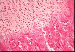
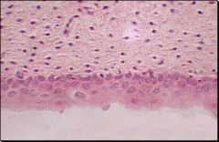
What type of tissue is this?
Identify the following: Bone Cartilage Chondrocytes |

Connective Tissue- hyaline cartilage.
|
|
|
How does bone appear in the embryo?
|
Hyaline cartilage
Development- cartilage replaced by bone Hyaline cartilage remains for joint articulation |
|
|
What are the cells of cartilage called?
|
Chondrocytes
|
|
|
What does cartilage consist of?
|
Dense network of fibers in
gel-like intercellular Firm & flexible |
|
|
What is the function of elastic cartilage and where is it found?
|
v. flexible. returns to original
position Found in auricle of ear |
|
|
Where is hyaline cartilage found?
|
Nose, larynx and bronchi or
repiratory sys. |
|
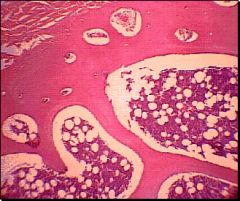
What type of tissue is this?
Identify the following: Spongy bone Compact bone Muscle Marrow |

Connective tissue- Bone
|
|
|
What causes hardness of bone & the sm elasticity?
|
Hardness = Calcium Phosphate
Elasticity = organic collagen fibers |
|
|
How are bone cells nourished?
|
Sm. tubular canals
|
|
|
What are the two forms of bone?
|
Spongy- extremely porous
Compact- solid |
|
|
How is muscle tissue attached to bone?
|
Short tendons attach to compact bone
|
|
|
What fills the spaces of the spongy bone?
|
Marrow
|
|

What type of tissue is this?
Identify the following: Striations Connective tissue cell nucleus Skeletal muscle cell nucleus |
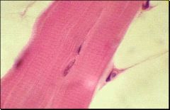
Skeletal Muscle Tissue
|
|
|
What type of muscle is under voluntary control?
|
Skeletal Muscle
|
|
|
Describe muscle cells in the skeletal muscle.
|
Myofibers- long, multinucleared,
parallel bundles |
|
|
Which types of muscles have "transverse striation"?
|
Cardiac and Skeletal
|
|
|
What causes striation?
|
Actin and Myosin protein
filaments- utilize cellular energy for muscle contraction. |
|
|
Where is smooth muscle found?
|
Walls of viscera & blood vessels
|
|
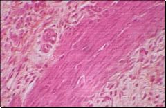
What type of tissue is this?
Identify the following: Connective tissue Smooth muscle |

Smooth muscle Tissue
|
|
|
Why do smooth myofibers form junctions?
|
Spread excitation & contraction from one cell to another
|
|
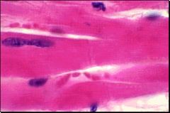
What type of tissue is this?
Identify the following: Intercalated disc Muscle cell nucleus |

Cardiac Muscle Tissue
|
|
|
What type of tissue is found in the myocardium?
|
Cardiac
|
|
|
What are the intercalated discs?
|
Specialized junctions between
cardiac cells. Allow spread of excitation |
|
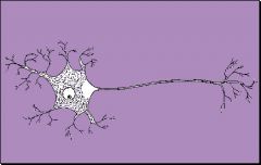
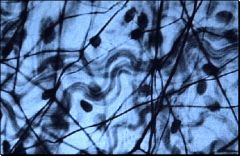
What type of tissue is this?
Identify the following: Cell body Dendrites Axon Terminals |

Nervous Tissue- Neuron
|
|
|
What is the function of a neuron?
|
Communication
|
|
|
What is the function of a neuron cell body?
|
Synthesize proteins and neurotransmitters
|
|
|
What is the function of neuron dendrites?
|
Chemical Reception
|
|
|
What is the function of a neuron axon?
|
Conduction of nerve impulses over long distances
|
|
|
What is the function of the neuron terminals?
|
transmissive segment
contacts dendrites of neighboring neuron (synapses) |
|
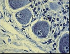
What type of tissue is this?
Identify the following: Cell bodies of neurons Nuclei of neurons Nuclei of support cells Axon Ganglion Nerve |

Ganglion Nervous Tissue
|