Reading...
![]()
Play button
![]()
Play button
![]()
Use LEFT and RIGHT arrow keys to navigate between flashcards;
Use UP and DOWN arrow keys to flip the card;
H to show hint;
A reads text to speech;
64 Cards in this Set
- Front
- Back
|
What does the perforating tarsal artery pass through?
(what does it branch off from?) |
Vascular canal of hock to plantar surface
(branches off of dorsal pedal a. to join medal/lateral plantar aa. @ deep plantar arch) |
|
|
TERMINAL ARCH WHERE?
|
INSIDE P3
|
|
|
What is the largest artery of the foot?
|
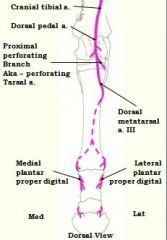
Dorsal metatarsal artery 3
|
|
|
primary retractor of pelvic limb?
|
biceps femoris m.
|
|
|
What arteries have a sigmoid anastomosis in the hindlimb?
|
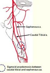
Caudal tibial artery and Saphenous artery
|
|
|
- Prime mover for advancement of hind limb, and flexor of hip
|
Iliopsoas m.
|
|
|
The anastomotic branches of what arteries forms the deep plantar arch?
|

Perforating tarsal artery
Medial plantar artery Lateral plantar artery these anastomose to form *deep plantar arch* on plantar side of hock joint |
|
|
The cranial tibial a. becomes the ?
|

cranial tibial a. becomes the dorsal pedal a. (in front of hock) then becomes dorsal metatarsal artery; largest artery of the foot.
dorsal metatarsal artery = great metatarsal a. |
|
|
Major extensor of the hip joint
|
Middle gluteal muscle
|
|
|
What is the main blood source to the mammary glands?
|
External pudendal artery
|
|
|
attachments to body?
|
Bony attachment (Synovial / diarthrodial) to the body, unlike forelimb.
|
|
|
Superficial gluteal muscle shows ___ and ____ parts
|
cranial and caudal parts
|
|
|
Hind limb Bears ____ of the body weight.
|
45%
|
|
|
innervated by the cranial gluteal nerve
innervated by the caudal gluteal nerve. |
All including CRANIAL part of the superficial gluteal m. innervated by the cranial gluteal nerve!!!!!.....
except caudal head of the superficial gluteal m. which is caudal gluteal nerve. |
|
|
Superficial gluteal attaches to
|
to the third trochanter of the femur.
|
|
|
All gluteal muscles are covered by
|
gluteal m. covered by tough gluteal fascia.
|
|
|
Middle gluteal muscle
blends with what muscle for rearing up on hind limbs? |
longissimus m.
|
|
|
Hamstring muscles possess ____ heads
|
vertebral
|
|
|
Subjected more or less injuries and lameness than forelimb??
|
LESS
|
|
|
When the limb is bearing weight what is the action of the hamstring muscles?
|
If limb bearing weight,
Extend the stifle and hock joints!! Minimize overextension of the stifle and rotation of the patella |
|
|
When the limb is not bearing weight what is the action of the hamstring muscles?
|
Flex stifle joint
|
|
|
Do the hamstring muscles have vertebral and pelvic heads?
|
YES
|
|
|
Pelvic heads attach to
|
to the ischum bone / ischiatic tuberosity
|
|
|
What nerve innervates the hamstring muscles?
|
All innerv. by branches of Sciatic nerve (aka ischiatic n)
|
|
|
pelvic heads of hamstring muscles innervated by ?
vertebral heads of hamstring muscles innervated by ? |
Sciatic nerve
caudal gluteal n. (check this) |
|
|
Hamstring muscles : name them
|
Semitendinosus m., semimembranosus m. and biceps femoris muscle are part of it.
|
|
|
Other muscles (aside from hamstring m.) resp. for extending stifle?
|
quadriceps group
|
|
|
quadriceps group innervated by
|
femoral nerve innervates quadriceps group
|
|
|
therefore problem with femoral nerve means?
|

Any disorder of the femoral nerve leads to inability to extend the stifle joint and weight bearing
|
|
|
What nerve innervates crus ( the caudo-medial muscle group ) (which are flexors of the digits and extensors of the hock joint)?
|
Tibial nerve
|
|
|
What nerve innervates the cranio-laterla mucles group (extensors of the digits and flexors of the hock joint)?
|

Common peroneal nerve (deep peroneal nerve)
|
|
|
What is located at the most lateral point of the hip?
|
Trochanteric bursa
|
|
|
Main artery of hindlimb
|
Femoral artery
|
|
|
What is the main artery of the hindlimb?
|
Femoral artery
|
|
|
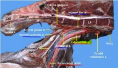
pudendal epigastric trunk comes off?
|
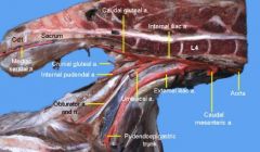
external iliac a.
|
|
|
internal pudendal a. and umbilical a. come off?
|

internal iliac a.
|
|
|
external pudendal a. comes off?
|
pudendal epigastric trunk (confirm this)
|
|
|
What artery passes out through inguinal canal ?
|
external pudendal a.
(that's why it's called "external", b/c goes outside abdominal cavity) |
|
|
Name all branches from which obdurator a. derives:
|
Internal iliac a. > caudal gluteal a. > cranial gluteal a. > obdurator a.
|
|
|
Where is the trochanteric bursa located?
|
Between the cranial part of the greater trochanter and aponeurotic attachment of the accessory gluteal muscle
|
|
|
Femoral artery branches:
|
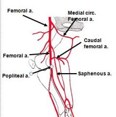
1. Saphenous artery
2. Popliteal artery: * femoral becomes popliteal at caudal aspect of stifle |
|
|
What is the further continuation of the popliteal artery in the hindlimb in order?
|
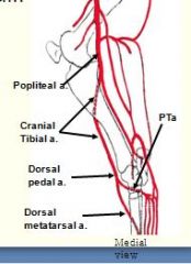
Cranial tibial artery
- becomes Dorsal pedal artery in front of hock - then becomes Dorsal metatarsal artery 3 |
|
|
What artery branches off the dorsal pedal artery?
|
Perforating tarsal artery (enters vascular canal to anastomose with deep plantar arch)
~DPA continues to become dorsal metatarsal III a. |
|
|
What is the distal border of the medial and lateral plantar arteries?
|
Fetlock
|
|
|
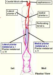
What does the saphenous artery divide into?
|
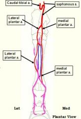
Medial plantar artery
Lateral plantar artery |
|
|
Where do the medial and lateral plantar metatarsal arteries come from?
|

Deep plantar arch -> medial and lateral plantar metatarsal arteries
|
|
|
What does the dorsal metatarsal artery divide into just proximal to the fetlock joint?
|

Medial plantar proper digital artery
Lateral plantar proper digital artery |
|
|
The dorsal and plantar branches of the digital arteries anastomose and form what?
|

Terminal arch
B/c dorsal metatarsal a. III divides just proximal to the fetlock joint into the medial and lateral plantar proper digital arteries (on plantar side). Then, these proper digital arteries wrap around oppos. sides and anastomose WITHIN third phalanx. |
|
|
What are the most important and prominent superficial veins?
(What is main drainage of pes?) |
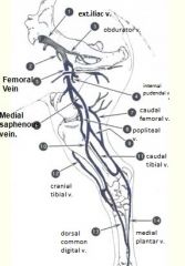
Dorsal metatarsal vein II <- main drainage of pes
Medial saphenous vein Lateral saphenous vein Femoral vein dorsal metatarsal v. -> Medial saphenous v. -> saphenous v. -> Femoral vein |
|
|
What forms the venous plexus in the hindlimb?
|
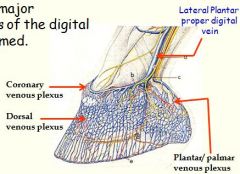
Highly developed network of veins in the hoof dermis
|
|
|
What lymphocenter drains the hindlimb?
Is this the main lymphatic drainage of hind limb? |
Popliteal lymphocenter <- main lymphatic drainage of hind limb
|
|
|
Where do the efferents of the popliteal lymphocenter go to?
|
Deep inguinal nodes
~so popliteal lymphocenter feeds deep inguinal nodes |
|
|
What are the main nerves of the hindlimb?
Distal hindlimb? |

Femoral nerve
Obturator nerve Sciatic nerve (Tibial, Common peroneal) distal hindlimb is innervated mainly by the tibial and common peroneal nn. |
|
|
What is the distal hindlimb mainly innervated by?
|
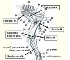
Tibial nerve (caudal)
Common peroneal nerve (cranial) - both branch off from sciatic n. |
|
|
Where does the common peroneal nerve divide into superficial and deep peroneal nn.?
|
At distal part of lateral aspect of stifle
|
|
|
What two tendons does the superficial peroneal nerve lie between?
|
Long digital extensor tendon
Lateral digital extensor tendon |
|
|
The prime mover that advances the pelvic limb:
|
iliospoas m.
|
|
|
when does femoral a. become popliteal a.?
|

when caudal femoral a. splits off
(saphenous a. is first branch, then 2nd branch is where have caudal femoral a. and popliteal a.) note: ca. femoral a. is just landmark for start of popliteal a., but also ca. femoral a. later merges w/saphenous a. |
|
|
Located superficially (just below skin) in the plantar grooves, along the digital flexor tendons.
|
medial and lateral plantar arteries
(these arteries come off saphenous a. on plantar side) note: grooves are on medial and lateral side |
|
|
trochanteric bursa is located:
|
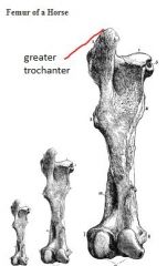
between cranial part of greater trochanter of the femur and under the tendon of insertion of the accessory gluteal muscle
|
|
|
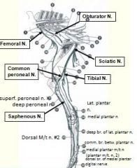
Name a set of nerves supplying the plantar pes.
|
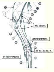
Medial and lateral plantar nn.
|
|
|
ext. obdurator m. innervate by ?
internal obd. m. innervated by ? |
* external obturator m. innervate by obdurator nerve
- ext. obturator m. ADDucts limb * internal obturator. m. innervated by sciatic n. - internal obturator m of medial thigh aBducts the thigh |
|
|
Is DDF part of reciprocal apparatus?
Is DDF part of stay apparatus? |
No (according to Holli)
Yes (according to Pasquini's book) |
|
|
Why is dorsal metatarsal 2 vein have "2" in name?
|
because it runs along 2nd metatarsal bone on medial side!
|