Reading...
![]()
Play button
![]()
Play button
![]()
Use LEFT and RIGHT arrow keys to navigate between flashcards;
Use UP and DOWN arrow keys to flip the card;
H to show hint;
A reads text to speech;
513 Cards in this Set
- Front
- Back
|
What are the different divisions of the mediastinum?
|
*Superior
*Inferior (Anterior, middle, posterior) |
|
|
What are the subdivisions of the inferior mediastinum?
|
Anterior
Middle Posterior |
|
|
What are the boundaries of the superior mediastinum?
|
Extends from the superior thoracic aperature to the transverse thoracic plane (sternal angle to IV disc between T4/T5)
|
|
|
What are the boundaries of the anterior mediastinum
|
Between the pericardium and sternum
|
|
|
What are the boundaries of the middle mediastinum
|
Occupied by the heart
|
|
|
What are the boundaries of the posterior mediastinum?
|
Between the pericardium and anterior portion of the vertebral bodies (T5-T12)
|
|
|
What are the boundaries of the inferior mediastinum
|
Goes from the transverse thoracic plane down to the diaphragm
Subdivided into the anterior, middle and posterior portions |
|
|
What is a mediastinum in general
|
A septum between two parts of an organ or a cavity (division)
|
|
|
What is the mediastinum in the thorax?
|
A division between the viscera of the lungs
Central compartment of the thoracic cavity |
|
|
What thoracic viscera does the mediastinum contain
|
All of them except for thel ungs
|
|
|
Is the mediastinum more moveable in children or in adults?
|
It is highly moveable in youth and less so with age (lots more restriction develop)
|
|
|
What are the hollow visceral structures that the mediastinum contains?
|
*Connective tissue
*Nerves *Fat *Lymphatic vessels *Blood *Lymph nodes *Espohagus |
|

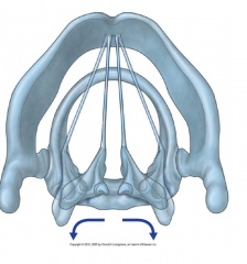
What is the red arrow pointing at?
|
The superior thoracic aperature
|
|
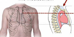
What is the red line indicating?
|
Transverse Thoracic Plane
|
|

What is the area above the red dotted line?
|
Superior mediastinum
|
|

What is the area below the red dotted line?
|
The inferior mediastinum
|
|
|
What are the arteries that can be found in the superior mediastinum?
|
*Aortic arch and branches
*Brachiocephalic trunk and veins *Superior vena cava *Left common carotid artery *Left subclavian artery |
|
|
What are hte nerves in the superior mediastinum?
|
*Vagus nerve
*Left recurrent laryngeal nerve and left pulmonary plexus (from the left vagus n.) *Cardiac plexus of nerves and esophageal plexus (from the right vagus nerve) *Phrenic nerve |
|
|
WWhat are the "visceral" structures found in the superior mediastinum
|
*Trachea
*thymus *Esophagus *Thoracic duct |
|
|
Where does the vagus nerve go to?
|
The heart
|
|
|
Is the vagus nerve sympathetic or parasympathetic
|
Parasympathetic
|
|
|
What cranial nerve is the vagus nerve
|
CN 10
|
|
|
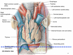
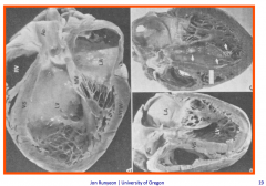
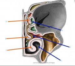
What does the superior mediastinum look like?
(Flip over for picture) |
|
|

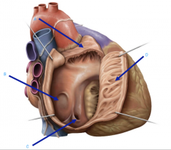
What is A?
|
Trachea
|
|

What is B?
|
Left Laryngeal Nerve
|
|

What is C?
|
Esophagus
|
|

What is D?
|
Phrenic Nerve
|
|
What is E?
|
Vagus Nerve
|
|

What is F?
|
Azygos Vein
|
|

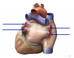
What is A?
|
Right Pulmonary Artery
|
|
What is B?
|
Left pulmonary artery
|
|

What is C?
|
Right and left main bronchi
|
|
|
How are the right and left vagus nerves different?
|
The left vagus nerve has the left laryngeal nerve which comes off the vagus nerve as a branch that bends upwards again.
|
|
|
Where does the laryngeal nerve go?
|
Goes to the larynx
(So if something wrong with it, there may be issues with voice production) |
|
|
Where does the laryngeal nerve wrap around?
|
Wraps around the arch of aorta
|
|
|
What is an anuerism
|
Expansion
|
|
|
What can happen of the arch of aorta gets an anurism (expansion)
|
It can get bigger and that pushes on and stretches the laryngeal nerve which can cause it to potentially get damaged
|
|
|
Is the ascending aorta big?
|
Yes, it can be around 3.5 cm - 5.5 cm
|
|
|
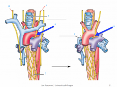
What comes off of the aortic arch?
|
*Brachicephalic trunk (it has the right subclavian artery and the right common carotid artery)
*Left common carotid artery *Left subclavian artery |
|
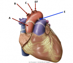
What is A?
|
Aorta (Ascending aorta)
|
|

What is B?
|
Brachiocephalic trunk
|
|

What is C?
|
Common Carotid artery
|
|

What is D?
|
Subclavian artery
|
|

What is E?
|
Ligamentum arteriosum
|
|
|
What does the anterior mediastinum house?
|
Lymphatic, fat and nerve
|
|
|
What does the posterior mediastinum house?
|
*Esophagus
*Vagus nerve (Esophageal plexus) *Thoracic sympathetic trunks *Lower thoracic splanchnic nerves *Thoracic aorta and its branches *Thoracic duct *Azygos, hemiazygos and accessory hemiazygos veins |
|
|
Is ther ea lot of space for the anterior mediastinum?
|
No
|
|
|
What is the function of the pericardium?
|
*Lubricate
*Protects against spreading infection *Limits size of heart (limits acute distension); protects against overfilling of heart chambers and outstretching of cardiac muscle *Diastolic coupling |
|
|
What is technically considered the base of the heart?
|
Left atrium
|
|
|
What sort of wall does the left atrium
|
Smooth walled
|
|
|
Does the left atrium have an auricle?
|
Yes
|
|
|
Does the left atrium have pectinate muscles?
|
Yes
|
|
|
What openings does the left atrium have?
|
pulmonary semilunar valve
Mitral valve |
|
|
What are the walls of the right atrium like?
|
Smooth and thin posterior wall
Rough and muscular anterior wall |
|
|
What divides the rough and musuclar anterior wall from the smooth and thin posterior wall of the right atrium?
|
Sulcus terminalis
|
|
|
Does the right atrium have pectinate muscles?
|
Yes
|
|
|
Where is the fossa ovalis located?
|
In the right atrium
|
|
|
What does the right atrium have openings for?
|
*Superior vena cava
*Inferior vena cava *Coronary sinus |
|
|
What valve does the right atrium have leading to the right ventricle?
|
Tricuspid valve
|
|
|
Does the right atrium have an auricle?
|
Yes
|
|
|
What forms the apex of the heart?
|
The left ventricle
|
|
|
What chamber of the heart performs the majority of the work
|
Left ventricle
|
|
|
What valve connects the left ventricle to the left atrium?
|
Mitral valve
|
|
|
Does the left ventricle have pectinate muscles?
|
No
|
|
|
Does the left ventricle have auricles?
|
No
|
|
|
Does the left ventricle have trabeculae carnae?
|
Yes
|
|
|
How many papillary muscle groups does the left ventricle have? And what are they called?
|
2
Anterior/posterior papillary muscles |
|
|
What is the left ventricle opening for?
|
For the aortic valve
|
|
|
Does the left ventricle have thick or thin walls?
|
Thick
|
|
|
What surface of the heart does the right ventricle make up?
|
Anterior and inferior surface of the heart
|
|
|
Does the right ventricle have trabeculae carnae?
|
Yes
|
|
|
What is a special feature in the right ventricle that the left ventricle does not have?
|
Conus arteriosus
|
|
|
What valve connects the right ventricle with the right atrium?
|
Tricuspid valve
|
|
|
What is the right ventricle an opening for?
|
Pulmonic valve
|
|
|
How many papillary muscle groups does the right ventricle have? And what are their names?
|
3 papillary muscle
Anterior, posterior and septal |
|
|
What special feature does the right ventricle have?
|
*Moderator band (or septomarginal trabecula)
Helps connect the anterior papillary muscle to the interventricular septum |
|
|
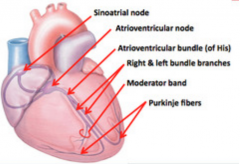
What are the two nodes of the conducting system?
|
SA and AV nodes
(Pacemaker tissue( |
|
|
Where is the SA node located/
|
Right atrium
|
|
|
Where is the AV node located?
|
Near coronary sinus opening
|
|
|
When an impulse is initiated at the SA node, what are the chambers that contract?
|
Left and right atrium
|
|
|
Why does the SA node not cause direct ventricular contraction?
|
Because of the fibrous skeleton of the heart which acts as an electrical insulator between the atria and ventricles which facilitates their independent contraction
|
|
|
What is the full electrical signal pathway
|
SA node initiates signal
Sends down to AV node AV right and left bundles (which stimulate the IVS, papillary muscles, walls of their respective ventricles and also the moderator band for the right bundle branch) Then sent down to the Purkinje fibers |
|
|
What does the left bundle branch stimulate? (Which muscles)
|
*Interventricular septum
*Anterior papillary muscle *Posterior papillary muscle *Left ventricualr wall |
|
|
What does the right bundle branch stimulate?
|
Muscles of the:
*IVS *Anterior papillary muscle through septomarginal trabecula and the right ventricular wall |
|
|
What does the right coronary artery attach to?
|
AV node (most people) and SA node in ~60%
|
|
|
What does the left coronary artery attach to?
|
SA node in ~40$ of people
AV bundle is supplied largely by LCA |
|
|
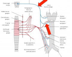
Where do the postsynaptic sympathetic fibers travel?
|
Original from the thoracic and cervical ganglia of the sympathetic fibers originate in upper thoracic and cervical ganglia of sympathetic trunks and travel to the heart after passing through the cardiac plexus and finally innervates the SA and AV nodes, heart muscle and coronary arteries. In addition, indirect sympathetic stimulation stems from adrenal gland (via blood stream)
|
|
|
What does sympathetic stimulation of the heart cause?
|
*Increase heart rate
*Increase force of contraction of heart *dilation of coronary arteries (via the beta-2 receptors on coronary vessels) |
|
|
Where are the presynaptic fibers of the parasympathetic supply
|
Bundles of axons from the vagus nerve which originates in the medulla oblongata. These fibers travel throughout the cardiac plexus to innervate the intrinsic ganglia (postsynaptic cell bodies) of the heart wall, which project the majority of their fibers to the Sa and AV nodes
|
|
|
Where is the postsynpatic fibers of the parasympathetic supply located
|
In the heart wall...mostly the wall of the rigt atrium and the interatrial septum
|
|
|
What is the general effect of parasympathetic stimulation of the heart?
|
Slows down heart rate
|
|
|
What does the middle mediatinum contain?
|
The heart, pericardium, phrenic nerve, vagus nerve and the roots of the great vessels (Ascending aorta, pulmonary trunk and superior vena cava)
|
|
|
The pericardium provides which of the following functions?
a) Lubrication b) Diastolic coupling c) Limits size of heart (distension) d) All of the above |
All of the above
|
|
|
What are the three layers of the pericardium?
|
1) Fibrous (outer) layer
2) Connective tissue (dense irregular) 3) Parietal/visceral layer |
|
|
What is the serous pericardium
|
Parietal and visceral layer of the pericardium
|
|
|
What type of epithelium is the serous membrane?
|
Simple squamous
|
|
|
Where is the parietal layer?
|
Lines the internal surface of the fibrous pericardium
|
|
|
What is the visceral layer of the periocardium?
|
On the external surface of the heart
|
|
|
What is between the visceral and parietal layer of the pericardium?
|
Pericardial fluid
|
|
|
Where is the fibrous layer of the pericardium found?
|
Outermost layer; outside of the parietal layer
|
|
|
What does the fibrous layer of the pericardium do?
|
Keeps the heart open
|
|
|
What type of epithelium is the mucus membrane?
|
Simple columnar
|
|
|
What type of epithelium is the cutaneous membrane?
|
Stratified squamous
|
|
|
Does the serous membrane make fluid?
|
Yes, it makes a small amount of fluid
|
|
|
What is the fluid made from the serous membrane used for?
|
For lubrication function
|
|
|
Where is the simple squamous epithelium found?
|
Lining the visceral and parietal layer
|
|
|
All epithelium sits directly on what?
|
Connective tissue
|
|
|
What is the order of layers for a blood vessel
|
Lumen
Simple squamous epithelim Connective tissue Muscle Connective tissue |
|
|
What layer of the pericardium connects to the great vessels as they enter and exit the heart
|
The Fibrous layer
(The pericartdium helps anchor to the great vessel) |
|
|
What type of epithelium is the endocardium?
|
Simple squamous
|
|
|
What layer of the heart is a continuation of blood vessel lining?
|
Endocardium
|
|
|
What are the functions of the pericardium?
|
*Mechanical protection (protects against excessive movement)
*Lubrication *Prevents the spread of infection and malignacy (cancer) *Limits acute distention (prevent rapid overfilling; so it doesn't get too big) *Diastolic coupling |
|
|
Where does pericarditis occur/
|
Can be anywhere but typically on the visceral and parietal layer
|
|
|
What does pericarditis cause?
|
An increased pericardial fluid production (There is a blood flow into the cavity)
|
|
|
What can pericarditis cause?
|
Increased pericardial fluid production
|
|
|
What happens if there is an increased amount of fluid
|
The fluid will build up which will increase the pressure and since the fibrous tissue itself is not very elastic, the fluid will compress the heart muscle which is cardiac tamponade (heart compresison)
|
|
|
If there is increased fluid what will that do to the pressure
|
Cause an increase in pressure
|
|
|
Can the cardiac tamponade or heart compression be audible with a stethoscope
|
Yes, therei s a not clear or distince heart beat, sounds like two pieces of sand paper rubbing together
|
|
|
What must be done when there is pericarditis?
|
Must be drained (to get the fluid out)
|
|
|
What can happen with the cardiac tamponade or heart compresison?
|
There can be thickening, scarring or calcification
|
|
|
There is cardiac tamponade
(The gray stuff around is all the built up pericardial fluid) |
|
|
What is the procedure to get fluid out of the pericardial cavity called?
|
Pericardiocentesis
|
|
|
How does pericardiocentesis work?
|
Get a needle inserted through the fifth or sixth intercostal space
|
|
|
What allows for the pericardiocentesis?
|
Cardiac notch of the left lung
|
|
|
What layers must the needle go through to allow for the pericardicentesis?
|
Fibrous and parietal layer of the pericardium
|
|
|
What are the two pericardial sinuses?
|
*Transverse pericardial sinus
*Oblique pericardial sinus |
|
|
What is the cardiac bypass sinus?
|
Transverse pericardial sinus
|
|
|
Where can the transverse pericardial sinus be found?
|
Behind the pulmonary trunk and anterior aorta and in front of the superior vena cava
|
|
|
Where can the oblique pericardial sinus
|
*Behind the heart
*In the cul-de-sac in the pericardial caivty that is posterior to the base of the heart *Bound by the perciardial reflections surrounding the pulmonary veins and IVC and pericardium overlying the esophagus |
|
|
Anyne with heart problems will have some degree what type of symptoms?
|
Fatigue
Shortness of breath Chest pressure |
|

What is 1?
|
SVC
|
|

What is 2?
|
Right atrium
|
|
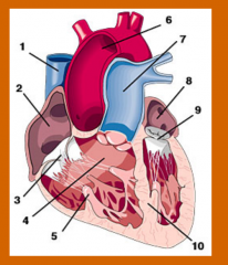
What is 3?
|
Tricuspid Valve
|
|

What is 4?
|
Right ventricle
|
|

What is 5?
|
Papillary muscle
|
|

What is 6?
|
Ascending aorta and arch
|
|

What is 7?
|
Pulmonary trunk
|
|

What is 8?
|
Left atrium
|
|

What is 9?
|
Mitral valve
|
|

What is 10?
|
Interventricular septum
|
|
|
The right atrium forms the ______ border of the heart
|
Right
|
|
|
The right atrium receives _____ blood
|
Venous
|
|
|
Where does right atrium receive venous blood from?
|
SVC
IVC Coronary sinus Anterior cardiac vein Thesbian vein |
|
|
What is the interior of the right atrium like, what kind of posterior and anterior wall does it have?
|
*Smooth, thin-walled posterior wall
-Rough, muscular anterior wall |
|
|
What does the sulcus terminalis of the right atrium do?
|
Seperates the smooth and rough wall of the right atrium
|
|
|
Does the right atrium have pectinate muscles?
|
Yes
|
|
|
What chamber of the heart has the fossa ovalis?
|
Right atrium
|
|
|
What openings can be seen in the right atrium?
|
Coronary sinus
IVC SVC Anterior caridac veins Thebesian veins |
|
|
The right atrium has the orifice of what valve?
|
Tricuspid valve
(In the anterior aspect of the RA) |
|
What is A pointing to?
|
Auricle
|
|

What is B pointing to?
|
Fossa Ovalis
|
|

What is C pointing to?
|
Orifice of the coronary sinus
|
|

What is D pointing to?
|
Musculi Pectinati
|
|
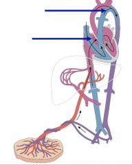
Explain the schematic of fetal circulation
|
The pulmonary trunk floods directly into the aorta because the umbilicial vein is already bring in oxygenated blood to the heart so they don't need to go to the lung; so that is why there is the foramen ovalis there to just allow the blood to flow between the left and right atrium.
The pulmonary trunk and aorta together push blood to the body |
|
|
What is the role of the placenta
|
Nutriet to the fetus
|
|
As soon as the baby is born what happens to the circulation?
|
As soon as the baby is born there is a rapid changei n the pressure of the system. Whichi s caused by the baby taking its first breath. There is a change in blood flow and blood flow pressure. That portion of the pulmonary trunk collapses and the blood is force out through the pulmonary arteries. The shunt collapses and fuses which is where the ligamentuam arteriosum is created
|
|
|
How much of the general population is patent foramen ovale foudn in?
|
Up to 25% of the genral population
|
|
|
What is patent foramen ovale (PFO)?
|
When the foramen ovale does not close allo f the way between the left and right atrium to allow for the fossa ovalis to be created
|
|
|
What happens to patients with patent foramen ovale?
|
They have a slight decrease in oxygened blood content because some of that blood will be going straight into the left atrium and bypassing the lung
|
|
|
What chamber forms the base of the heart?
|
The left atrium
|
|
|
The left atrium receives what type of blood?
|
Oxygenated blood
|
|
|
Where does the left atrium receive blood from?
|
The pulmonary veins
|
|
|
What is the walls of the left atrium like?
|
Most smooth-walled (will see some pectinate muscles, but much more smooth)
|
|
|
How many pulmonary veins enter the left atrium?
|
Four pulmonary veins enter the posterior wall
|
|
|
What is the left atrium have an orifice for?
|
The mitral valve
|
|
What are these arrows pointing to?
|
The left and right superior and inferior pulmonary veins
|
|
|
The right ventricle forms the _______ surfaces of the heart
|
Anterior and inferior
|
|
|
What is the trabeculae carneae?
|
Thick band of muscle
|
|
|
What does the trabeculae carneae do?
|
Allows for contraction
|
|
|
Is there trabeculae carneae found in the right ventricle?
|
Yes
|
|
|
What is the conus arteriosus?
|
The transition zone between the right ventricle and the pulmonary trunk
|
|
|
What valve connects the right ventricle with the right atrium?
|
Tricuspid valve
|
|
|
What does the right ventricle have an orifice of?
|
The pulmonic valve
|
|
|
How many papillary muscles does the right ventricle have and what are they called?
|
Anterior
Posterior Septal (On septal walls) |
|
|
Where is the septomarginal trabecula or moderator band found?
|
In the right ventricle
|
|
|
The pulmonary trunk is the arterial continuation of what?
|
The right ventricle
|
|
|
What does the pulmonary trunk split into?
|
The right and left pulmonary arteries
|
|
|
What does the pulmonary trunk do?
|
Conducts de-oxygenated blood to the lungs for oxygenation
|
|
|
What does the left ventricle of the heart form?
|
The apex of the hert
|
|
|
The right ventricle performs more work than the left ventricle, true or false?
|
False
(Left ventricle performs more work because it is pumping blood to the whole body so goes against a lot more resistance) |
|
|
What is the valve that is found between the left ventricle and the left atrium called?
|
The mitral valve
|
|
|
Does the left ventricle have trabeculae carneae?
|
Yes
|
|
|
How many papillary muscles does the left ventricle have and what are they called?
|
2
Anterior, posterior |
|
|
What orifice does the left ventricle have?
|
Orifice of the aortic valve
|
|
|
What pathway does blood move thorugh the various blood vessels?
|
Heart -> Elastic arteries -> Muscular arteries -> Arterioles -> Capillaries -> Venules -> Medium veins -> Large veins -> Heart
|
|
|
What are the three layers of a blood vessel?
|
Tunica intima
Tunica media Tunica externa or adventitia |
|
|
What layer lines the blood vessel?
|
Tunica intima
|
|
|
What is the structure of the tunica intima?
|
*Composed of endothelium (simple squamous epithelium) and underlying connective tissue (basement membrane)
*Varying amount of elastic fibers in basal lamina |
|
|
What is the function of the tunica intima
|
Smooth layer to reduce friction of passing blood components; creates a selectively permeable filter to blood components; secrets vasoconstrictors and vasodilators
|
|
|
What is the structure of the tunica media?
|
*Layer of smooth muscle
*Elastin sheets in the artereis *thickest layer of an artery |
|
|
What is the thickest layer of an artery
|
Tunica media
|
|
|
What is the function of the tunica media?
|
Muscle that allows for vasoconstriction or vasodilation and elastin that allows the arteries to stretch and recoil
|
|
|
What is the structure of the tunica externa or adventita like?
|
Composed of conenctive tissue rich in collagen and elastic fibers
Lymphatic vessels, arterioles (nutritive arties to outer portion of vessel wall) and nerves travel within the adventita Thickest layer in veins |
|
|
What is the thickest layer in veins?
|
Tunica externa or adventitia
|
|
|
What is the function of the tunica externa or adventita?
|
To protect and anchor vessels
|
|
|
Elastic arteries have thicker what layer?
|
Tunica intima
|
|
|
What do elastic arteries have?
|
*Thicker tunica intima
*Sub-endothelial layer containing elastic fibers *Larger lumen than do other arteries |
|
|
Where are the elastic fibers of the elastic arteries found?
|
In tunica intima and tunica media layers
|
|
|
Elastic arteries are high or low resistance pathways?
|
Low
|
|
|
What are the fasting conducting of the artereis?
|
Elastic arteries
|
|
|
Why are elastic arteries allows to act as pressure reservoirs?
|
Because they can expand and recolil and minimize blood pressure changes during the cardiac cycle
|
|
|
What are examples of elastic artereis?
|
Aorta
Brachiocephalic trunk Carotids Pulmonary trunk |
|
|
What is the thickest layer of the muscular arteries
|
Tunica media
|
|
|
Which has more smooth muscle, elastic or muscular arteries?
|
Muscular
|
|
|
What do the muscular arteries do?
|
Distribute and regulate blood flow to different regions of the body
|
|
|
What do arteries do?
|
Control the flow of blood to capillary beds
|
|
|
What vessels are the primary contributor to blood pressure?
|
Arterioles
|
|
|
Arteries are large or small?
|
Small
|
|
|
Do arterioles have a narrow or large lumen?
|
Narrow
|
|
|
Does the arterioles have a relatively thick or thin muscular wall?
|
Thick
|
|
|
Are arteries or veins more subject to greater pressure?
|
Artereis
|
|
|
Between arteries and veins, which one actively control blood pressure?
|
Arteries
|
|
|
Which has a thicker tunica media, arteries or veins?
|
Artereis
|
|
|
Which has more elastic and collagen fibers, arteries to vein? Why?
|
Arteries,
To stretch and recoil and resist the greater pressure |
|
|
Which is considered the "round" vessel, arteries or veins?
|
Arteries
|
|
|
Are veins subject to more or less pressure than artereis?
|
Less
|
|
|
Compared to artereis, do veins have relativey thin or thick tunica media layers?
|
Thin
|
|
|
What is the thickest layer in a vein?
|
Tunica externa
|
|
|
Which appear "collapsed" in dissection, veins or arteries?
|
Veins
|
|
|
Do veins have thinner walls than artereis?
|
Yes
|
|
|
Do veins have larger lumen than arteries?
|
Yes
|
|
|
Between venules and veins which are more analogus to capillaries?
|
Venules
|
|
|
The smallest venule will just have a layer of what?
|
Smooth muscle around them
|
|
|
What type of venule will have all three tunics?
|
Largest
|
|
|
What do venules provide a site for?
|
For WBCs to exit the circulation
|
|
|
Are valves present in venules?
|
No
|
|
|
Will all veins have all three tunics?
|
Yes
|
|
|
Which of the tunicas of the veins is the thickest?
|
Tunica externa
|
|
|
Do veins have large lumens?
|
Yes
|
|
|
ARe valves present in veins?
|
Yes
|
|
|
What is an anastomosis?
|
Connection or joint
|
|
|
What is an anastomosis in the circulatory system?
|
Connection between vessels
|
|
|
What is an artery to artery connection called?
|
Arterio-arterial anastomosis
|
|
|
What are arterio-venous anastomoses?
|
Connections between arterioles and venules
|
|
|
What do arterio-venuous anastomoses do?
|
Shunt blood away from capillary beds, completely or partially, thereby controlling the amount of blood entering a given capillary bed
|
|
|
Are the cardiac or skeletal muscles striated?
|
Both of them are
|
|
|
How many nuclei does the skeletal and the cardiac muscle have?
|
Cardiac muscle is single nucleated
Skeletal muscle is multi-nucleated |
|
|
What has gap junctions, skeletal or cardiac muscle?
|
Cardiac
|
|
|
What does the gap junctions do?
|
Allows for there to be an electrical connection, so if one fires; adjacent fires will fire along with it. They all contract and pull on each other
|
|
|
Where does the cardiac muscle have its gap junctions
|
On all muscle of heart so that they can contract and squeeze together as an unit
|
|
|
Why does the skeletal muscle have multi-nucleated?
|
Because the skeletal muscle is larger than the cardiac muscle and cna be very ong cells so needs lots of nuclei
|
|
|
Where are the intercalated discs found?
|
In cardiac muscle
|
|
|
What do the intercalated discs do?
|
Connects the branching cells of cardiac muscle (like desmosomes)
|
|
|
Where does the base of the heart face?
|
Faces posteriorly toward T6-T9
|
|
|
What chambers of the heart make up the base of the heart?
|
Left atrium mainly; and a little bit of the right atrium
|
|
|
Where is the apex of the heart
|
5th intercostal space in the mid clavicular line
|
|
|
Is the apex of the heart motionless or not during the cardiac cycle?
|
Motionless (It is held firm)
|
|
|
Where are the maximal heart sounds found?
|
Apex of the heart
|
|
|
What is the PMI
|
Point of maximal intensity
|
|
|
Where can the PMI be found
|
Apex of the heart
|
|
|
The apex of the heart is the inferolateral portion of the _______
|
Left ventricle
|
|
|
What is the left ventricle attached to?
|
Diaphragm
(Which is why it remains motionless during contractions) |
|
|
What are the four cardiac surfaces
|
1) Anterior
2) Diaphragmatic 3) Right pulmonary 4) Left Pulmonary |
|
|
What does the anterior surface of the heart contain?
|
Right ventricle
|
|
|
What does the diaphragmatic surface of the heart contain?
|
(Resting on the ventricle)
*Left ventricle *Some right ventricle |
|
|
What does the right pulmonary surface of the heart contain?
|
Right atrium
|
|
|
What does the left pulmonary surface of the heart contain?
|
Left ventricle
|
|
|
What are the cardiac borders?
|
1) Right
2) Inferior 3) Left 4) Superior |
|
|
What does the right cardiac border contain?
|
Right atrium from SVC to IVC
|
|
|
What does the inferior cardiac border contain?
|
Right ventricle
Some left ventricle |
|
|
What does the left cardiac border contain
|
Left ventricle
Some left auricle |
|
|
What does the superior cardiac border contain?
|
1) Right and left atrium and auricle
2) Ascending aorta 3) Pulmonary trunk 4) SVC enters the heart |
|
|
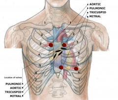
What auscultation can you hear at the right upper sternal border?
|
Aortic
|
|
|
What auscultation positions is the left upper sternal border?
|
Pulmonic
|
|
|
What auscultation positions can be heard from the left lower sternal border?
|
Tricuspid
|
|
|
What auscultation can be heard from the LV apex?
|
Mitral
|
|
|
What causes the lub tone?
|
AV valve close
|
|
|
What causes the dub tone?
|
Semilunar valve closure
|
|
|
How many sounds are heard for both lub and dub
|
Two sounds each
(So two sounds for lub and two for dub) |
|
|
What is a diagram for the site of auscultation of valves?
|
|
|
|
During systole what are the state of the valves?
|
The semilunar valves are open
The atrioventricular valves are closed |
|
|
What are some problems that can occur to the aortic valve?
|
*Bicuspid aortic valve
*Calcified aortic valve |
|
|
What can the bicuspid aortic valve cause?
|
Has two valves instead of three, can become stinotic later 4th-5th decade of life
|
|
|
What can happen because of a clacified aortic valve?
|
Calcium chunks, can't open very well; get stiff
|
|
|
What is the function of the cardiac valves?
|
Maintain unidirectional flow and prevent back flow
|
|
|
What happens if the valve does not close al lthe way?
|
Blood would go out that way but some will also go back to where it came from.
|
|
|
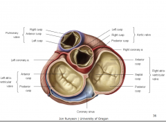
How many cusps does the tricuspid valve have? And what are they called?
|
3
Anterior Posterior Septal |
|
|
Where can the mitral valve be found?
|
Posterior to the sternum at the 4th costal cartilage
|
|
|
How many cusps does the mitral valve have? What are they called?
|
2
Anterior Posterior |
|
|
What kind of arrangement does cardiac muscle have?
|
Spiral
When contracting, it wringing out the blood |
|
|
What kind of plane do valves sit in?
|
Transverse plane
|
|
|
Why are valves held open?
|
Held open because of the carilaginous support of valves (so they don't collapse in).. They are attached to a cartilaginous ring which allows for structural integrity and for the valve to keep its shape
|
|
|
What kind of layer is the atria siting on? And what does that do?
|
Sittingo n a fibrocartilaginous layer
Holding the skeleto of the valves intact and alos providing electrical insulation |
|
|
How are ventricles able to contract if the atria has that fibrocartilaginous layer?
|
There are fibers that run through and link the atria to the ventricles. There is a built in pause at the bundle of his that allows for a delay which allows the chambers to contract in succession and not all together.
|
|
|
Where is the apex of the conus arteriosus?
|
Left third costal cartilage
|
|
|
How many cusps does the pulmonary valve have?
|
3
Anterior Right Left |
|
|
What accounts for the different in wall thickness between the pulmonic and aortic valve?
|
The tunica media layer
Because it is made of smooth muscle. Aortic valve is able to have a thicker tunica media |
|
|
Where can the aortic valve be found?
|
Posterior to the left side of the sternum at the 3rd ICS (intercostal space)
|
|
|
How many cusps does the aortic valve have?
|
3
right -Gives rise to the right coronary artery Left-Gives rise to the left coronary artery Posterior cusp |
|
|
Image of all of the valves
|
|
|
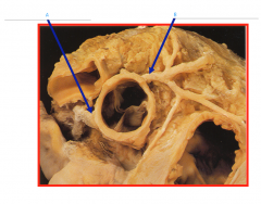
What is A and B connected to?
|
Aorta
|
|

What is A?
|
Left coronary artery
|
|

What is B?
|
Right coronary artery
|
|

What is 1?
|
Right coronary artery
|
|

What is 2?
|
Right marginal artery
|
|

What is 3?
|
Posterior descending artery
|
|
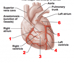
What is 4?
|
Anterior descending artery
|
|

What is 5?
|
Circumflex artery
|
|

What is 6?
|
Left coronary artery
|
|
|
When is coronary blood flow the lowest, why?
|
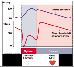
During ventricular systole, aortic valve cusps occlude coronary flow.
Because systole cusps are going to flap open, so the valve cusps occlude the coronary blood flow |
|
|
What is the advantage of having coronary blood flow be the lowest during ventricular systole?
|
Because the increase in blood pressure in the ventricle and the arch of aorta. The arch is the biggest artery and the strongest and has the thickest media of all of the vessels; while coronary arteries are smaller, not as much stretch and muscle. So without the cusps closing off the entrance they would allow increase blood flow into the body through those tiny arteries in the heart which is bad, so this protects them.
|
|
|
What is considered the left main artery?
|
Left coronary artery
|
|
|
What does the left coronary artery come off from?
|
The left sinus of Valsalva (aortic valve)
|
|
|
What does the left coronary artery travel through?
|
Left atrioventricular groove
|
|
|
What are the branches of the left coronary artery?
|
*Left anterior descending
*Left circumflex |
|
|
What does the right coronary artery comes from?
|
The right (anterior) sinus of valsalva
|
|
|
What does the right coronary artery travel through?
|
Right atrioventricular sulcus
|
|
|
What are the branches off the right coronary artery?
|
*Right atrial
*SA nodal (60%) -> Pectinate muscles *Marginal branches *Posterior descending artery |
|
|
Where does the left ventricle typically get most of its blood from.
|
Left coronary artery
|
|
|
What does right coronary dominance mean?
|
Left ventricle gets most of its blood from the right coronary artery.
(Opp for left coronary dominance and codominant) |
|
|
A 37 year old patient with severe chest pain, shortness of breath, and congestive heart failure was admitted to the local hospital. His ECG reveals a myocardial infarction in the area of the apex of the heart. Which of the following is most likely occluded?
A. Right marginal artery B/ Right coronary artery at its origin C. Anterior interventricular artery D. Posterior interventricular artery E. Circumflex branch of left coronary artery |
C. Anterior interventricular artery
|
|
|
What coronary artery occlusion is called the "The Widow Maker"
|
Occlusion of left anterior descending coronary artery
|
|
|
What coronary arteries are most likely to occlude?
|
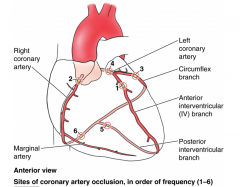
For sites 1-3; 85% of occlusions occur there
|
|
|
What is a pulmonary embolism?
|
Blood clot in pulmonary circuit (such as when blood pools can cause clots) then clot will dislodge and will move up the femoral vein into the inferior vena cava (which is going from small to bigger) then it moves through the heart; the pulmonary trunk and the vessels get smaller and will then reach smallest vessel and stop when can feel intense pain
|
|
|
What is aortic dissection
|
The aorta has opened up; blood is leaking out of the aorta and rapidly fills the pericardium with blood causing the cardiac tamponade
|
|
|
What is the difference between stinotic and incompetant valves?
|
Stinotic: Narrowing of opening of valve caused by stiffening of valve cusps (narrowing(
Incompetant: Doesn't close; fluid going the wrong way |
|
|
What valve failure will lead to sudden death?
|
If left AV valve fails, ventricle contract and blood from the left ventricle will go into the left atrium and will start building up there and that can start pulling out which can lead to sudden death
|
|
|
Which valve is the least important?
|
Pulmonary semilunar valve; if it fails, can still get some blood to the lungs, so inefficient, can still live on
|
|
|
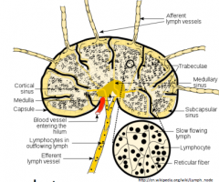
What are the smallest lympahtetic vessels?
|
Capillary
|
|
|
What do lymphatic capillaries carry?
|
Lymph
|
|
|
Where can lymphatic capillaries be found?
|
In most vascular veds, (with notable exception of the CNS, bone and teeth(
|
|
|
What lymphatic vessels collect lymph from the lymphatic capillaries?
|
Superficial and deep lymphatic vessels
|
|
|
How do lymphatic vessels accomplish progressively draining lymph into larger vessels?
|
By passing it through many lymph nodes
|
|
|
What do large lymphatic vessels drain into?
|
Larger lymphatic trunks
|
|
|
What do lymphatic trunks converge to form?
|
The thoracic duct and right lymphatic duct (which drain directly to the venous system)
|
|
|
Where does the right lymphatic duct empty into?
|
The right subclavian vein
|
|
|
Where does the right lymphatic duct receive lymph from?
|
Right upper extremity, thorax, head and neck
|
|
|
Where does the thoracic duct empty into?
|
The left subclavian vein
|
|
|
Where does the thoracic duct receive lymph from
|
From the rest of the body (left upper extremity, thorax, and head; all of abdomen, pelivs and both lower limbs)
|
|
|
What are the three tonsils?
|
*Palatine tonsil
*Pharyngeal tonsil *Lingual tonsil |
|
|
Where is the palatine tonsil found?
|
Posterolateral oropharynx
|
|
|
What does the palatine tonsils also called?
|
Tonsils
|
|
|
Where can the pharyngeal tonsils be found?
|
Roof and posterior wall of nasopharynx ("Adenoids")
|
|
|
Where can the lingual tonsils be found?
|
The root of the tongue
|
|
|
Where is the tubal tonsil found?
|
Lymphoid tissue around opening of auditory tube
|
|
|
What are the functions of a lymph node?
|
*Maturation of new lymphocytes
*Filtration of lymph *Initiate immune response |
|
|
Where are the three clusters of lymph nodes located?
|
*Axillary
*Cervical *Inguinal |
|
|
Where is the thymus located?
|
Anterior and superior to the heart , just posterior to the manubrium of the sternum, within the superior mediastinum
|
|
|
When is the thymus most active?
|
In childhood
|
|
|
What is the thymus progressively replaced by as we age?
|
Fat
|
|
|
What is the thymus the site for?
|
Site of formation and maturation of t-lymphocytes, an important component of the immune system
|
|
|
Which of the following can be found at the left 5th intercostal space in the mid-clavicular line?
A. The apex of the heart B. The point of maximal impulse (PMI) C. The base of the heart D. Both A and B E. Both B and C |
D. Both A and B
|
|
|
Male smoker presents empysema and now complaints of lower extremity swelling, now scrotal swelling, nausea and anorexia.
Exam is notable for a barrel chest, elevated jugular venous pressure, enlarged liver and edema (swelling) of the lower extremities all the way to the scrotum. What cardiac valve is most likely to be contributing to his symptoms? |
Right AV valve
(Blood would pool in the body and the ventricles would have to work harder which leads to hypertrophy) |
|
|
What are the forms of cardiomyopathy
|
1) Dilated cardiomyopathy
2) Hypertrophic cardiomyopathy 3) Restrictive cardiomyopathy |
|
|
What is hypertrophy of the heart caused by too much work caused by?
|
1) Increased blood pressure (can't take a break from high blood pressure)
2) Incompetent valve 3) Stinotic valve 4) Asteroscylerosis (Hardening of arteries) |
|
|
What does the ventricular wall seem like and what able blood in a hypertrophic cardiomyopathy?
|
The ventricular wall is so thick that there is barely any room for blood.
There is a lot of muscle to push the blood, but not enough blood to be pushed through so the muscle gets bigger and bigger with less and less blood being able to be pushed through |
|
|
What do you see in a dilated cardiomyopathy?
|
Big space, relatively thin wall, heart flow is failing
|
|
|
When is heart failing (what type of cardiomyopathy)
|
Dilated
|
|
|
What are the causes of dilated cardiomyopathy
|
End result of hypertrophic stage (works so much that it starts to fail)
|
|
|
Which of the following is unique to the left atrium
A) Auricle B) Foramen Ovale C) Receives oxygen rich blood D) All of the Above E) Only A and B |
C) Receives oxygen rich blood
|
|
|
What is an aneurism?
|
Just swelling but no rip (if it was ripped would be called "dissected")
|
|
|
Which of the followign are potential signs & symptoms of an aortic aneurism?
A. Difficulty swallowing B) High Blood Pressure C) Chest Fullness D) All of the Above E) A and C only |
D. All of the above
|
|
|
What causes the difficulty in swallowing during aortic aneurism
|
Because close proximity to esophagus and also because it it pinch the left laryngeal nerve
(The laringeal nerve goes after the vagus nerve and straight to the larynx) which goes behind the aorta -> Can have a hoarse voice |
|
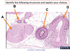
What is A?
|
Nerve
(There is no lumen, and also has connective tissue sheath) |
|

What is B?
|
Arteriole
(Because round lumen and thick muscular wall) |
|

What is C?
|
Artery
(Muscular artery, bigger muscular wall) |
|

What is D?
|
Vein
(Smaller vein, will run with the muscular artery) |
|
|
What ratio is there between veins and arteries
|
Veins and arteries have a 1:1 ratio
There is a vein for every artery |
|

What does the precapillary sphincters do?
|
Control blood flow
|
|
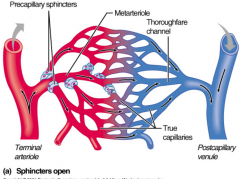
What do the capillary beds do?
|
where gas and nutrient exchange occurs and waste must be picked up
|
|
|
The diameter of a capillary is about the size of what?
|
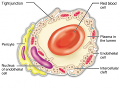
A red blood cell
|
|
|
How can a red blood cell move through if it is a larger than a capillary?
|
It can fold up
|
|
|
Do capillaries have an outer layer?
|
No
|
|
|
How does the endothelium of a capillary differ from the other blood vessels?
|
More porous and permeable
|
|
|
How does pressure drive fluid flow through the capillaries?
|
There is pressure exerted by the fluid in the capillaries (if there is an increase in pressure that pushes more fluid out), also calculate oncotic pressure and hydrostatic pressure into that. If it is positive net pressure then fluid goes out and if it is negative then fluid comes in.
|
|
|
What is the oncotic pressure?
|
Pressure exerted by proteins
The proteins don't leak out, they stay the same and act on the osmotic pressure and pull fluid into the venous end |
|
|
How do materials cross the capillary walls?
|
1) Directly across the endothelial membrane
2) Intercellular clefts 3) Fenestrations 4) Pinocytsis (Vessicle fusion) |
|
|
What causes the leaky capillaries?
|
Gaps between the endothelial cells
|
|
|
What can cause capillaries to become even leakier?
|
Pollen (allergies)
The cold |
|
|
What happens to the excess fluid leaking out from the capillaries?
|
Taken up by the lymphatic system
|
|
|
What is the function of the lymphatic system?
|
*Circulation - Return fluid and nutrient
*Immune - Produce WBCs, antibodies, filters *Digestive - Absorption and transportation of fats |
|
|
How much fluid leaks out in a day?
|
2-3 L /day
(lose around 10% of fluid) |
|
|
Is getting proteins back into the blood important?
|
Yes
|
|
|
Where are the vessels of the lymphatic system located?
|
In all areas with a blood supply except in the central nervous system
|
|
|
What is the function of the vessels of the lymphatic system?
|
Absorb and transport lymph fluid
|
|
|
Is it true that anywhere with capillary beds will also have lymph vessels?
|
Yes
|
|
|
What do the lymph capillaries begin a?
|
Blind end tubes
|
|
|
Are circulatory capillaries or lymph capillaries larger and more permeable?
|
Lymph capillaries
|
|
|
How are the endothelial cells for the lymph capillaries arranged
|
Arranged like shingles on a roof
|
|
|
What keeps the lymph capillaries open under high pressures?
|
Anchoring filaments
|
|
|
How does fluid flow in lymph capillaries?
|
Following pressure gradient
Moving back into the venous system by following the pressure gradient |
|
|
Describe the movement of lymph through the lymph vessel?
|
Pressures have the shingles open and pushes in the fluid. Then it accumulates in the first segment and that slowly stretch the diameter and causes the myogenic reflex (stretch and open the Ca++ channels which causes a contraction)
The smooth muscle then contract; pressure increases and then it is able to move into the next chamber (it acts like a valve) |
|
|
Do lymph vessels have valves that move all the way up?
|
Yes
|
|
|
Which has thinner walls, lymph vessels or veins?
|
Lymph
|
|
|
What are the main vessels of the lymphatic vessels compared to veins
|
Similar to veins but the walls are thinner and contain more valves
|
|
|
How is lymph flow determined by?
|
*Interstitial fluid pressure
*Lymphatic pump *Skeletal muscle pump *Movement *Atriovenous pump *External Compression |
|
|
How does the lymphatic pump work?
|
Smooth muscle stretches then contracts
|
|
|
How can external compression hep with lymph flow
|
Get swelling because there is fluid out of the capillaries and the lymph is overloaded; so if add a squeeze then can give more interstitial fluid to drive lymph up
|
|
|
What is the specialized macrophage in spinosum layer in the integumentary system?
|
Langerhan Cells
|
|
|
Where do the langerhan cells originate in?
|
In bone marrow
|
|
|
How do the langerhan cells move?
|
Langerhan cells originate in the bone marrow, and then move from the marrow and to the skin (still immature) and then wait till exposed to pathogen. When the pathogen comes in, the cell engulfs them. Then send signals to migrate through connective tissues and taken up in terminal end of lymphatic system, for immune response
|
|
|
How many veins are traveling with artery
|
Multiple
|
|
|
Are there also lymphatic vessels with the veins and arteries?
|
Yes
|
|
|
How do you encourage lymphatic drainage if you have a swollen ankle due to an ankle sprain?
|
*Passive or active ROM (movement)
*Use muscle pump (flex gastrocnemius) *Decrease capillary hydrostatic pressure - elevates foot *Intermittent compression *Put ice packs (might slow response because helps with the pain so can move ankle which is key to flow and get walking to use the skeletal muscle pump) |
|
|
As pressure increases, the lymph flow ______
|
Increases
|
|
|
Why is lymph flow plateau at an increased in the pressure, why
|
Because have an increased interstitial pressure to point where lymph flow stops -> Vessel collapses (too much pressure causes collapses) because compressed the vessels
|
|
|
Why does lymph flow increase when interstitial pressure increase up until 2 mmHg but above 2 mmHg it does not increase at all and instead plateaus?
|
Because although flow is increased by interstitial pressure, that same pressure compresses larger lymph vessels and impedes flow
|
|
|
What is a lymphatic massage like?
|
*Light touch
*Not deep tissue massage Intermittent gentle compression |
|
|
What are the four functions of the lymphatic system?
|
*Return of excess filtered fluid (2-3 L/day) [Roughly lose 10% by capillaries, so needs to return]
*Return of filtered protein (essential) [if not returned; will die] *Transport of Absorbed Fat *Defense against disease [immune response] |
|
|
The thoracic duct travels _______
A) Between the hemi azygos and azygos veins B) Anterior to the vertebral column C) From the cisterna chyli to the left venous angle D) All of the above E) Only B and C |
D. All of the above
|
|
|
All of the lymph vessels move up to where?
|
Collection place sitting on the abdominal cavity deep in the abdominal cavity called Cisterna chyli
(Then from there, thoracic duct travels up and makes a bend then drains into the left venous angle) |
|
|
The thoracic duct collects lymph from ________ of the body inferior to the diaphragm, and from ________ side of the body superior to the diaphragm
A) Both sides, the left side B) The right side, both sides C) Both sides, the right side D) The left side, both sides |
A. Both sides, the left side
|
|
|
Is the cisterna chyli on the right or leftside of the anterior portion of the vertebral column
|
Right
|
|
|
How does the thoracic duct travel
|
Goes to the cisterna chyli and then moves up and bends over into the vneous angle on the left subclavian. Then it catches it and then drains back into the vein because it gets processed before getting to the blood by the lymph nodes.
|
|
|
Why can the lymph just go straight into the superior vena cava when the lymph is picking up waste products, proteins and fats?
|
Because lymph nodes can filter
|
|
|
What do lymph nodes do?
|
*Filter lymph (macrophages destroy microorganisms and debris [mechanical and biological filter])
*Immune system (Lymphocytes are activated and mount and attack against antigens) |
|
|
What is the efferent and afferent nerve into and out of the lymph nodes like?
|
Fewer efferent vessels, causing flow of lymph to stagnate allowing lymphocytes and macrophages more time to carry out function.
Many afferent in and only 1 out; slows down system and that increase in residence time of lymph in nodes to help the cells take action (Slows down) |
|
|
Where are lymph nodes located?
|
*Cervical nodes
*Axillary nodes *Inguinal nodes *Deep nodes (Right in cisternea chilea) *Pericranial ring (base of head) *Tracheal nodes (Nodes related to trachea and bronchi) |
|
|
What are the three areas of lymph nodes that the physician can check?
|
*Cervical
*Axillary *Inguinal |
|
|
What organ is an enlarged lymph node?
|
Spleen
|
|
|
Where is the spleen found?
|
On the left under ribs 9-11
Upper left quadrant (From when divide the belly button into four sections) Inferior to the diaphragm, posterior/lateral to the stomach, deep to ribs 9-11, lateral to the left kidney |
|
|
What is the upper left quardrant?
|
When divide the belly button into four sections
|
|
|
If have mono what will happens to the spleen?
|
Will enlarge enough that it can be palpated
|
|
|
What are the functions of the spleen
|
*Lymphocyte proliferation and immune surveillance and response
*'Cleanses' the blood of aged cells and platelets and debris *Stores breakdown products of RBSc (ie iron) for later reuse *Stores blood platelets *Site of fetal erythrocyte production (normally ceases after birth) *Contains lymphocytes, macrophages, and huge numbers of erythrocytes |
|
|
What happens to the spleen during diving?
|
Spleen shrinks during diving and can release RBCs, increasing Hb concentration and extending dive times
|
|
|
What are the spleen of Japanese Ama divers like?
|
Able to contract and shrink their spleens by about 20% at depths at 20-30 m
This helps to increase the hemoglobin and hematocrit which increases the oxygen storage capaciy |
|
|
Which of the following is present in lymph?
A) Interstitial fluid B) Macrophages C) Lymphocytes D) All of the above |
D) All of the above
|
|
|
What is considered lymph?
|
Anything entering lymph vessel will be lymph
|
|
|
Where do you expect the thymus to be?
|
Superior mediastinum
|
|
|
What are the structures and organs of the lymphatic System
|
*Lymphocytes
*Bone marrow *Thymus *Tonsils *Nodes *Spleen |
|
|
What is the tonsil basically?
|
Basically lymph tissue with epithelium on top
|
|
|
What does the tonsil do for the lymph system?
|
To capture invaders and destroy them, slows things down like nodes
|
|
|
Where are the tonsils of the pharynx?
|
*Palatine tonsil (At posterior end of the oral cavity)
*Lingual tonsils (Grouped at the base of the tongue) *Pharyngeal tonsil (Adenoids) (In posterior wall of the nasopharynx) *Tubal tonsils (Surrounding the openings of the auditory tubes into the pharynx) |
|
|
What covers the tonsil mass invaginates?
|
Epithelial tissue
|
|
|
What does epithelial overlying tonsil mass invaginates form?
|
Tonsillar crypts
|
|
|
What do the tonsillar crypts do?
|
Trap and destroy bacteria and particulate matter
And also so macrophages and lymphocytes can do their business |
|
|
Tonsilitis is the infection and inflammation of which tonsil?
|
Palatine
|
|
|
Can tonsilitis be acute or chronic or both?
|
Both
|
|
|
What is tonsillectomy a removal of?
|
Palatine tonsils and sometiems the pharyngeal tonsil
|
|
|
When are tonsillectomies required?
|
When tonsils are so large they obstruct breathing or swallowing
Or if chronically infected |
|
|
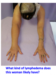
What is lymphedema?
|
Accumulation of lymph in interstitial space
|
|
|
Is lymphedema common?
|
Yes, very common
|
|
|
Why is lymphedema dangerous?
|
It is progressive if untreated (self-exaserbatting)
Causes stress on the cardiovascular system Physically or emotionally damagin |
|
|
What is primary lymphedema?
|
Congenital or hereditary developmental abnormality
|
|
|
What is secondary lymphedema?
|
Caused by a known injury or insult to the lymphatic system (trauma)
|
|
|
Secondary because only isolated to one side
(Most likely because of breast cancer; the masterectomy would remove the lymph nodes so all of the lymph will sit in the arm and cause problems with the lymph systems) |
|
|
What are the symptoms of lymphedema?
|
*Swelling
*Loss of ROM *Difficult fitting into your clothes *Tightness of rings, watch or bracelet *Chronic skin infections *Tightness and pitting of skin (unhealthy skin) *Pitting of the skin (No cure) |
|
|
Is there a cure for lymphedema?
|
No
|
|
|
What does therapy for lymphedema aim for?
|
To reduce swelling and maintain reduction
|
|
|
What is complete decongestive therapy for lymphedema?
|
*Mobilize accumulated protein rich fluid (using manual lymph drainage (MLD))
*MLD mainpulates the anchoring filaments of the lymphatic capillaries to cause opening and movement of the lymph, so more fluid can be picked up *Proper skin care *Compression garments |
|
|
What is elephatiasis
|
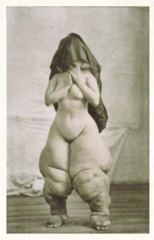
"Lymphatic filariasis)
Mosquito borne bug little worms form the mosquito has eggs laid that collect in lymph vessels, waste product of leg gets collected in lymph and causes toxity and damage. Giant lower extremities. |
|
|
Why would it be bad to lance when you have elephantiasis
|
Because you are introducing a wund to the area without lymph flow not going to heal which is an issue
|
|
|
What is the treatment for elephantiasis?
|
*Skin care
*Parasite treatment (through antibiotics) *Complete decongestive therapy (work on lymph flow) |
|
|
What is the main function of respiratory system?
|
Gas exchange
|
|
|
What are secondary functions of the respiratory system?
|
*Filtration (Filter the air we breathe)
*Air traphic control-so no food in the air system *Speech and voice resonance (need air moving out to make sound) *Smell |
|

What the orange arrow
|
External nares
|
|
What is the blue arrow?
|
Boundary of the internal nares
|
|
|
What does the internal nares open and communicate with?
|
The pharynx
|
|
|
What are turbinates covered with?
|
Respiratory epithelium (pseudostratified epithelium)
And olfactory epithelium (to get smells also act as resonating chambers) |
|
|
What are pseudostratified epithelium loaded with?
|
Goblet cells
|
|
|
What do goblet cells do?
|
Produce mucus
|
|
|
What are mucus produced from?
|
Plasma that leaks out of the capillaries (that is warm)
|
|
|
What does the turbinates do when air comes into the nose?
|
Turbinates creates a "turbin" which swirls the air around and increase the surface area (add all these together and you create a reat system to warm and humidify and filter the air)
|
|
|
What do the internal structures of the upper airway specialized for (function wise)
|
1) Warming, moistening and filtering air
2) Receiving olfactory stimuli 3) Sinuses modify speech sounds (act as large resonating chambers) |
|
|
What is the function of the nasal septum?
|
Divide the nasal cavity into right and left
|
|
|
What are the function of the inferior, middle and superior nasal turbinates?
|
*Increase turbulence and surface area of the air coming down
*Warm and humidify air *Centrifugal filtration |
|
|
What are the meati?
|
Space between the turbinates
|
|
|
What is the carina?
|
Point of bifurcation of the trachea
|
|
|
An 83-year-old man with a typical coronary ciruclation has been suffering from an embolism of the marginal branch of the right coronary artery. This condition would result in ischemia to which of the following areas of the heart?
A) Anterior portion of left atrium B) Anterior interventricular region C) Posterior portion of left ventricle D) Posterior portion of right ventricle E) Posterior interventricular region |
D) Posterior portion of right ventricle
|
|
|
A 12-year-old boy was admitted to Sacred Heart Hospital with a known history of heart problems. His left ventricular hypertrophy was most likely to result from which of the following?
A) Stenosis of pulmonary trunk B) Stenosis of the aorta C) An abnormally small left AV opening D) Incompetent pulmonary semilunar valve E) An abnormally large right AV opening |
B) Stenosis of the aorta
|
|

What is A?
|
Superior turbinate
|
|
What is B?
|
Middle turbinate
|
|

What is C?
|
Inferior turbinate
|
|

What is D?
|
Superior meatus
|
|

What is E?
|
Middle meatus
|
|

What is F?
|
Inferior meatus
|
|
|
What is the function of the nasal septum?
|
Divide nasal cavity into right and left
|
|
|
What epithelium covers the turbinates?
|
Pseudostratified columnar ciliated epithelium
|
|
|
What does the turbinates do when air comes rushing in?
|
Air comes rushing in and wrapping around the turbinates
Epithelium loaded with goblet cells create mucus and the mucus humidify the air and the sticky part filters the air and the air is sent around through a centrifugal filter (the outside of the water moves the fastest, so the heavy, bigger particles are on the outside of the turn so they stick to the mucus) by the time it reaches the alveoli, will have warm and clean air |
|
|
How does humdiity dictate mouth breathing?
|
Respiratory system in tropics makes it so allow for more mouth breathing because everything is already very humid and moist
|
|
|
Does water loss increase or decrease when get to higher altitude?
|
Decrease
Because of the increase of pressure which decreases the water vapor pressure so gives up lots more water and also breathe a lot more when increase the altitude |
|
|
Where does the pharynx begin
|
Internal nares
|
|
|
Where does the pharynx end?
|
Cricoid cartilage of the larynx
|
|
|
Is the nasopharynx for air, food or both?
|
Air
|
|
|
Is the oropharynx for air, food, or both?
|
Both
|
|
|
Is the laryngopharynx for air, food or both?
|
Both
|
|
|
What structure closes the nasopharynx during swallowing?
|
Soft palate and uvula (it rises while swallowing so food doesn't go up the nose)
|
|
|
What are the boundaries of the oropharyx
|
Uvula -> Upright epiglottis
|
|
|
What are the boundaries of the laryngopharynx?
|
Epiglottis to the top of the cricoid cartilage
|
|
|
What prevents food from entering the larynx?
|
Epiglottis tips down as larynx moves up
Ventricular folds contract to prevent food entering trachea |
|
|
As you swallow, what prevents food from entering the larynx?
|
Thyroid moves upward with the larynx and the epiglottis tilts down to cover the entry into the trachea and the ventricular and vocal folds also protect the hole
|
|
Explain this swallowing process
|
A) Initiate swallowing, tongue on the hard palate, uvula sealed off nasopharynx and food moving to back of oropharynx
B) As larynx moves up, epiglottis shifts down and it starts to slide down the top of trap door C) Then slides down the recesses of the sides and acts as roof and rolls int othe esophagus |
|
|
Is swallowing voluntary, involuntary or both
|
Both
|
|
|
How does milk come out of nose while drinking it as a child
|
As air comes back up from laughing (like the movement of the diaphragm) you have fluid in your oropharynx and that breaks the seal of the uvula. So stuff starts flowing out both ways (mouth and nose) because of the blasted air
|
|
|
What is a glottis?
|
Ture vocal cords and vocal opening (vocal apparatus)
|
|
|
What is the rima glottidis
|
The vocal opening, created when vocal cords have moved apart
|
|
|
Does the thyroid cartilage go all the way around?
|
No
|
|
|
What type of cartilage is the thyroid cartilage
|
Hyaline cartilage
|
|
|
Where is the thyroid cartilage found
|
The anterior wall of the larynx
|
|
|
What does the thyroid cartilage anchor?
|
Anterior anchor of vocal cords and ligaments
|
|
|
What is the layman's term for the thyroid cartilage
|
Adam's apple
|
|
|
What does the arytenoid cartilage anchor?
|
Anchor the vocal folds posteriorly
|
|
|
Where are the corniculate cartilage sit?
|
Artoiculate with arytenoid cartilages
|
|
|
What do arytenoid and corniculate cartilages together?
|
Open and close glottis and produce sounds
|
|
|
Where is the cuneiform cartilages found?
|
Within the tissue folds between the arytenoid cartilages and the epiglottis
|
|
|
What is the cuneiform cartilages?
|
As elastic structure around laryngeal inlet attach to cuneiform, is reinfrocement within the tissue allow for vigorous and robust attachments. Best attachment is thickening in connective tissue to add some ssupport.
|
|
|
What causes the vocal cords to become slack vs taut
|
Tilt of cricoid cartilage
|
|
|
What causes the size of the vocal opening to increase or decrease?
|
Movement of the arytenoid cartilage (abduction and adduction)
|
|
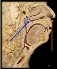
Explain the components of this nerve
|
The long one is the vagus nerve which is very important in thoracic and abdominal mainly visceral
Top one is afferent (sensory) Both one is innervate muscle of larynx (primary motor) |
|
|
Which of the following would likely produce a voice with a high pitch?
A) Anatomically thick vocal cords B) Anatomically short vocal cords C) Forceful movement of air across the vocal cords D) Vocal cords with very little tension applied to them E) None of the descriptions above would produce a voice with a high pitch |
B) Anatomically short vocal cords
|
|
|
How does thickness have to do with pitch?
|
Thicker is lower pitch
|
|
|
How does length have to do with pitch
|
Longer has to do with lower pitch
|
|
|
How does tension have to do with pitch?
|
Increased tension ahs to do with increased pitch
|
|
|
What does volume change for vocal sounds?
|
Change amount of air through the voice box
|
|
|
What is anther function of glottis?
|
For abdominal pressure; increases the stiffness of torso by holding breath and bringing down. Take in the deep breathe and seal both ends.
|
|
|
What is the epithelial tissue of larynx
|
Larynx for passing air and food, so top of epiglottis is stratified squamous to protect against hte fold
|
|
|
What is epiglottis primarily used for?
|
Defect food
|
|
|
Vocal fold can close to stop food from going in also, true or false
|
True
|
|
|
Show division of upper and lower respiratory tract
|
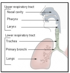
|
|
|
When someone says that food went down the wrong pipe, where most likely is food?
|
Food behind epiglottis
Vagus nerve has affernet response, response is cough, the phrenic nerve causes efferent diaphragmatic and afferent has to do with larynx) |
|
|
What is the trachea extending from
|
Larynx to level of T4-T5, T7 erect T7
|
|
|
Why is the cartialge C-Shaped
|
Expansion of the esophagus when swallowing (allows for opening of esophagus)
|
|
|
What is the type of epithelium for trachea?
|
Pseudostrafiied ciliated stratified epithelium
|