Reading...
![]()
Play button
![]()
Play button
![]()
Use LEFT and RIGHT arrow keys to navigate between flashcards;
Use UP and DOWN arrow keys to flip the card;
H to show hint;
A reads text to speech;
20 Cards in this Set
- Front
- Back
|
What closes the inferior thoracic aperture?
|
The diaphragm
|
|
|
List the abdominal viscera
|
GI: caudal end of esophagus, stomach, small and large intestines, liver, pancreas, and gallbladder
Spleen, kidneys &ureters, suprarenal gland, major neurovascular structures |
|
|
Describe the role of the abdominal wall in breathing
|
The abdominal relaxes to allow expansion of the thoracic cavity and inferior displacement of abdominal viscera during contraction of the diaphragm and contracts to aid expiration, especially forced expiration
|
|
|
Describe how intra-abdominal pressure can be increased and the expulsive functions they aid in.
|
Contraction of abdominal wall muscles can dramatically increase intra-abdominal pressure when the diaphragm is in a fixed position. Air is retained in the lungs by closing valves in the larynx of the neck. This pressure assists in (a) micturition, (b) parturition - childbirth, (c) defecation, (d) emesis - vomiting, and (e) coughing/sneezing.
|
|
|
Name the bony components of the abdominal wall
|
-The five lumbar vertebrae and intervertebral discs
-Superior expanded parts of the pelvic bones -Bony components of the inferior thoracic wall including the costal margin, rib XII, the end of rib XI and the xiphoid process |
|
|
Name the three muscles lateral to the vertebral column that are part of the posterior abdominal wall.
|
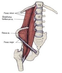
(a) Quadratus lumborum
(b) Iliacus & (c) Psoas major: distal ends pass into thigh and act as hip flexors |
|
|
Name the anterior and lateral muscular components of the abdominal wall and describe the orientation of the layers.
|
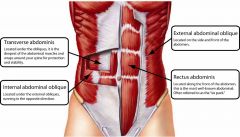
Lateral: transversus abdominis is oriented horizontally
internal oblique is angled upwards external oblique is angled downwards (like pockets) Anterior: rectus abdominis spans distance between inferior thoracic wall and pelvis |
|
|
What are aponeuroses?
|
Flat tendinous sheets derived from muscles of the lateral wall that contribute to structural continuity in the abdomen.
|
|
|
Describe the extend of the ventral and dorsal mesenteries.
|
The ventral mesentery exists for proximal regions of the gut tube, while the dorsal mesentery exists along the entire length of the system.
|
|
|
What are the two types of peritoneum (think pleura) and what structure does the peritoneum become a component of?
|
PARIETAL PERITONEUM lines the abdominal wall
VISCERAL PERITONEUM covers suspended organs Peritoneum reflects off the abdominal wall to become a component of the mesenteries that suspend the viscera. |
|
|
Define intraperitoneal
|
Structures, such as elements of the GI system, that are suspended from the abdominal wall by mesenteries.
|
|
|
Define retroperitoneal
|
Structures that are not suspended in the abdominal cavity by mesentery and that lie between the parietal peritoneum and abdominal wall.
|
|
|
Where are neurovascular structures unpaired and why?
|
Large vessels, nerves, and lymphatics are associated with the posterior abdominal wall along the median axis of the body in the region where, during development, the peritoneum reflects off the wall as the dorsal mesentery, which supports the developing gut tube. As a consequence, branches of the neurovascular structures that pass to parts of the GI system are unpaired, originate from the anterior aspects of their parent structures, and travel in mesenteries or pass retroperitoneally in areas where the mesenteries secondarily fuse to the wall.
|
|
|
What is the name of the muscular extensions anchoring the diaphragm to the anterolateral surface and what is the lowest point of attachment on the right and left side.
|
Each attachment is called a CRUS; extend to vertebra LIII on the right and LII on the left.
|
|
|
Describe the median arcuate ligament
|
Crosses the aorta and is continuous with the crus on each side
|
|
|
Describe the medial and lateral arcuate ligaments
|
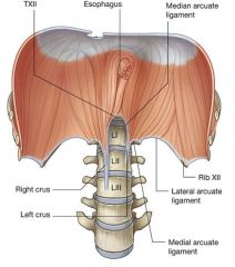
Cross muscles of the posterior abdominal wall and attach to vertebrae, the transverse processes of vertebra LI and rib XII, respectively.
|
|
|
Name the bony elements that form the pelvic inlet.
|
-sacrum posteriorly
-pubic symphysis anteriorly -distinct bony rim on the pelvic bone laterally on each side |
|
|
In what cavity is the bladder found?
|
The bladder expands from the pelvic cavity into the abdominal cavity; the uterus does likewise during pregnancy.
|
|
|
Name the structures that pass through the aperture formed by the inferior margin of the abdominal wall and the pelvic bone?
|
The majory artery and vein of the lower limb, femoral nerve, lymphatics, the distal ends of psoas major and iliacus muscles
|
|
|
The primitive foregut gives rise to what part of the GI tract? What is unique about the mesentery of the foregut?
|
Distal end of the esophagus, the stomach, and the proximal part of the duodenum. The foregut is the only region suspended by both ventral and dorsal mesentery.
|