![]()
![]()
![]()
Use LEFT and RIGHT arrow keys to navigate between flashcards;
Use UP and DOWN arrow keys to flip the card;
H to show hint;
A reads text to speech;
363 Cards in this Set
- Front
- Back
|
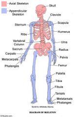
How many regions is the skeleton divided into:
Name the regions: |
2 regions
|
|
|
Axial Skeleton |
forms the central supporting axis of the body; Includes:
|
|
|
Appendicular Skeleton |
includes the bones of the:
|
|
|
|
|
|
How many bones are in the skeleton? |
206
At birth there are about 270, and even more bones form during childhood |
|
|
Sesamoid Bones |
|
|
|
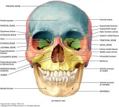
What are the cranial Bones of the skull in the Axial Skeleton? |
*the skull has 22 bones total, 8 cranial bones and 14 facial* |
|
|
What are the Facial bones; part of skull; of the Axial Skeleton? |
*14 facial bones total* |
|
|
What bones are in the auditory occicles? |
6 bones total |
|
|
What bones make up the vertebral column? |
Vertebral column has a total of 26 bones. |
|
|
What bones make up the Thoracic Cage? |
*plus 25 bones of thoracic vertebrae* |
|
|
What bones make up the Pectoral girdle in the Appendicular Skeleton? |
*Pectoral Girdle total 4 bones* |
|
|
What bones make up the Upper limbs? |
*Upper Limbs total of 60 bones* |
|
|
Hip Bones |
2 |
|
|
What bones make up the Lower Limbs? |
*Lower Limbs have total of 60 bones* |
|
|
What are the articulations of bones |
Condyle - A rounded knob that articulates with another bone (occipital condyles of skull).
Facet - A smooth, flat, slightly concave or convex articular surface (articular facets of the vertebra)
Head - The prominent expanded end of a bone, sometimes rounded (head of femur) |
|
|
What are the extensions and projections of bones? |
Crest - a narrow ridge (iliac crest of pelvis
Epicondyle - expanded region superior to a condyle, (medial epicondyle of femur)
Protuberance - bony outgrowth or protruding part (chin)
Spine - sharp, slender, or narrow process (spines of mandible)
Trochanter - two massive processes unique to femur
Tuberosity - rough elevated surface (tibial tuberosity) |
|
|
What are the Depressions of bones? |
Fossa - shallow, broad, or elongated basin (mandibular fossa)
Sulcus - groove for a tendon, nerve or blood vessell (intertubercular sulcus of the humerus) |
|
|
What are the Passages and cavities of the bones? |
Canal - tubular passage or tunnel in a bone (auditory canal of the skull)
Fissure - slit through a bone (orbital fissure behind the eye)
Foramen - hole through a bone, usually round (foramen magnum of the skull)
Meatus - opening into a canal (external meatus of the ear)
Sinus - air filled space in a bone (frontal sinus of the forehead) |
|
|
|
|
|
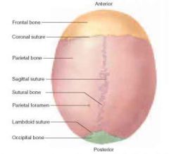
The Skull |
|
|
|
Sutures |
visible seams on the surface of the skull |
|
|
Name the sutures of the skull? |
|
|
|
Name the cavities of the skull? |
|
|
|
Name the sinuses? |
Sinuses are named for the in which they occur.
|
|
|
Cranial Bones |
those that enclose the brain
they compose the cranium (braincase) |
|
|
Dura Mater |
called a meninges the thickest and toughest of the three in the skull. |
|
|
Foramen Magnum |
A large hole in the cranium where the spinal cord meets the brain. |
|
|
Cranium |
a rigid structure with a foramen magnum, where the spinal cord meets the brain.
consists of 2 major parts:
calvaria (skullcap) - not a single bone but dome of top of skull
base - floor of the cranial cavity |
|
|
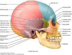
Frontal Bone |
extends from the forehead back to prominent coronal suture, which crosses the crown of the head from right to left and joins the frontal bones to parietal bones. |
|
|
Parietal bones |
form most of the cranial roof and part of its walls. |
|
|
Temporal Bones |
forms the lower wall and part of the floor of the cranial cavity. |
|
|
Occipital Bone |
forms the rear of the skull and much of its base. |
|
|
Sphenoid bone |
a complex shape with a thick median body and outstretched greater and lesser wings.
Moth like shape. |
|
|
Sella Turcica |
in the body of the sphenoid bone, is a pair of sphenoid sinuses and have a saddlelike surface feature.
Has a deep pit called the hypophyseal fossa which houses the pituitary gland. |
|
|
Ethmoid Bone |
is an anterior cranial bone located between the eyes.
Three major portions:
perpendicular plate - thin median plate of bone that forms the superior 2/3 of nasal septum
cribriform plate - forms roof of nasal cavity; has a median blade called the crista galli, an attachment point for the duramater.
labyrinth - large mass on each side of perpendicular plate. |
|
|
Facial Bones |
|
|
|
Maxillae |
|
|
|
Palate |
|
|
|
The Palatine Bone |
|
|
|
Zygomatic Bones |
|
|
|
Lacrimal bones |
|
|
|
Nasal Bones |
|
|
|
Inferior nasal concha |
|
|
|
The Vomer |
forms the inferior half of the nasal septum.
resemblance to the blade of a plow |
|
|
The mandible |
the strongest bone of the skull.
the only one that can move significantly. |
|
|
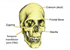
Mandibular Condyles |
an oval knob that articulates with the mandibular fossa of the temporal bone
forms the temporomandibular joint |
|
|
Ramus |
an anatomical branch, as in a nerve or in the pubis.
posterior portion of mandible |
|
|
Hyoid Bone |
a slender U shaped bone between the chin and larynx. |
|
|
Fontanels |
spaces between the unfused cranial bones. |
|
|
How many fontanels are there and what are their names? |
4 sites
*most fontanels ossify by the time an infant is 1 yr. old, the largest one, can still be palpated 18 - 24 after birth* |
|
|
Vertebral column (spine) |
|
|
|
Beyond the age of 3 years, the vertebral column is slightly ______________, with four bends called the ____________________. |
|
|
|
What is the most obvious feature of a a vertebra? |
it is the body (centrum) - a mass of spongy bone and red bone marrow covered with a thin shell of compact bone.
weight bearing portion of vertebra |
|
|
What are the characteristics of vertebra and are they all the same? |
All vertebra have same characteristics |
|
|
|
|
|
Thoracic Cage |
|
|
|
Sternum (breastbone) |
a bony plate anterior to the heart.
subdivided into three regions:
|
|
|
The Ribs |
|
|
|
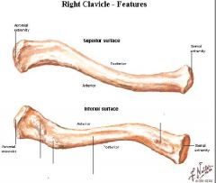
the Clavicle |
|
|
|
|
|
|
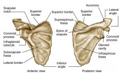
The scapula |
the three sides of the triangle are:
|
|
|
The Upper Limb |
divided into four segments containing a total of 30 bones per limb:
|
|
|
The Humerus |
has a hemispherical head that articulates with the glenoid cavity of the scapula |
|
|
The Radius |
has a distinctive discoidal head at its proximal end. |
|
|
The Ulna |
at the proximal end of the ulna is a deep C shaped trochlear notch, posterior side of notch if formed by a prominent olecranon - the bony point where you rest your elbow on a table. |
|
|
The Carpal Bones (hands) |
arranges in two rows of four bones each.
proximal row starting at the lateral (thumb side) are:
scaphoid lunate triquetrum pisiform
distal row:
trapezium trapezoid capitate hamate |
|
|
Metacarpal Bones |
Bones of the palm. |
|
|
Phalanges |
Identified by: proximal middle distal |
|
|
Pelvic Girdle |
consist of a complete ring composed o three bones:
2 - hip (coxal) bones 1 - sacrum
supports the trunk on the lower limbs and encloses and protects the viscera of the pelvic cavity - mainly the lower colon, urinary bladder, internal reproductive organs.
|
|
|
The Pelvis |
The pelvis is a bowl-shaped structure composed of
as well as their ligaments and muscles. |
|
|
Adult hip bone forms by the fusion of three childhood bones. What are they? |
|
|
|
Lower Limb |
adapted for weight bearing and locomotion
divided into 4 regions containing a total of 30 bones.
femoral region - thigh, from hip to knee, contains femur. patella, a sesmoid bone, at junction of femoral and crural regions.
crural region - leg proper, from knee to ankle, 2 bones, medial tibia, lateral fibula
tarsal region (tarsus, heal) - ankle, union of crural with the foot. tarsal bones treated as part of foot.
pedal region (pes) - foot, composed of: 7 - tarsal bones 5 - metatarsals 14 - phalanges (in toes).
|
|
|
Femur |
the longest and strongest bone of the body
measuring about 1/4 of one's height. |
|
|
Tibia |
Thick strong on medial side only weight bearing bone of the crural region |
|
|
Fibula |
slender lateral strut that helps to stabilize the ankle. |
|
|
The Ankle and Foot |
Tarsal bones of ankle are arranged in proximal and distal groups.
Calcaneus - forms the heel; largest tarsal bone
Hallux - great toe, contains only 2 bones
Metatarsals - I through V from medial to lateral. |
|
|
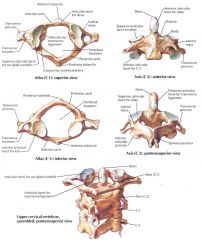
C2 vertebra is called? |
The Axis |
|
|
C1 vertebra is called? |
The Atlas |
|
|
Which of these is not a paranasal sinus?
|
B - Temporal |
|
|
Which of these is a facial bone?
|
E - lacrimal |
|
|
Which of these cannot be palpated on a living person?
|
A - crista galli
|
|
|
All of the following are groups of vertebrae except for _______, which is a spinal curvature.
|
E - sacral |
|
|
Thoracic vertebrae do not have -
|
A - transverse foramina |
|
|
The tubercle of a rib articulates with
|
E - transverse process of a vertebrae |
|
|
The disc-shaped head of the radus articulates with the ________ of the humerus
|
C - capitulum |
|
|
All of the following are carpal bones except the ___________, which is a tarsal bone.
|
B - cuboid |
|
|
The bone that supports your body weight when you are sitting down is?
|
E - ischium |
|
|
Which of these is the bone of the heel?
|
B - calcaneus |
|
|
Gaps between the cranial bones of an infant are called _____? |
fontanels |
|
|
The external auditory canal is a passage in the ____ bone. |
Temporal bone |
|
|
Bones of the skull are joined along lines called? |
Sutures |
|
|
The ______bone has greater and lesser wings and protects the pituitary gland |
sphenoid bone |
|
|
A herniated disc occurs when a ring called the _____ cracks. |
anulus fibrosis |
|
|
The transverse ligament of the atlas hold the _____ of the axis in place. |
dens |
|
|
The sacroiliac joint is formed where the ______ surface of the sacrum articulates with that of the ilium. |
auricular |
|
|
The _____ processes of the radius and ulna form bony protuberances on each side of the wrist. |
Styloid processes |
|
|
The thumb is also known as the _______ and the great toe is also know as the _________ |
Pollex ( thumb) Hallux ( great toe) |
|
|
The ________arch of the foot extends from the heel to the great toe. |
medial longintudinal |
|
|
True or False.....
not everyone has a frontal sinus
|
True
|
|
|
the pisiform bone and patella are both sesmoid bone. |
true
|
|
|
Joint (articulation) |
Example: glenohumeral joint - where glenoid cavity of scapula meets the humerus.
Four major categories: Bony Fibrous Cartilaginous Synovial |
|
|
Arthrology |
the science of joint structure, function, and dysfunction |
|
|
Kinesiology |
the study of musculoskeletal movement |
|
|
Biomechanics |
deals with a broad variety of movements and mechanical processes in the body, including the physics of blood circulation, respiration, and hearing. |
|
|
Classification of Joints |
Type of tissue or components: 1. Synostosis - Bone - Bone (no movement, cranial) 2. Synarthosis - Fibrous - bound by collagen, (sutures (fibers short), Gomphoses (fibers short), syndesmosis (longer fibers)more movable 3. Synchondroses - Cartilage (symphyses) 4. Synovial - (bone,fibrous, cartilage, fluid) |
|
|
Sutures |
are immobile or only slightly movable fibrous joints that closely bind the bones of the skull to each other.
classified as: serrate - wavy lines (coronal, sagittal, lambdoid sutures) lap (squamous suture) - overlapping beveled edges. (squamous suture between temporal and parietal bones.) Plane (butt) sutures - have straight nonoverlapping edges. (between the palatine processes of the maxillae in roof of mouth. |
|
|
Gomphoses |
attachment of a tooth to its socket. |
|
|
Syndesmosis |
|
|
|
Cartilaginous Joints |
also called an amphiarthrosis or amphiarthroidial joint.
two bones linked by cartilage.
two types of cartilagionous joints: Synchondrosis Symphyses - |
|
|
Synchondrosis |
- a joint in which the bones are bound by hyaline cartilage.
Example: temporary bone between the epiphysis and diaphysis of long bone. |
|
|
Symphyses |
two bones are jonied by fibrocartilage.
Example: pubic symphysis, the right and left pubic bones are joined by the cartilaginous interpubic disc. also the joint between the bodies of two vertebrae. |
|
|
Synovial Joint |
the most familiar type of joint. Also called a diarthrosis or diarthrodial joint.
covered with articular cartilage, a layer of hyaline cartilage, 2 to 3 mm thick. separated by a narrow space, joint (articular) cavity, containing a slippery lubricant called synovial fluid.
joint (articular) capsule encloses the cavity and retains fluid. outer fibrous capsule, inner synovial membrane
|
|
|
|
|
|
Meniscus |
a disk of cartilage between the articulating end of the bones in a joint that acts as a cushion .
a crescent-shaped fibrous cartilage between the bones at certain joints, esp at the knee.
|
|
|
Tendon |
a strip or sheet of tough collagenous connective tissue that attaches a muscle to a bone.
often most important structures stabilizing a joint. |
|
|
Ligament |
collagenous connective tissue that attaches one bone to another. |
|
|
Bursa |
a fibrous filled sac filled with synovial fluid, located between adjacent muscles, where a tendon passes over a bone, or between bone and skin.
bursae cushion muscles, help tendons slide more easily over the joints. |
|
|
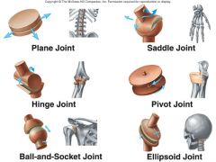
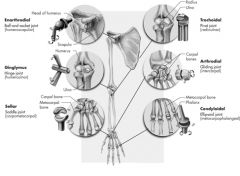
Classes of Synovial Joints |
Ball and socket joints - monaxial; scapula and head of humerus (humeroscapular) Condylar (ellipsoid) joint - biaxial; meta carpal and phalanx. (metacarpophalangeal) Saddle joint - biaxial; carpal bones (trapeziometacarpal) Plane (gliding) joint - biaxial; carpal bones (intercarpal) Hinge joint - monaxial; humerus and ulna (humeroulnar) Pivot joint - monaxial; radius and ulna (radioulnar) |
|
|
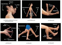
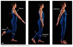
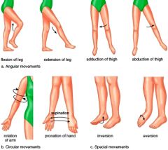
Flexion |
a movement that decreases a joint angle.
Example: bending of elbow or knee. |
|
|
Extension |
a movement that straightens a joint and generally returns a body part to the zero position. |
|
|
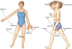
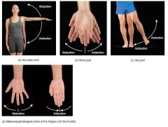
Abduction |
is the movement of a body part in the frontal plane away from the midline of the body.
Example: moving the feet apart to stand spread- legged. |
|
|
Adduction |
is movement in the frontal plane back toward the midline.
**some joints can be hyperadducted, as when you stand with ankles crossed, or fingers crossed** |
|
|
Hyperabduct |
if you raise your arm high enough to cross slightly over the front or back of your head. |
|
|
|
|
|
Elevation |
movement that raises a body part vertically in the frontal plane. |
|
|
Depression |
lowers a body part in the same plane |
|
|
Protraction |
the anterior movement of a body part in the transverse (horizontal) plane. |
|
|
Retraction |
is posterior movement.
Example: your shoulder retracts when you reach in front of you to push a door open. |
|
|
Circumduction |
one end of an appendage remains fairly stationary the other end makes a circular motion. |
|
|
Rotation |
applies to any bone turning around a fixed axis.
In joint movements, a movement in which a bone spins on its longitudinal axis. |
|
|
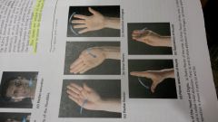
Supination |
of the forearm is a movement that turns the palm to face anteriorly or upward; in anatomical position. |
|
|
Pronation |
causing the palm to face posteriorly or downward. |
|
|
|
|
|
|
|
|
Lateral Flexion |
tilting the head or trunk to the right or left of the midline |
|
|
Right rotation or left rotation |
when the chest or the face turns to the right or left of the forward-facing zero position. |
|
|
Ulnar flexion |
tilts the hand toward the little finger |
|
|
radial flexion |
tilts it toward the thumb |
|
|
|
|
|
|
|
|
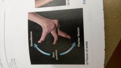
Dorsiflexion |
a movement in which the toes are elevated as one might do in applying toenail polish. |
|
|
Plantar Flexion |
movement of the foot so the toes point downward |
|
|
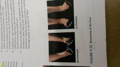
Inversion |
a foot movement that tips the soles medially, somewhat facing each other |
|
|
Eversion |
a movement that tips the soles laterally, away from each other |
|
|
Opposition |
a movement of the thumb in which it touches any fingertip of the same hand |
|
|
Reposition |
Return to zero position |
|
|
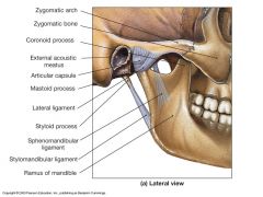
Temporomandibular jaw joint (TMJ) |
is the articulation of the condyle of the mandible with the mandibular fossa of the temporal bone |
|
|
|
|
|
Lateral Ligament |
prevents posterior displacement of the mandible |
|
|
sphenomandibular ligament |
on the medial side of the joint extens from the sphenoid bone to the ramus of the mandible |
|
|
glenohumeral joint humeroscapular |
shoulder joint; is where the hemispherical head of the humerus articulates with the glenoid cavity of the scapula |
|
|
Glenoid Labrum |
a ring of fibrocartilage around shoulder joint capsule |
|
|
What tendons form the rotator cuff? |
|
|
|
Coxal Hip Joint |
point where the head of the femur inserts into the acetabulum of the him bone. |
|
|
tibiofemoral Knee joint |
the largest and most complex diarthrosis of the body.
Primarily a hinge joint. |
|
|
Meniscus |
absorb the shock of the body weight jostling up and down on the knee and prevent the femur from rocking from side to side on the tibia |
|
|
Talocrural ankle joint |
includes two articulations:
|
|
|
Internal and external rotation of the humerus are made possible by a __________ joint.
a. pivot b. condylar c. ball and socket d. saddle e. hinge |
C - ball and socket |
|
|
which of the following is the least movable?
a. a diarthrosis b. a synostosis c. a symphysis d. a synovial joint e. a condylar joint |
B - a synostosis |
|
|
Which of the following movements are unique to the foot?
a. dorsiflexion and inversion b. elevation and depression c. circumduction and rotation d. abduction and adduction e. opposition and reposition |
A - dorsiflexion and inversion |
|
|
Which of the following joints cannot be circumducted?
a. carpometacarpal b. metacarpophalangeal c. glenohumeral d. coxal e. interphalangeal |
E - interphalangeal |
|
|
Which of the following terms denotes a general condition that includes the other four?
a. gout b. arthritis c. rheumatism d. osteoarthritis e. rheumatoid arthritis |
C - rheumatism |
|
|
In the adult, the ischium and pubis are united by?
a. a synchondrosis b. a diarthrosis c. a synostosis d. an amphiarthrosis e. a symphysis |
C - amphiarthrosis |
|
|
In a second-class lever, the effort _____
a. is applied to the end opposite the fulcrum b. is applied to the fulcrum itself c. is applied between the fulcrum and resistance d. always produces an MA less than 1.0 e. is applied on one side of the fulcrum to move a resistance on the other side. |
A - is applied to the end opposite the fulcrum |
|
|
Which of the following joints has anterior and posterior cruciate ligaments?
a. the shoulder b. the elbow c. the hip d. the knee e. the ankle |
D - the knee |
|
|
To bend backward at the waist involves ______ of the vertebral column.
a. rotation b. hyperextension c. dorsiflexsion d. abduction e. flexion |
B - hyperextension |
|
|
The rotator cuff includes the tendons of all of the following muscles except?
a. the subscapularis b. the supraspinatus c. infraspinatus d. the biceps brachii e. the teres minor |
D - the biceps brachii |
|
|
The lubricant of a diarthrosis is called _____? |
a synovial joint |
|
|
A fluid-filled sac that eases the movement of a tendon over a bone is called a/an _______? |
bursa |
|
|
A ______ joint allows one bone to swivel on another. |
pivot |
|
|
________is the science of movement. |
kinesology |
|
|
The joint between a tooth and the mandible is called a/an ______ |
gomphosis |
|
|
In a ______________suture, the articulating bones have interlocking wavy margins, somewhat like a dovetail joint in carpentry. |
Serrate |
|
|
In kicking a football, what type of action does the knee joint exhibit? |
Extension |
|
|
The angle through which a joint can move is called it _______? |
Range of motion (ROM) |
|
|
The menisci of the knee are functionally similar to the _______ of the temporomandibular joint. |
articular disc |
|
|
At the ankle, both the tibia and fibula articulate with what tarsal bone? |
Talus |
|
|
True or False?
More people get rheumatoid arthritis than osteoarthritis? |
False
*Osteoarthritis occurs in almost everyone after a certain age; rheumatoid arthritis is less common |
|
|
A doctor who treats arthritis is called a kinesiologist. |
False
**A kinesiologist studies joint movements; a rheumatologist treats arthritis. |
|
|
Synovial Joints are also known as synarthroses. |
False
Synovial joints are diarthroses and amphiarthroses, but never synarthroses. |
|
|
There is no meniscus in the elbow joint. |
True |
|
|
Reaching behind you to take something out of your hip pocket involves hyperextension of the shoulder. |
True |
|
|
The anterior cruciate ligament normally prevents hyperextension of the knee. |
True |
|
|
The femur is held tightly in the acetabulum mainly by the round ligament. |
False
The round ligament is somewhat slack and probably does not secure the femoral head. |
|
|
The knuckles are diarthroses |
True |
|
|
Synovial fluid is secreted by the bursae |
False
**Synovial fluid is secreted by the synovial membrane of the joint capsule and fills the bursae. |
|
|
Unlike most ligaments, the periodontal ligaments do not attach one bone to another |
True |
|
|
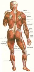
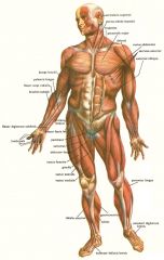
Muscular System Skeletal Muscles |
There are about 600 muscles in the human muscular system, which together account for about 40% of a person's body weight.
Skeletal Muscles are also called voluntary muscles.
|
|
|
The study of the muscular system is called? |
Myology |
|
|
What does the word "muscle" mean? |
"little mouse" |
|
|
What are the functions of the Muscles? |
Movement
Stability
Control of body openings and passages
Heat production - the skeletal muscles produce as much as 85% of one's body heat, which is vital to the functioning of enzymes and all metabolism.
Glycemic Control - skeletal muscles absorb, store, and use a lare share of one's glucose and play a highly significant role in stabilizing its blood concentration. |
|
|
Sizes of Muscles |
|
|
|
Muscle Tissue Characteristics are? |
|
|
|
Types of Skeletal Muscle |
Striation - a voluntary muscle
Smooth Muscle - Involuntary
Cardiac Muscle - autonomic |
|
|
What are the connective tissues of muscle? |
|
|
|
|
|
|
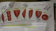
Muscle Shapes |
|
|
|
Muscle Compartment |
a group of functionally related muscles enclosed and separated from others by connective tissue fascia.
a compartment also contains the nerves and blood vessels that supply the muscle group |
|
|
What are the types of muscle attachments? |
Indirect - the ;muscle ends conspicuously short of its bony destination, and the gap is bridged by a fibrous band or sheet called a tendon.
Direct - fleshy attachement; there is so little separation between muscle and bone to the naked eye, the red muscular tissue seems to emerge directly from the bone. Example: margins of the brachialis. |
|
|
Retinaculum |
a band of connective tissue that muscles pass under.
Example: one of these covers each surface of the wrist like a bracelet. |
|
|
Origin and Insertion |
Origin - immovable; the bony site of attachement at the relatively stationary end of a bone.
Insertion - movable; the attachment site at its more mobile end. |
|
|
What are the functional groups of muscles? |
prime mover - agonist; the muscle that produces most of the force during a particular joint action. **flexing elbow - agonist is the brachialis**
synergist - a muscle that aids the prime mover. ** biceps brachii aids brachialis**
antagonist - a muscle that opposes the prime mover; it relaxes to give the prime mover almost complete control over the action. **triceps in antagonist for brachialis**
fixator - a muscle that prevents a bone from moving; so primary can move. **the rhomboids that hold the scapula in place** |
|
|
Intrinsic Muscle |
is entirely contained within a particular region, having both its origin and insertion there. |
|
|
Extrinsic Muscle |
acts upon a designated region but has its origin elsewhere. |
|
|
Innervation |
refers to the identity of the nerve that stimulates the muscle. |
|
|
Spinal Nerves |
99% in the peripheral nervous system
Emerge through the intervertebral foramina, Innervate muscles below the neck |
|
|
Cranial Nerves |
arise from base of brain, emerge through the skull foramina, and innervate muscles of the head and neck. |
|
|
Muscular System Blood supply |
receives about 1.24L of blood per minute at rest - which is about 1/4 of the blood pumped by the heart.
Working muscles have a great demand for glucose, fatty acids, and oxygen. |
|
|
Muscles of Facial Expression |
|
|
|
Glossus |
has to do with tongue Mouth |
|
|
Temporalis |
elevation, retraction, and lateral and medial excursion of the mandible
Innervates - mandibular nerve origin - temporal lines/temporal fossa of cranium insertion - coronoid process and anterior border of mandibular ramus |
|
|
Masseter |
Elevation of the mandible, with smaller roles in protraction, retraction and lateral medial excursion
Origin - Zygomatic arch Insertion: lateral surface of mandibular ramus and angle. Innervates - Mandibular nerve |
|
|
Digastric |
depresses mandible when hyoid is fixed; opens mouth widely, as ehen ingesting food or yawning; elevates hyoid when madible is fixed.
Origin - mastoid notch of temporal bone Innervates - facial nerve, mylohyoid nerve |
|
|
Geniohyoid |
Hyoid has to do with neck.
depresses mandible when hyoid is fixed. |
|
|
Sternocleidomastoid |
Unilateral action tilts head slightly upward and toward the opposite side; looking over shoulder.
common action rotating head left or right.
Superficial neck muscle |
|
|
Trapezius |
Extends and laterally flexes neck
Superficial neck muscle |
|
|
Diaphragm |
prime mover of inspiration (2/3 of air intake); contracts in preparation for sneezing, coughing, crying, laughing, and weight lifting.
Innervation: Phrenic Nerve |
|
|
External Intercostals |
elevate and protract ribs 2-12, expanding thoracic cavity and creating a partial vacuum causing inflow of air.
Innervation : Intercostal nerves |
|
|
Internal Intercostals |
In inspiration, aids in elevating the ribs, expanding thoracic cavity; in inspiration.
Innervation: Intercostal nerves |
|
|
Muscles of Anterior Abdominal Wall |
External Abdominal Oblique - supports abdominal viscera against pull of gravity; stabilizes vertebral column during heavy lifting.
Internal Abdominal Oblique
Transverse Abdominal
Rectus Abdominis - flexes lumbar region of vertebral column, producing forward bending at the waist. |
|
|
Muscles of the back |
|
|
|
Erector Spinae |
extension and lateral flexion of vertebral column.
group of muscles on each side of the spinal column is a large muscle mass that extends from the sacrum to the skull. These muscles are primarily responsible for extending the spinal column to maintain erect posture. |
|
|
|
|
|
Pectoralis Minor |
serratus anterior
draws scapula laterally and forward around the chest wall, rotates scapula, depresses apex of shoulder |
|
|
Serratus Anterior |
draws scapula laterally and forward around chest protracts scapula prime mover in all forward -reaching and pushing actions |
|
|
Trapezius |
stabilizes scapula and shoulder during arm movements elevates and depresses apex of shoulder; rotates and retract scapula |
|
|
Rhomboideus Minor and Rhomboideus Major |
Retracts scapula and braces shoulder, fixes scapula during arm movements |
|
|
Pectoralis Major |
Flexes, adducts, and medially rotates humerus , as in climbing or hugging; aids in deep inspiration. |
|
|
Latissimus Dorsi |
Adducts and medially rotates humerus; extends the shoulder joint as in pulling on oars of boat |
|
|
Deltoid |
Anterior fibers flex and medially rotate arm; involved in swinging arm. |
|
|
Teres Major |
Extends and medially rotates humerus; contributes to arm swinging |
|
|
Rotator Cuff Muscles are? |
|
|
|
Flexion Muscles of forearm |
Brachialis - prime mover of elbow flexion
Biceps Brachii - rapid or forceful supination of forearm; synergist in elbow flexion
Triceps Brachii - Extends elbow; long head extends and adducts humerus. primary extensor |
|
|
Pollicis |
means thumb |
|
|
Minimi |
fifth digit, pinkie |
|
|
Primary flexor muscles of the hip |
Iliacus - flexes thigh at hip when trunk is fixed; Psoas |
|
|
Gluteus Maximus |
extends thigh at hip as in stair climbing |
|
|
Muscles in Knee and leg |
|
|
|
Hamstrings are composed of what muscles |
Antagonists to quadracep femoris muscle group |
|
|
Gastrocnemius and Soleus |
Plantar flexes foot
Antagonist is the tibilis anterior |
|
|
Which of the following muscles is the prime mover in spitting out a mouothful of liquid?
a. platysma b buccinator c. risorius d. masseter e. palatoglossus |
B - buccinator |
|
|
Each muscle fiber has a sleeve of areolar connective tissue around it called?
a. the fascia b. the endomysium c. the perimysium d. the epimysium e. the intermuscular septum |
B - endomysium |
|
|
Which of these is not a suprahyoid muscle?
a. genioglossus b. geniohyoid c. stylohyoid d. mylohyoid e. digastric |
A - genioglossus |
|
|
Which of these muscles is an extensor of the neck?
a. external oblique b. sternocleidomastoid c. splenius capitis d. iliocostalis e. latissimus dorsi |
C - splenius capitis |
|
|
Which of these muscles of the pelvic floor is the deepest?
a. superficial transverse perineal b. bulbospongiosus c. ischiocavernosus d. deep transverse perineal e. levator ani |
E - levator ani |
|
|
Which of these actions is not performed by the trapezius?
a. extension of the neck b. depression of the scapula c. elevation of the scapula d. rotation of the scapula e. adduction of the humerus |
E - adduction of the humerus |
|
|
Both the hands and feet are acted upon by a muscle or muscles called:
a. the extensor digitorum b. abductor digiti minimi c. flexor digitorum profundus d. abductor hallucis e. flexor digitorum longus |
B - abductor digiti minimi |
|
|
Which of the following muscles does not extend the hip joint?
a. quadriceps femoris b. gluteus maximus c. biceps femoris d. semitendinosus e. semimembranes |
A - quadriceps femoris |
|
|
Both the gastrocnemius and _______muscles insert on the heel by way of the calcaneal tendon.
a. semimembranosus b. tibialis posterior c. tibialis anterior d. soleus e. plantaris |
D - soleus |
|
|
Which of following muscles raises the upper lip?
a. levator palpebrae superioris b. orbicularis oris c. zygomaticus minor d. masseter e. mentalis |
C - zygomaticus minor |
|
|
The _________of a muscle is the point where it attaches to a relatively stationary bone |
Origin |
|
|
A bundle of muscle fibers durrounded by perimysium is called a/an |
fascicle |
|
|
The ___ is the muscle that generates the most force in a given joint movement |
Prime mover agonist |
|
|
The three large muscles on the posterior side of the thigh are commonly known as the ____ muscles. |
hamstring |
|
|
Connective tissue bands called _____ prevent flexor tendons of the forearm and leg from rising like bowstrings |
flexor retinacula |
|
|
A muscle that works with another to produce the same or similar movement is called a/an |
synergist |
|
|
The abdominal aponeuroses converge on a median fibrous band on the abdomen called the ____ |
linea alba |
|
|
A circular muscle that closes a body opening is called a/an ______ |
sphincter muscle |
|
|
A muscle somewhat like a feather, with fibers obliquely approaching its tendon from both sides, is called a/an ______ muscle. |
bipennate |
|
|
True or False.....
Cutting the phrenic nerves would paralyze the prime mover of respiration |
True |
|
|
The orbicularis oculi is a sphincter |
True |
|
|
The origin of the sternocleidomastoid muscle is the mastoid process of the skull |
False
the mastoid process is its insertion |
|
|
To push someone away from you, you would use the serratus anterior more than the trapezius |
True |
|
|
Curling the toes employs the quadratus plantae |
True |
|
|
The scalenes are superficial to the trapezius |
False
The trapezius is superficial to the scalenes |
|
|
Exhaling requires contraction of the internal intercostal muscles. |
False
Normal exhalation does not employ these muscles. |
|
|
Hamstring injuries often result from rapid flexion of the knee. |
False
They result from rapid extension of the knee not flexion. |
|
|
The tibialis anterior and tibialis posterior are synergists. |
False
They are on opposite sides of the tibia and act as antagonists. |
|
|
Muscle Cell definition |
A muscle cell is essentially a device for converting the chemical energy of ATP into the mechanical energy of movement. |
|
|
Universal Characteristics of Muscle |
|
|
|
Skeletal Muscle |
Skeletal muscle may be defined as voluntary striated muscle that is usually attached to one or more bones.
A skeletal muscle exhibits alternating light and dark transverse bands, or striations
extra ordinary length, skeletal muscle cells are usually called muscle fibers or myofibers. |
|
|
The Muscle Fiber |
the plasma membrane of a muscle fiber is called the sarcolemma.
its cytoplasm is called the sarcoplasm |
|
|
Sarcolemma |
plasma membrane in muscle fiber |
|
|
Sarcoplasma |
Cytoplasm of muscle fiber |
|
|
Myofibrils |
long protein cord in the cytoplasm...
each is a bundle of parallel protein microfilaments called myofilaments. |
|
|
Myofiber |
the muscle cells |
|
|
Glycogen |
carbohydrate in cytoplasm |
|
|
myoglobin |
red pigment, stores oxygen |
|
|
myoblasts |
stem cells that fuse in embryonic development to produce each fiber. |
|
|
Satellite Cells |
myoblasts that remain as unspecialized between the muscle fiber and endomysium |
|
|
Sarcoplasmic Reticulum (SR) |
forms a network around each myofibril |
|
|
Terminal cisternae |
dilated end-sacs which cross the muscle fiber from one side tot he other |
|
|
Transverse (T) tubules |
in the sarcolemma tubular infoldings which penetrate through the cell and emerge on the other side. |
|
|
Triad |
T tubule and the two terminal cisternae associated it. |
|
|
Myofilaments |
Thick filaments - made of several hundred molecules of a protein called myosin.
Thin filaments - composed of two intertwined strands of a protein called fibrous (F) acton. Also another protein called tropomyosin, and this tropomyosin has a smaller calcium-binding protein called troponin bound to it.
Elastic Filaments - are made of a huge springy protein called titin. |
|
|
Myosin + Acton |
Contractile proteins;
they do the work of shortening the muscle fiber. |
|
|
Tropomyosin + Troponin |
Regulatory protein;
they act like a switch to determine when the fiber can contract and when it cannot. |
|
|
Titin |
|
|
|
Dystrophin |
|
|
|
Striations |
Striated muscle has dArk "A bands" alternating with lIghter "I bands".
A stand for anistropic I stand for Isotropic. *refers to the way the bands affect polarized light. |
|
|
Striation Bands |
|
|
|
Sarcomere |
the functional contractile unit of the muscle
each muscle contains
the distance from one Z disc to the next |
|
|
Motor Neurons and Motor Units |
Skeletal muscles are served by nerve cells called somatic motor neurons, whose cell bodies are in the brainstem and spinal cord.
Their axons, called somatic motor fibers lead to the skeletal muscles. |
|
|
Motor Unit |
Since the muscle fibers behave as a single functional unit, one nerve fiber and all the muscle fibers innervated by it are called a motor unit.
Effective muscle contraction usually requires the activation of several motor units at once. |
|
|
Small Motor Units |
where fine control is needed
fine movements of the eye and hand |
|
|
Large Motor Units |
where strength is more important .
powerful movements of the gluteal or quadriceps muscles. |
|
|
Neuromuscular Junction |
the point where a nerve fiber meets its target cell is called a synapse.
Then the target cell is a muscle fiber, the synapse is called a neuromuscular junction (NMJ), or motor end plate
Each terminal branch of the nerve fiber within the NMJ forms a separate synapse with muscle fiber. |
|
|
Synaptic Knob |
At each synapse, the nerve fiber ends in a bulbous swelling. |
|
|
Synaptic cleft |
A narrow space that separates the knob from directly touching the muscle fiber. |
|
|
Schwann Cell |
A cell that envelops the entire junction and isolates it from the surrounding tissue fluid. |
|
|
Synaptic vesicles |
spheroidal organells in the synaptic knob which are filled with a chemical acetycholine (ACh).
|
|
|
Acetylcholinesterase (AChE) |
both the sarcolemma and that part of the basal lamina in the cleft contain this enzyme.
It breaks Acetylcholine down after it has stimulated the muscle cell. |
|
|
Electrically excitable cells |
because their plasma membranes exhibit voltage changes in response to stimulation muscle fiber and neurons are regarded as these type of cells |
|
|
Polarized |
Charged like a little battery |
|
|
Depolarization |
changing the plasma membrane briefly to a positive charge |
|
|
Repolarization |
When the plasma membrane turns negative again from the loss of the positive potassium ions. |
|
|
Behavior of Skeletal Muscle Fibers |
|
|
|
Rigor Mortis |
the hardening of the muscles and stiffening of the body that begins 3 to 4 hours after death. |
|
|
Threshold |
minimum voltage necessary to generate an action potential in the muscle fiber and produce a contraction |
|
|
Twitch |
At threshold or higher, a stimulus thus causes a quick cycle of contraction and relaxation |
|
|
Latent Period |
a delay between the onset of the stimulus and the onset of the twitch |
|
|
Recruitment
Multiple motor unit (MMU) summation |
the process of bringing more motor units into play |
|
|
Treppe |
pattern of increasing tension with repetitive stimulation |
|
|
Isometric contraction |
contraction without a change in length |
|
|
Isotonic Contraction |
contraction with a change in length but no change in tension |
|
|
Concentric contraction |
a muscle shortens as it maintains tension |
|
|
Eccentric Contraction |
a muscle lengthens as it maintains tension |
|
|
Muscle contraction |
All muscle contraction depends on ATP and calcium; no other energy source can serve in its place. |
|
|
ATP |
the supply of it depends on the availability of oxygen and organic energy sources such as glucose and fatty acids. |
|
|
Anaerobic fermentation |
enables a cell to produce ATP in the absence of oxygen |
|
|
Aerobic respiration |
produces far more ATP and no lactic acid, but it requires a continual supply of oxygen. |
|
|
Phosphate transfers
Immediate Energy |
short, intense exercise, myoglobin in a muscle fiber supplies oxygen for a limited amount of aerobic respiration at the outset. These 2 enzymes produce ATP
Myokinase - transfers P1 from one ADP to another, converting to ATP Creatine kinase - obtains P1 from a phosphate-storage molecule, creatine phosphate (CP) |
|
|
Short term Energy |
pathway from glycogen to lactic acid, called the glycogen-lactic acid system, produces enough ATP for 30 to 40 seconds of maximum activity. |
|
|
Long term Energy |
after 40 seconds the respiratory and cardiovascular systems "catch up" and deliver oxygen to the muscles fast enough for aerobic respiration to meet most of the ATP demand |
|
|
Muscle fatigue |
the progressive weakness and loss of contractility that results from prolonged use of the muscles. |
|
|
Oxygen debt |
the difference between the resting rate of oxygen consumption and the elevated rate following an exercise. |
|
|
To make a muscle contract more strongly, the nervous system can activate more motor units. This process is called: a. recruitment b. summation c. incomplete tetanus d. twitch e. treppe |
A - recruitment |
|
|
The functional unit of a muscle fiber is the ______, a segment from one Z disc to the next. a. myofibril b. I band c. sarcomere d. neuromuscular junction e. striation |
C - sarcomere |
|
|
Before a muscle fiber can contract, ATP must bind to? a. a Z disc b. myosin head c. tropomyosin d. troponin e. actin |
B - myosin head |
|
|
Before a muscle fiber can contract, Ca2 must bind to? a. calsequestrin b. myosin head c. tropomyosin d. troponin e. actin |
D - troponin |
|
|
Which of the following muscle proteins is not intracellular? a. actin b. myosin c. collagen d. troponin e. dystrophin |
C. - collagen |
|
|
Smooth muscle cells have ______, whereas skeletal muscle fibers do not. a. sarcoplasmic reticulum b. tropomyosin c. calmodulin d. Z discs e. myosin ATPase |
C - calmodulin |
|
|
ACh receptors are found mainly in a. synaptic vesicles b. terminal cisternae c. thick filaments d. thin filaments e. junctional folds |
E - junctional folds |
|
|
Single-unit smooth muscle cells can stimulate each other because they have: a. a latch-bridge b. diffuse junctions c. gap junctions d. tight junctions e. cross-bridges |
C - gap junctions |
|
|
A person with a high VO2max
|
experiences less muscle fatigue during exercise than someone with a low VO2max. |
|
|
Slow oxidative fibers have all of the following except what? a. abundance of myoglobin b. abundance of glycogen c. high fatigue resistance d. red color e. high capacity to synthesize ATP aerobically |
B - abundance of gylcogen |
|
|
The minimum stimulus intensity that will make a muscle contract is called? |
threshold |
|
|
A state of prolonged maximum contraction is called? |
complete tetanus |
|
|
Parts of the sarcoplasmic reticulum called _____lie on each side of T tuble. |
terminal cisternae |
|
|
Thick myofilaments consist mainly the protein? |
myosin |
|
|
The neurotransmitter that stimulates skeletal muscle is _____? |
acetycholine |
|
|
Muscle contains an oxygen-binding pigment called? |
myoglobin |
|
|
The ______of skeletal muscle play the same rolse as dense bodies in the smooth muscle |
Z disc |
|
|
A state of continual partial muscle contraction is called? |
muscle tone |
|
|
_______is an end product of anaerobic fermentation that causes muscle fatigue. |
lactic acid |
|
|
True or False.......
Each motor neuron supplies just one muscle fiber. |
False
A motor neuron may supply 1,000 or more muscle fibers; a motor unit consists of one motor neuron and all the muscle fibers it innervates. |
|
|
To initiate muscle contraction, calcium ions must bind to the myosin heads |
False
Calcium binds to troponin, not to myosin |
|
|
Slow oxidative fibers are relatively resistant to fatigue. |
True |
|
|
Thin filaments are found in both the A bands and I bands of striated muscle. |
True |
|
|
Thin filaments do not change length when a muscle contracts. |
True |
|
|
Smooth muscle lacks striations because it does not have thick and thin myofilaments. |
False
Thick and thin filaments are present but not arranged in a way that produces striations |
|
|
If no ATP were available to a muscle fiber, the excitation stage of muscle action could not occur |
True |
|
|
For the first 30 seconds of an intense exercise, muscle gets mos of its energy from lactic acid |
False
A muscle produces most of its ATP during this time by anaerobic fermentation, which generates lactic acid; it does not consume lactic acid |
|
|
A muscle must contract tot he point of complete tetanus if it is to move a load |
False
Under natural conditions, a muscle seldom or never attains a complete tetanus. |
|
|
the hands have more phalanges than the feet |
false -
each hand and foot has 14 phalanges |
|
|
as an adaptation of pregnancy, the females's pelvis is deeper than the males. |
false -
The female pelvis is wider and shallower than the male's. |
|
|
there are more carpal bones that tarsal bones. |
True |
|
|
in a living person you can palpate the muscles in the infraspinous fossa but not those of the subscapular fossa? |
True |
|
|
the lumbar vertebrae do not articulate with any ribs and therefore do not have any transverse processes. |
false- The lumbar vertebrae have transverse processes but no transverse costal facets. |
|
|
the most frequently broken bone is the humerus. |
False
the most frequently broken bone is the clavicle |
|
|
in strict anatomical terminology, the words arm and leg both refer to regions with only one bone. |
false - Arm refers to the region containing only the humerus; Leg refers to the region containing the tibia and fibula. |
|
|
if you rest your chin on your hands and elbows on table, the olecranon of the ulna rests on the table. |
True |