Reading...
![]()
Play button
![]()
Play button
![]()
Use LEFT and RIGHT arrow keys to navigate between flashcards;
Use UP and DOWN arrow keys to flip the card;
H to show hint;
A reads text to speech;
12 Cards in this Set
- Front
- Back
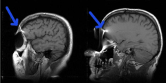
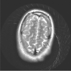
What artifact is this?
|

Susceptibility artifact
Common in T2*, GRE, Echo planar sequences Occur near boundaries between tissues with different magnetic susceptibility, and when metals are present. Remedy: remove metals and try to use non gradient echo sequences Increase bandwidth, matrix, slice thickness |
|
|
What are 3 classes of substances that contribute to magnetic susceptibiltiy?
|
Diamagnetic -small internal magnetic field opposite direction of external field (most tissues/wood)
Paramagnetic- small internal magnetic field parallel direction of external field (deoxygenated blood) Ferromagentic - strong (steel |
|

What artifact is this?
|
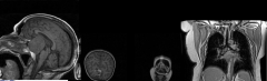
Aliasing (wrap around artifact)
occurs in phase encoding (2D: occurs in 1 direction; 3D: occurs in 2 directions) Field of view too small for excited tissue Remedy: Orient the thinnest axis of anatomy along the phase-encoding direction Enlarge FOV Exclude all anatomy from the top few slices (3D head exam) Use spatial saturation bands to null tissue outside FOV |
|

What is this artifact?
|
Ghosting (Nyquist ghost)
Phase encoding Results from motion Also results from sequences that require rapid on and off of phase gradient (EPI sequences) This causes Nyquist ghost with anatomy shifted 1/2 field of view Pulsatile flow Remedy: Apply eddy current correction Switch PE and FE directions Apply saturation bands Motion resistant sequences respiratory/cardiac gating ask patient to be still. |
|

What is this artifact?
|
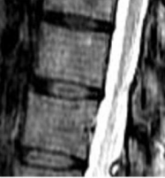
Truncation Artifact
Occurs 2/2 nature of Fourier reconstruction which are most inacureate around sharp boundaries (CSF/tissue, Cysts/edema) - results in alternating bright and dark lines (ringing) Decreases with increased matrix Pre and post reconstruction filters |
|
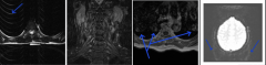
What is this artifact?
|

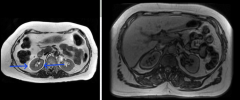
Chemical Shift Type 1(left) and Type 2 (right)
Type 1 Misregistration of fat and water tissue resulting in shift in the Frequency encoding direction (small receiver bandwidth) Type 2 Fat and water in the same voxel negate each other resulting in no signal at fat/water interfaces (india ink) Remedy: Type 1: Increase bandwidth Type 2: Image in phase 4.6 msec (1.5T) and 2.3 msec (3T) or lengthen TE greater than 30 msec and apply shimming over FOV |
|

What is this artifact?
|

Spikes in K-space
Noise spikes during acquisition produces corrugated sinusoidal pattern. NOT in phase or frequency encoding direction Remedy: Repeat |
|

What is this artifact?
|
Zippers
Form of noise artifact related to spurious RF signal from malfunctioning equipment or breach in MR faraday cage Remedy: Close scan room door Chech for RF emissions from equipment in scan room. Find RF leak |
|

What is this artifact?
|
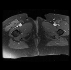
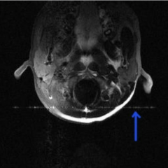
Fat saturation failure
Occurs when a fat sat pulse fails to supress due to resonant frequency of fat not being uniform in the image, poosrly shimmed magnetic field, anatomy distant from isocenter Remedy: Apply shimming within ROI Move anatomy to isocenter For finger, have patient hold them together. |
|
What is this artifact?
|

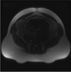
Dielectric effect
In large patients where torso = or > RF pulse size, causes central signal void, worse with higher field strength Remedy: Use dielectric pads Engage multichannel transmission |
|

What is this artifact?
|
Zipper artifact in a noncartesian readout (like filtered backprojection)
|
|
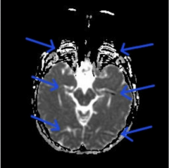
What is this artifact?
|

T2 shine through
Inherent T2 in DWI |