Reading...
![]()
Play button
![]()
Play button
![]()
Use LEFT and RIGHT arrow keys to navigate between flashcards;
Use UP and DOWN arrow keys to flip the card;
H to show hint;
A reads text to speech;
44 Cards in this Set
- Front
- Back
|
Neurons or...and _____ are the types of cells that make up the nervous system
|
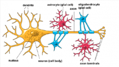
nerve cells
glial cells- provide support for neurons |
|
|
How many neurons and glial cells does brain have?
|
100 billion neurons and 1 trillion glial cells
|
|
|
how many synapses per neuron? how many in total?
|
5000 synapses that act as input!
10^5 in total |
|
|
brain is ___ of body weight, but consumes __ of body's ___ and ___ of its ___
|
Brain is only 2% of body weight, but consumes 25% of body's oxygen and 70% of its glucose.
|
|
|
What are the zones of the Neuron?
|
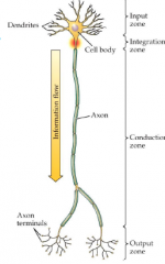
4 Zones.
1. Input Zone 2. Integration Zone 3. Conduction Zone 4. Output Zone |
|
|
Input Zone
|
neuron receives input from other cells (electrical signal/neurotransmitter) through the region of DENDRITES
|
|
|
Integration Zone
|
the region of the cell body or soma where the INPUTS (signals) are combined and transferred.
|
|
|
Conduction Zone
|
Every neuron has ONE axon that leads away from the cell body to the terminals to TRANSMIT the electrical impulse
*the combined signal (threshold reached) is the output which is conducted down the axon that transmits this output |
|
|
Output Zone
|
region of the axon terminals at the end of the axon that (transmits the signal (OUTPUT) to other neurons)
Communicates activity to other cells. |
|
|
What is the Neuron Doctrine?
|
19th Century idea proposed by Cajal that the fundamental unit of the brain is the neuron, or nerve CELL.
|
|
|
Camillo Golgi
|
Use the Silver or Golgi stain to visualize the fundamental unit of the brain, the neuron.
He proposed the RETICULAR THEORY stating that everything in the Nervous System, such as the brain, is made up a single interlinking network, rather than many integrated units. |
|
|
Santiago Cajal
|
Also visualized the neuron with the Golgi Stain but proposed the NEURON DOCTRINE that states that the brain is made up a of many integrated units
|
|
|
Location of neuronal communication?
|
across synapses at the dendritic spines of postsynaptic neuron
|
|
|
What is a synapse?
|
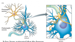
A gap between presynaptic axon terminal and postsynaptic dendritic spine.
Each synapse has three components : 1. Presynaptic Membrane: on the axon terminal of presynaptic neuron. 2. Synaptic Cleft- gap that separates the two membranes. 3. Postsynaptic Membrane: on the dendrite or cell body of the postsynaptic neuron. So synapse includes the output, space, and input region |
|
|
Steps in Synaptic Communication
|
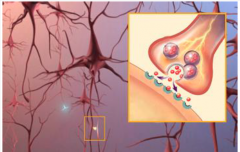
1. Threshold reached, and signal conducted down axon to axon terminals.
2. Electrical signal causes release of synaptic vesicles filled with neurotransmitter by fusion with presynaptic membrane. into the synaptic cleft. 3. Neurotransmitters in synaptic cleft act as keys to postsynaptic neurotransmitter receptors on postsynaptic membrane. 4. Activated post membrane neurotransmitter receptors open to allow ion flow through postsynaptic channels (either receptors or separate channels) into the postsynaptic dendritic spine causing graded potentials that will sum at the axon hillock to potentially pass threshold and excite a signal. |
|
|
What are the glial cells and the types?
|
Glial cells provide support for the neurons and neuronal activity
1. Astrocytes 2. Oligodendrocytes 3. Microglial cells 4. Ependymal Cells 5. Schwaan and Satellite Cells |
|
|
Astrocyte
|
Found in CNS: Star shaped cells with many processes (arms) that receive neuronal input and monitor activity
|
|
|
Oligodendrocytes
|
Provide myelination to CNS cells
|
|
|
Microglial cells
|
or microglia are small cells that remove debris from injured cells in the CNS
|
|
|
Ependymal Cells
|
ependymal cells create the thin epithelium-like lining of the ventricular system of brain and spinal cord and is involved in production of cerebrospinal fluid.
|
|
|
Schwaan Cells
|
provide myelination to PNS cells
|
|
|
Satellite Cells
|
cells that cover surface of nerve cell bodies in PNS cells
|
|
|
Myelination
|
the process by which glial cells (oligodendrocytes and Schwaan cells) wrap axons in a fatty sheath or myelin for insulation and speed conduction.
|
|
|
Multiple Sclerosis
|
A disease that causes demyelination
|
|
|
difference between oligodendrocyte and Schwaan cell?
|
Oligo in CNS and can wrap many segments of axons and of different neurons
Schwaan in PNS and wraps only one segment of one neuron. |
|
|
what is a neuronal circuit?
|
functional entity of interconnected neurons that is able to regulate its own activity using a feedback loop
|
|
|
Anatomical Division of Nervous System
|
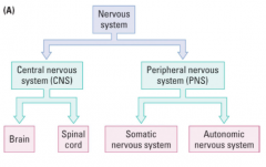
* all the nerve fibers radiating out beyond the brain and spinal cord as well as all the neurons outside the brain and spinal cord form the PNS
so PNS defined as neurons and nerve processes outside the CNS |
|
|
what encases the CNS
|
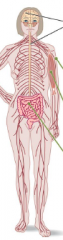
skull encases brain,
and vertebra encase spinal cord |
|
|
What are the divisions of the ANS, and what is it?
|
ANS stands for Autonomic Nervous System, is a branch of the PNS that is not voluntary and is divided into
1. Sympathetic Nervous System- controls the flight or fight response 2. Parasympathetic Nervous System- active during relaxed, non-dangerous situations. 3. Enteric Nervous System- controls digestions |
|
|
Define Homeostasis in terms of neuroscience
|
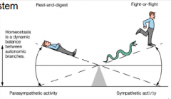
is the dynamic balance between the autonomic branches ( sympathetic and parasympathetic nervous systems)
from rest, digest to flight or flight |
|
|
Somatic Nervous System
|
the branch of the PNS that encases voluntary control of body movements through skeletal muscles
|
|
|
Types of Directional Neuronal information flow
|
1. Sensory/Incoming (Afferent Pathways) - sensory information coming into the CNS
2. Motor/ Outgoing (Efferent Pathways)- information leaving the CNS processing center is CNS |
|
|
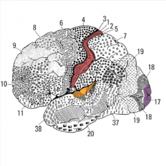
Brain Anatomy : brain surface
|
The brain is split into two cerebral hemispheres. The outermost layer of these hemispheres is the cerebral cortex. The cerebral cortex is convoluted (more surface area). The raised regions are called gyri (gyrus) and the farrows are called sulcus (sulci)
|
|
|
Where is the central sulcus and sylvan fissure?
|
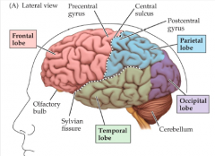
Central sulcus separates the Frontal and Parietal lobe, and the pre central gyrus and post central gyrus .
The Sylvian Fissure separates the Temporal Lobe from the Frontal and Parietal Lobes. |
|
|
What are the four lobes of the brain and their functions
|
Frontal - functions in impulse control, decision making, and contains the motor cortex
Parietal - contains the somatosensory cortex (processes pain, touch and temperature) Occipital- functions in visual processing, visual cortex Temporal - functions in auditory processing and memory |
|
|
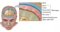
What are meninges?
|
The 3 membranes ( the dura mater, arachnoid membrane and pia mater) that line the skull and vertebral column, surrounding and protecting the brain and spinal cord.
|
|
|
layers of the meninges from skull to brain
|
|
|
|
where does the Cerebrospinal Fluid circulate and what is it's function?
|
in spinal cord- the central canal
in brain - in ventricles and subarachnoid space of meninges Function of CSF- 1. provides an exchange medium between the blood and brain. 2. acts as shock absorber so CSF found around and inside the brain. |
|
|
what is Ventricular System and how many are there?
|
the ventricular system is a series of chambers in the brain filled with cerebrospinal fluid
the two lateral ventricles, and the third and fourth ventricles. |
|
|
Types of matter in the brain and where is each found?
|
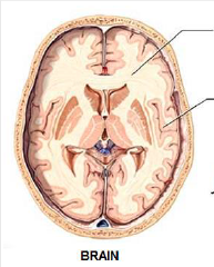
"types" of matter represents the color of the two types of brain tissue.
1. White Tissue (matter) - consists of mostly axons with white MYELIN sheaths 2. Grey Tissue (matter)- contains mostly cell bodies and dendrites, which LACK MYELIN cerebral cortex- grey inside- mostly white so connections are inside the network |
|
|
what is the neocortex
|
neocortex, or cortex is part of the cerebral cortex and has 6 distinct layers distinguished by
1. type of neuron in layer 2. pattern of dendrites or axons. |
|
|
what is the cerebral cortex
|
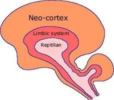
the outermost layer of convoluted grey matter that covers the internal parts of the cerebrum (brain)
|
|
|
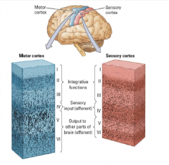
What are the functions of the layers of the cerebral cortex?
|
layer 4- receives sensory input (afferent pathways)
layers 1-3: integrative funcitons layers 5/6: outputs to other parts of brain (efferent) * this means the grey matter does the processing and white matter communicates this information to different parts of the brain. why the lowest layers are output ,near the white matter |
|
|
What is an application of the different layers of the neocortex?
|
Cytoarchitectonic Maps- Brodmann defined brain areas by characteristics and organization of cells
|