Reading...
![]()
Play button
![]()
Play button
![]()
Use LEFT and RIGHT arrow keys to navigate between flashcards;
Use UP and DOWN arrow keys to flip the card;
H to show hint;
A reads text to speech;
53 Cards in this Set
- Front
- Back
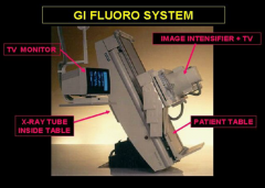
GI Fluoro System
|
II above pt.
xray tube below pt and shielded = less scatter radiation |
|
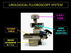
GU fluoro system
|
II below pt closer to kidneys and bladder= less blur
xray tube above and not shielded= more scatter radiation |
|
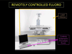
Remote controlled system = GU systems + remote
|
...
|
|
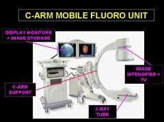
C-arm mobile unit
|
...
|
|
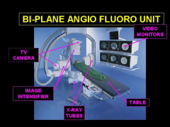
Biplane Angio fluoro unit
|
...
|
|
|
which focal spot is used during fluoroscopy
which focal spot is used for "digital spot" imaging? |
fluoro= small FS
Spot imaging= large FS because of greater tube current needed |
|
|
What is F#?
How does an aperture work? |
f number= aperture size.
Aperture limits light to camera lens. The smaller the aperture, the larger the F# = more radiation needed to maintain brightness = less quantum mottle but higher patient dose. |
|
|
If F# doubles, how does patient dose change?
|
4x. Dose increase = the F# ratio squared
|
|
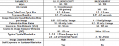
compare GI fluoro and Radiography
|
...
|
|
|
How does an II work?
|
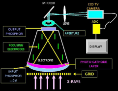
1. xrays pass through grid to filter scatter= 3-5x increase in pt dose.
2. filtered xrays pass through titanium entrance surface (strong because of II internal vaccuum) 3. xrays are captured by entrance phosphor and converted to light 4. emitted light contacts photocathode layer (contains antimony (Sb)) and emits electrons 5. ejected electrons accelerated across vaccuum towards output phosphor which are focused by focusing electrodes 6. focused electrons contact output phosphor which is then converted to light 7. reconverted light passes through aperture and is focused by group of lenses onto television camera 8. television camera (like CCD) converts light to electrical signal |
|
|
what is Flux Gain?
|
Mechanism by which output light from the output phosphor is increased.
Increasing voltage across the II tube between the photocathode and output phosphor = increased kinetic energy of electrons= more light emitted from output phosphor |
|
|
What is minification gain?
what is the magification gain if input surface is 300 and output diamter is 25 |
electrons from large photocathode are concentrated on output phosphor = more energy per unit area.
minification gain (Mx) = (area of input suface/ area of output surface)^2 300/25 = 12 12^2=144 |
|
|
What is brightness gain?
|
amount of increase in light emitted from image intensifier compared to direct exposure of phosphorescent screen
= Flux gain x Minification gain= several thousand times hence image intensifier |
|
|
what is conversion gain?
|
efficiency of II to convert xrays to light. Decreases with time.
2 remedies: 1. use larger apperature (low F#)= more image noise. 2 use more xrays = higher pt. dose replace when Gx drops more than 50% |
|
|
what is contrast ratio?
|
ratio of signal to no signal
place lead disc on input surface and measure signal around disc and behind disc. should be 10-20:1 |
|
|
How does FOV work during mag mode?
|
portion of body is selected and projected onto output phosphor. Appears magnified but the part of the body is actually minified less.
Therefore less energy/area = less light |
|
|
how does mag mode effect radiation dose?
" " Spatial resolution |
since less light emitted, ABC will increase different factors (kVp, mA, longer plse width, less beam filtration) ultimately increases dose 1.4-2x for each mag position.
Improved spatial resolution |
|
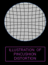
Pincushion Distortion
|
bending of staight lines at periphery of image
|
|
S-distortion
|
related to magnetic field of earth on accelerated electrons in II
S-shaped lines |
|
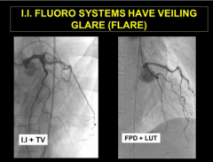
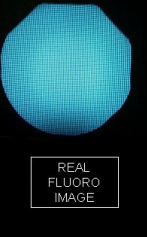
Flare artifact
|
...
|
|
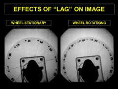
Lag artifact
|
...
|
|
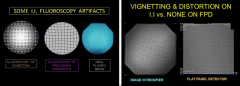
Vignetting artifact
|
distance from source to center is closter than from source to edge
|
|
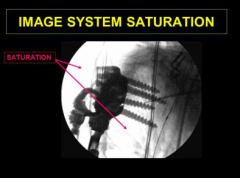
saturation
|
...
|
|
|
How do flat panel detectors (FPD) work?
|
1. Xray passes through grid
2. Interact with CsI needles (phosphor) which emit light 3. Light interacts with photodiode (not photocathode) which converts energy to free electrons which are stored as charge. (length between centers of detector elements= pitch) think fill factor. 4. Electronics convert charge into image. 4. |
|
|
What is the disadvantage of increasing number of detector elements?
|
Fill factor decreases because size of transistor is unchanged. The area available to sense light is relatively decreased/DEL which requires more radiation.
|
|
|
for a 30cm FOV and a 2000x2000 resolution screen, how big is a pixel/DEL?
|
answer 0.15mm
30cm= 300mm 300mm/2000=0.15mm |
|
|
What is binning
cost and benefit? |
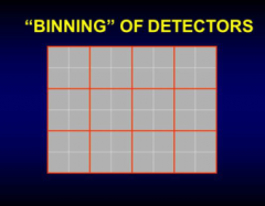
adding mathematical information from several DEL's to form a larger DEL
Decreased quantum mottle/less patient dose lower spatial resolution |
|
|
How do you calculate the spatial resolution of a FPD?
Question: FOV =20cm matrix=1000x1000 resolution in lp/mm? |
(1/ 2xDEL)
answer: 2.5 lp/mm 20cm=200mm DEL= 200mm/1000 pixels= .2mm/DEL Spatial resolution = 1/(2 x 0.2) |
|
|
Bad pixels
How does the system correct for bad pixels? |
bright or dark spots
interpolation |
|
|
persistence
|
=ghost
|
|
|
what is kell factor value (o.7)
For a 525 line system (480 lines) what is vertical spatial resolution for FOV of 11.5 cm |
1.46 lp/mm
480 x 0.7/ (115mm x 2) |
|
|
What is last frame hold?
Frame averaging? |
last image held on monitor= no additonal radiation
signal from last frame averaged with new frame=less quantum mottle at the expense of ghosting |
|
|
Cost/Benefit of edge enhancement?
Grayscale algorithm |
Sharper image but more noise
Enhances contrast and extends dynamic range by limiting saturation of white or black regions |
|
|
Does magnification improve spatial resolution on FPD?
|
no
|
|
|
Resolution of FPD is limited by what?
how about I.I.'s? Which system is limited by dynamic range? |
detector element size
Even though II's have better resolution, they are limited by television screen resolution II systems |
|
|
What is the effect of pixel binning on resolution?
"" frame averaging on resolution |
decreased
decreased due to blur |
|
|
What is the effect of pulsed fluoro on patient dose and blur?
|
both decreased
|
|
|
what happens to patient dose when you increase the SID?
|
increases. Not because of geometry but because the system will output more radiation to compensate for the inverse square law
|
|
|
In fluoroscopy, collumate, keep II as close to the patient as possible and only mag when needed
|
...
|
|
|
which ABC system delivers greater dose to the patient?
One which will increase kVp or mA first |
mA. Always try to increase kVp first
|
|
|
What is the effect of aperture size?
|
large apertures mean noisier image but less patient dose
|
|
|
What is the effect of pulse rate on dose and framerate?
|
low pulse rate means lower dose but lower frame rate
|
|
|
How does fluoro compare to spot image vs CT and DR
|
fluoro 0.02-0.03 uGy/image
Spot image 5-10 uGy/image DR 4-5 uGy/image CT 8-10 uGy/image |
|
|
What is federal regulation for skin dose?
|
87 mGy/min for normal use
174 mGy/min for High level |
|
|
What is cancer risk rate?
Conversion between skin entrance dose to Effective dose |
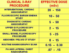
6% per Sv of effective dose
0.05-0.1 mSv/mGy Therefore effective dose for upper GI = 2-15 mSv, SBS = 5-25 mSv, BE=10-50mSv |
|
|
Estimate scattered radiation
Where is it greatest? |
0.1% (0.001) skin dose at 1meter
on the tube side |
|
|
How do you calculate effective dose to personnel?
|
(0.04 x external dosimter) + 1.5xInternal dosimeter
no more than 4.17 mSv per month or 50 mSv per year for body, 12.5 mSv/month or 150mSv/yr for eyes and 500mSv/yr for hands |
|
|
How much radiation is needed to induce cateracts?
|
2.5 Sv
|
|
|
What is low contrast discrimination in fluoroscopy
|
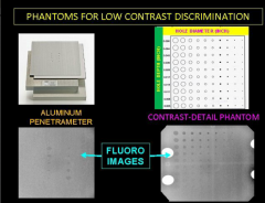
Penetrameter 2 sheets of aluminum 3/4 of an inch thick sandwich a leaded phantom
simulates the phantom being in the patient Another way is to drill multiple holes into an aluminum block of varying diameters and depths |
|
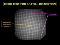
spatial distortion test
|
...
|
|
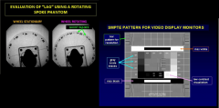
QC motion blur and display calibration
|
...
|
|
|
what is the purpose of an aperture
|
reduce quantum mottle
|
|
|
5-10 fluoro spot images=1 min of fluoro
|
...
|