Reading...
![]()
Play button
![]()
Play button
![]()
Use LEFT and RIGHT arrow keys to navigate between flashcards;
Use UP and DOWN arrow keys to flip the card;
H to show hint;
A reads text to speech;
43 Cards in this Set
- Front
- Back
|
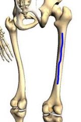
DERMATOMES. At middle of inguinal ligament and a ittle into the upper thigh
|
L1
|
|
|
Lateral aspect of the thigh
|
L2
|
|
|
The great toe
|
L4
|
|
|
The middle three digits (digits 2 -4 )
|
L5
|
|
|
Little toe
|
S1
|
|
|
Medial part of posterior thigh and calf
|
S2
|
|
|
Tap patellar tendon to test
|
L2-L4
|
|
|
Tap Achilles tendon to test
|
S1
|
|
|
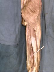
Does the great saphenous vein pass ANTERIOR or POSTERIOR to medial malleous?
|
The great saphenous vein passes ANTERIOR the medial malleous. This kinda makes sense: the great saphenous vein is huge and it basically passes in everything superficial. It runs is course in the superficial fascia for the entire length of the leg & it's just in front of everything
|
|
|
Does the great saphenous vein pierce the investing fascia?
|
No. As we discussed the great saphenous vein runs in the superficial area, the entire length of the leg.
|
|
|
The great saphenous vein joins the
|
Superficial epigastric vein.
Again, the great saphenous vein follows its "superficial" ways....It even only associated with superficial veins! |
|
|
The great saphenous vein becomes tributary to the
|
femoral vein
|
|
|
Arising from the dorsal venous arch on the dorsum of the foo, the small saphenous vein run its course on the __________ fascia. At the level of the popliteal fossa, It then pierces the investing fascia.
|
Arising from the dorsal venous arch on the dorsum of the foo, the small saphenous vein run its course on the SUPERFICIAL fascia.
But It can't take the heat like the GREAT saphenous vein for long, so at the level of the popliteal fossa, the small then pierces the investing fascia & finds refuge there. WIMP. |
|
|
Once the small saphenous vein pierces the investing fascia, it becomes tributary to the _________________ vein.
|
Once the SMALL saphenous vein pierces the investing fascia, it becomes tributary to the POPLITEAL vein.
|
|
Shown here is the Popliteal Vein.
These ______________ & _____________ veins come together near the popliteal fossa and form the origin of the popliteal vein in the popliteal fossa. |
These ______________ & _____________ veins come together near the popliteal fossa and form the origin of the popliteal vein in the popliteal fosssa.
|
|
|
The veins transform as usual--> Finish this
Anterior & Posterior Tibial Veins--> Popliteal Vein --> Femoral Vein ---> Above inguinal ligament-> ____________ Vein |
Anterior & Posterior Tibial Veins--> Popliteal Vein (in popliteal fossa) --> Femoral Vein ---> Above inguinal ligament-> EXTERNAL Iliac Vein
|
|
|
Let's keep going! Deep veins (popliteal vein, femoral vein, anterior + posterior tibial veins) take the blood from lower limb & return it to heart.
External Iliac Vein ---> ____________ Vein ----> Inferior Vena Cava ---> ____________ Atrium |
External Iliac Vein---> Common Illiac Vein ----> Inferior Vena Cava ---> Right Atrium
|
|
|
Great Saphenous Vein passes ANTERIOR or POSTERIOR to medial malleolus?
|
Great Saphenous Vein pass ANTERIOR to MIDDLE malleolus.
Like we said, Great Saphenous vein gotta be in front of everything. Yes, PLEASE note the Great Saphenous never NEVER pierces the Investing layer. |
|
|
Small saphenous vein passes ________________ to ____________ malleolus
|
SMALL saphenous vein passes POSTERIOR to LATERAL malleolus
|
|
|
Deep Vein Thrombosis is common in which deep veins?
|
Common in POPLITEAL and femoral veins (rmbr, popliteal vein is tributary to femoral vein)
|
|
|
Ok now this order:
Popliteal Nodes ---> _________________ Nodes ---> _______________ Nodes (Hint: Follows the same course as small saphenous vein) |
Popliteal Nodes ---> Deep Inguinal Nodes ---> External Iliac Nodes
|
|
|
Alright, back to the popliteal nodes: drain what area
|
Cutaneous & Subcutaneous portions of the LATERAL foot
|
|
|
Superficial Inguinal Nodes, which lies along the inguinal ligament) drain which areas:
|
Basically any area, for a BIKINI wax
The superficial thigh region The anterolateral abdominal wall (below the umblicus) The gluteal region The genital area (except glans) |
|
Which is the ONLYYYYYYY muscle that is completely surrounded by investing layer of fascia in the lower limb ( & prob in upper limb too)
(FOR ALL the other muscles in this superficial & investing layer of fascia is EXTERNAL to the muscles. ) |
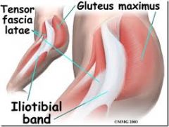
The tensor fascia lata muscle
On the cadaver, it's going to be only other thing, externally, that attaches to the gluteus maximus. The smaller tensor fasciae latae, like the gluteus maximus muscle, also attaches to the iliotibial tract. |
|
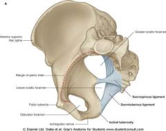
Note how the ligaments are arranged & make up the formation of the foramen
For instance, foramen above sacrospinous ligament is called ______________ The foramen b/w the sacrospinous & sacrotuberous ligament is called ______________ |

formation of the foramens
For instance, foramen above sacrospinous ligament is called GREATER sciatic foramen The foramen b/w the sacrospinous & sacrotuberous ligament is called LESSER sciactic foramen Note how the sacrotuberouss ligament dips downwards, while the sacrospinous ligament tries to keep the foramen looking like a real circle and just essentially shoots right across to the ischial spine from the body. |
|

Which ligament is this: sacrospinous or sacrotuberous ligament?
|
The sacrotuberus is the ligament dipping down from the coccyx (see answer in previous slide) so it's gotta be the sacrotuberous ligament. You don't really have to feel it ...but it'll probably feel bony f you're in lab.
|
|
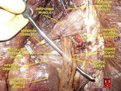
piriformis s labeled. I find it easiest to look for sciatic nerve & then look for the uppermost "slim" muscle above which the sciatic nerve juts out.
What vein, nerve, arteries are above the piriformis muscle & pass through the greater sciatic foramen |
Superior Gluteal VAN.
The superior gluteal artery, the largest branch of the INTERNAL ILIAC artery. This is all internal n the 'iliac' region-so that makes sense. |
|
Right gluteal region.
What nerve this pointing to? Which nerve accompanies it? |
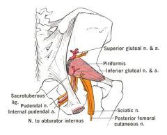
You know it's pudendal nerve b/c
-it's the most medial thing coming out b/w the piriformis & superior gemellus -Basically you're looking for a structure that the most medial item from sciatic nerve. Then follow structure down in lab to confirm that the pudendal nerve is passing down to the sacrotuberous ligament--Pudendal Nerve. Pudendal Nerve & Nerve to Oburator Internus |
|
Which muscle attaches to proximal tuberosity of this highlighted region.
|
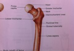
1. Linea Aspera On Femur
2. Tuberosity Proximal to Linea Aspera = Gluteus Tuberosity 3. Gluteal Tuberosty - Gluteus maximus |
|
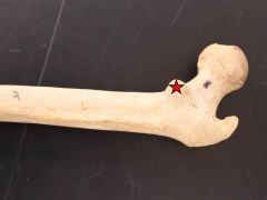
Insertion point of _________________ & ________________
|
Star= Lesser trochanter/ Lesser trochanter is most inferior from head of femur
Insertion point of Iliopsoas muscle. In the cadaver, the iliopsoas muscle is immediately medial to sartorius muscle. |
|
|
Fill in w/ extra or intra
The artery of head of femur + ligament carrying it (ligament of the femoral head are ________CAPSULAR & ___________ SYNOVIAL |
The artery of head of femur + ligament carryng it (ligament of the femoral head are INTRACAPSULAR & EXTRASYNOVIAL
‘extra-synovial’, means that it is not actually surrounded by synovial fluid, and hence it has a better blood supply. Artery of head of femur= branch of medial circumflex artery in Grant's dissector |
|
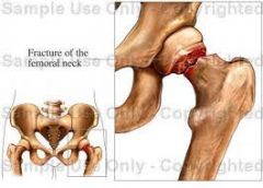
Doesn't that fracture of the femoral neck look darn painful ?! Yeah, this kind of fracture -- an intracapsular fracture--threatens the entire blood supply to the head of the femur (remember, how the branch of the medial circumflex artery)
|
Fracture everywhere else on the femur are JUST NOT that serious
|
|
|
Does medially rotating the femur stabilize or destabilize the femoral ligaments?
|
DESTABILIzes. When femor is medially rotated, the insertion points of ligaments are brought closer to the origin--shortens --> destabilizes ligament.
|
|
|
Does flexing the the femur stabilize or destabilize the femoral ligaments?
|
Destabilize the ligaments
|
|
|
Dislocation of hip joint:
Most of the time, the head of femur displaces ANTERIORLY or POSTERIORLY? |
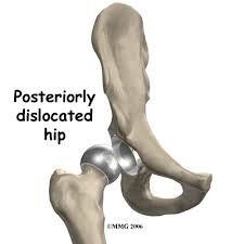
POSTERIORLY
|
|
|
Back to Upper Limb:
Most of the time, the clavicle tends to dislocate how? |
Generally, clavicle tends to dislocate anterior - superiorly.
|
|
|
Groovy Amina is baaaaaack!
Only 4 things that are INTRAcapsular intrasynovial are: What are they? everything else: intracapsular EXTRA synovial |
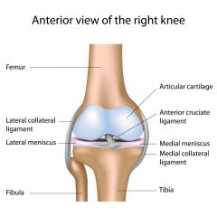
Glenoid labrum, acetabulum labrum, and the medial and lateral meniscis
|
|
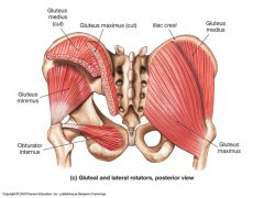
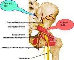
Inferior Gluteal Nerve innervates
Superior Gluteal Nerve innervates |
Inferior Gluteal Nerve innervates Gluteus Maximus. Helps you get of your butt.
Superior Gluteal Nerve innervates Gluteus Medius & Minmus. These muscle prevents pelvic drop & allow you to walk normally |
|
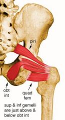
Focus on the muscle a below the piriformis for a minute.
All of these muscle serve to ____________ (medially or laterally) rotate the femur. Will this movement stabilize or destablize the femoral ligaments? The superior 2 muscles -- the gemellus superior & obturator internus--are innervated by _______________ The inferior 2 muscles-inferior gemellus & quadratis femoris-- are innervated by __________________________. |
All of these muscle serve to laterally rotate the femur. Destablize the femoral ligaments! (Look above)
The superior 2 muscles -- the gemellus superior & obturator internus--are innervated by Nerve to the Obturator Internus (which will run lateral to pudendal nerve) The inferior 2 muscles are innervated by nerve to the quadratus femoris. |
|
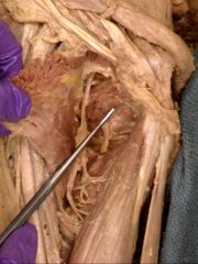
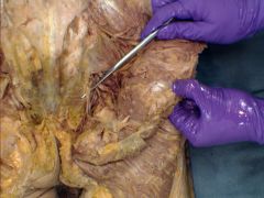
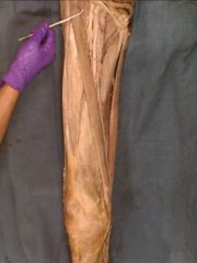
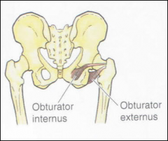
Deep to the adductor brevis muscle, identify the obturator externus muscle. (Well it's shown by probe)
Does the obturator externus swing POSTERIOR or ANTERIOR to the hip joint capsule? |
Note that this is the posterior view, so tendon is visible, but muscle dissapears from view.
Also, go back to the last slide if you didn't realize that you would need to reflect the quadratis femors muscle to actually find the obturator externus. |
|
|
Sciatic nerve divides into wthat 2 nerves?
|
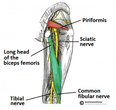
COMMON TIBIAL & FIBULAR NERVE
This makes sense it just divides into the region it innervates. Sticky 'i' keyboard so might me missing i's from now on |
|
|
JUST medially to the SCIATIC nerve you'll find the
|
posterior cutaneours nerve of the thigh
|
|
|
Where should you give a gluteal injection
|
UPPER lateral quadrant
|