![]()
![]()
![]()
Use LEFT and RIGHT arrow keys to navigate between flashcards;
Use UP and DOWN arrow keys to flip the card;
H to show hint;
A reads text to speech;
242 Cards in this Set
- Front
- Back
|
this is the portion of the external ear that functions to funnel sound |
auricle |
|
|
the boundary between the external ear and the middle ear is... |
tympanic membrane (eardrum) |
|
|
this is the structure that travels through the temporal bone and connects the middle ear with the pharynx; functions in pressure equilibrium |
Eustacian tube |
|
|
which portion(s) of the ear are filled with air? which portion(s) are filled with fluid? |
outer and middle inner |
|
|
what are the two muscles contained within the middle ear? |
tensor tympani stapidus |
|
|
the tensor tympani is innervated by the ____________ nerve while the stapidus is innervated by the ____________ nerve. |
trigeminal facial |
|
|
the tensor tympani attaches to which ossicle? |
malleus |
|
|
the stapidus attaches to what ossicle? |
stapes |
|
|
what is the purpose of the two muscles within the middle ear? |
reflexive - to dampen loud sounds |
|
|
two portions of the inner ear |
bony labyrinth membranous labyrinth |
|
|
the inner ear resides deep within what cranial bone? |
temporal
|
|
|
Cranial nerves 7 and 8 exit the skull via the ____________. |
internal acoustic canal |
|
|
this is the portion of the ear that contains the auditory and vestibular receptors |
membranous labyrinth of inner ear |
|
|
two portions of the bony labyrinth of the inner ear: |
cochlea vestibule |
|
|
two portions of the vestibule |
utricle saccule |
|
|
the membranous labyrinth is filled with ____________ |
endolymph |
|
|
the bony labyrinth is filled with ____________ |
perilymph |
|
|
the ____________ sits on top of the cochlear duct while the ____________ sits on bottom |
scala vestibuli scala tympani |
|
|
both the scala vestibuli and the scala tympani are filled with ____________ |
perilymph |
|
|
the scala vestibule and the scala tympani are continuous and contact each other at the ____________, the apex of the cochlea |
helicotrema |
|
|
the inner core of the cochlea is also known as the ____________. contains the spiral ganglia |
modialis |
|
|
the scala tympani contacts the ____________ to allow for the dispersal of increased pressure created by inner ear vibrations back into the middle ear |
round window |
|
|
the cochlear duct sits on top of the ____________ |
basilar membrane |
|
|
this is the membrane that separates the cochlear duct from the scala vestibuli |
vestibular membrane |
|
|
these are the auditory receptor cells for hearing |
hair cells |
|
|
the hair cells are found within the ____________ which rests on top of the ____________ |
Spiral organ of Corti basilar membrane |
|
|
the "hairs" of the hair cells are found within the ____________ |
tectorial membrane |
|
|
what is the organization of the hair cells? |
1 row inner 3-5 rows outer |
|
|
the cell body for the nerve that receives input from the hair cells of the hear is found where? |
spiral ganglion |
|
|
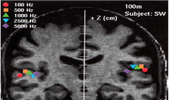
describe the tonotopic localization of the spiral organ of corti |
high frequencies heard at the base low frequencies heard at the apex |
|
|
how many neurons are involved in the auditory pathway? |
4 |
|
|
this is the first order neuron of the auditory pathway |

cochlear nerve (receives innervation from SOC) |
|
|
the first order neuron of the auditory pathway synapses at the ____________ found within the medulla |
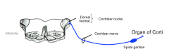
dorsal and ventral cochlear nuclei |
|
|
second order neurons in the auditory pathway form the ____________ which decussate in the caudal pons and ascend via the contralateral ____________. |
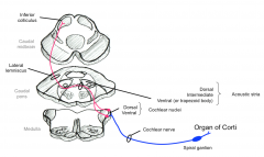
acoustic stria lateral lemniscus |
|
|
three portions of the acoustic stria |
dorsal intermediate ventral |
|
|
the ventral nucleus of the acoustic stria forms the ____________ as it decussates |
ventral/trapezoid body |
|
|
in the auditory pathway, secondary neurons from the dorsal and ventral auditory nuclei synapse onto the ____________ within the caudal midbrain |
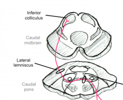
inferior colliculus |
|
|
from the inferior colliculus, third order neurons in the auditory pathway project to the ____________ via the ____________. |
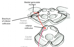
medial geniculate nucleus brachium of inferior colliculus |
|
|
from the medial geniculate nucleus, fourth order neurons in the auditory pathway pass through the ____________ and project to the primary auditory cortex |
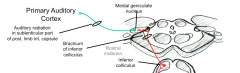
posterior limb of internal capsule (sublenticular) |
|
|
three accessory nuclei found within the auditory pathway; makes the pathway bilateral |
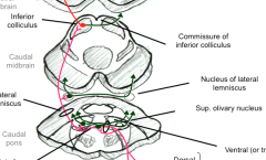
superior olivary nucleus nuclei of trapezoid body nuclei of lateral lemniscus |
|
|
what is the purpose of efferent signals back to the ear? |
to sharpen for selective attention to certain sounds to dampen loud sounds (through Tensor Tympani and stapidius muscles) |
|
|
this is a symptom of Bell's palsy (facial nerve) caused by an inability of the stapidius to respond to loud sounds; sound is too loud in the affected ear |
hyperacoussus |
|
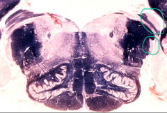
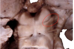
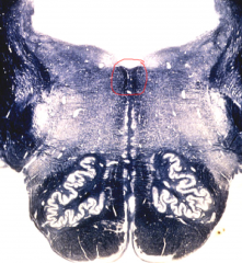
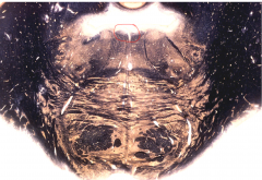
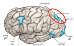
what are the circled areas? |
dorsal and ventral cochlear nucleus |
|
what lies just deep to the circled area? |
circled area = acoustic tubercle just deep = dorsal cochlear nucleus |
|
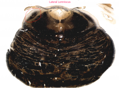
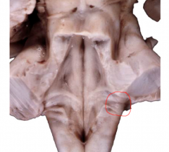
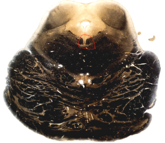
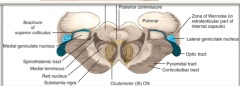
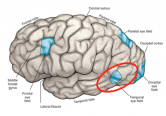
name the circled area |
lateral lemniscus |
|
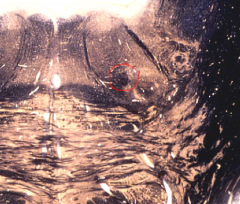
name the circled area |
superior olivary nucleus |
|
name the circled area |
brachium of the inferior colliculus |
|
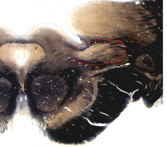
name the circled area |
medial geniculate nucleus |
|
|
where is the primary auditory cortex found? |
transverse temporal gyrus of Heschel |
|
|
how is the primary auditory cortex organized? |
low tones lateral, high tones medial |
|
|
where must a lesion be in the auditory pathway to produce unilateral deafness? |
proximal to cochlear nuclei (spiral organ, spiral ganglion, cochlear nerve, cochlear nuclei) anything distal = no hearing loss |
|
|
type of deafness that results from interference with the passage of sound waves through the external or middle ear; transmission through cranial bones can still occur |
conduction deafness eg. earwax, otitis media |
|
|
type of deafness that results from damage to receptor cells of the spiral organ or to the cochlear nerve; transmission of sound waves through air and cranial bones does not occur |
nerve (sensorineural deafness) |
|
|
this is the muscle that opens a slit in the pharynx allowing the Eustachian tube to equilibrate any pressure buildup in the middle ear |
tensor palati |
|
|
the body of hair cells sits in ____________ while the stereo cilia sit in ____________. |
perilymph endolymph (within the tectorial membrane) |
|
|
these are special capillaries that produce endolymph |
stria vascularis |
|
|
this is the tallest stereo cilium of hair cells |
kinocilium |
|
|
if the shearing force produced by sound waves causes the stereo cilia to bend towards the kinocilium, the hair cell is... |
activated |
|
|
in the auditory system, when a hair cell is activated, it releases ____________ which binds receptors on the dendrites of CN8 and causes an action potential |
glutamate |
|
|
which portion of the cochlea is wider: the base or the apex (helicotrema) |
the apex (width = 0.5 mm) width at base = 0.2 mm |
|
|
which hair cells constitute 95% of the afferent impulses in the auditory system? |
inner |
|
|
which hair cells receive 100% of the efferent impulses in the auditory system? |
outer |
|
|
two functions of the vestibular system |
keeps body on an even keel keeps eyes on target when the head is in motion |
|
|
3 sources that play a role in maintaining balance via the vestibular system (need 2/3) |
vision proprioception vestibular |
|
|
all vestibular activity is ____________ in nature |
reflexive |
|
|
this is the phenomenon that presents when the vestibular system is abnormal |
vertigo |
|
|
where are the 4 nuclei of the ventricular system found? |
floor of the fourth ventricle in caudal pons |
|
|
ascending impulses from the vestibular nuclei are involved in ____________ while descending impulses are involved in ____________. |
vestibulo-ocular reflex keeping body on even keel |
|
|
this disease causes degeneration of the dorsal columns of the spinal cord and can lead to a positive Romberg's sign |
pernicious anemia |
|
|
3 spots within the inner ear where the neuroepithelia for the ventricular system is found: |
utricle saccule ampulla of semicircular ducts |
|
|
the neuroepithelia from the utricle and saccule are chiefly involved with the ____________ system while the neuroepithelia of the semicircular ducts are associated with the ____________ system. |
vestibulospinal vestibulo-ocular |
|
|
within the utricle and saccule, the spot where the hair cells are located is the ____________. |
macula |
|
|
within the ampulla, the spot where the hair cells are located is the ____________ |
crista ampullaris |
|
|
the tectorial membrane is to the auditory system as the ____________ is to the vestibular system |
otolithic membrane |
|
|
these are little crystals that provide weight to the otolithic membrane of the vestibular system - cause inertia whenever head moves |
otoliths |
|
|
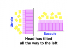
how are the utricle and saccule organized relative to each other in a standing upright position? |
at 90 degree angle |
|
|
in an upright position, which part of the vestibular system is oriented in the horizontal plane? |
macula of the Utricle |
|
|
in an upright positon, which part of the vestibular system is oriented in the vertical plane? |
macula of the saccule |
|
|
this is a condition where the otolith crystals become dislodged and can insert themselves into the crista ampularis causing excessive dizziness; usually happens at certain times of the day |
benign paroxysmal positional vertigo |
|
|
the cell bodies of first order neurons in the vestibular pathway lie in... |
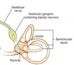
vestibular (scarpa's) ganglion |
|
|
from the vestibular nuclei in the pontomedullary junction, fibers descend via the ____________ and ____________ to influence balance and posture |
medial and lateral vestibulospinal tracts |
|
|
from the vestibular nuclei in the pontomedullary junction, fibers ascend via the ____________ to influence Cranial nerves 3, 4, and 6 |
medial longitudinal fasciculus |
|
|
in the crista ampullaris of the ampulla, this structure extends from one side of the duct to the other; changes in position cause deflections of this structure and create hair cell potentials |
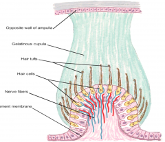
cupula |
|
|
in the vestibular system, the utricle and saccule respond to ____________ acceleration while the ampulla of the semicircular ducts respond to ____________ acceleration |
linear angular |
|
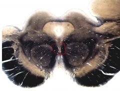
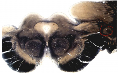
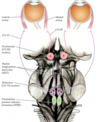
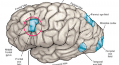
what is the circled structure? |
medial longitudinal fasciculus |
|
what is the circled structure? what cranial nucleus is it located near? |
MLF near the abducens nucleus |
|
what is the circled structure? what cranial nucleus is it located near? |
MLF near the trochlear nucleus |
|
what is the circled structure? what cranial nucleus is it located near? |
MLF near the oculomotor nucleus |
|
|
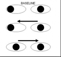
in the vestibule-occular reflex, if the head is turned to the right, what two CN nuclei are stimulated to keep the eyes on a target? |
left abducens (to lateral rectus) right oculomotor (to medial rectus) (left eye abducts, right eye adducts) |
|
|
with nystagmus, the slow phase is induced by the ____________ while the fast phase is induced by the ____________. |
slow phase = vestibulo-occular reflex fast phase = cerebral cortex |
|
|
how is nystagmus named? |
based on the direction of the fast phase |
|
|
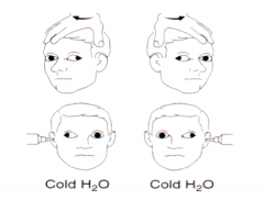
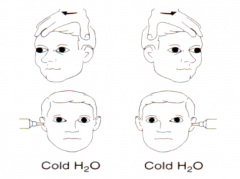
in normal individuals, what happens if you inject cool water into their ear? |
nystagmus to opposite side COWS |
|
|
in normal individuals, what happens if you inject warm water into their ear? |
nystagmus to same side COWS |
|
|
in comatose individuals, if their head is turned to one side or the other and the eyes turn in the opposite direction, this is referred to as the ____________ and is indicative of an intact vestibulo-ocular reflex |
doll's eye movement/oculocephalic reflex |
|
|
in comatose individuals with an intact brainstem, what happens if you inject cold water into their ear? |
eyes move slowly towards the stimulation (no fast resetting phase) |
|
where is the lesion? |
bilateral MLF lesion |
|
where is the lesion? |
low brainstem |
|
|
what ion enters the hair cell when stimulated? |
potassium |
|
|
endolymph has a high concentration of ____________ and a low concentration of ____________ |
high potassium low sodium |
|
|
perilymph has a high concentration of ____________ and a low concentration of ____________. |
sodium potassium |
|
|
how is it hypothesized that the bending of stereo cilia causes the influx of potassium into hair cells? |

tip-lengths between cilia open a "gate" |
|
|
what is the destiny of potassium after it enters the hair cell? |
filtered to the stria vascularis (site of endolymph production) where it is pumped back into the endolymph (maintains ion gradient) |
|
|
this condition is caused by a malfunction in the pump that pumps potassium into the stria vascularis; results in endolymph that lacks potassium; deafness and vertigo |
endolymphatic hydrops |
|
|
which structure is more important when lying down: the utricle or the saccule? |
the saccule |
|
|
this is the maneuver used to treat BPPV; designed to get otoconia back into the right spot |
Epple maneuver |
|
|
why do the hair cells within the semicircular ducts (in the crista ampularis) exhibit rapid adaptation? |
because of the friction of the walls of the duct, the fluid gets to rotating the same direction as the head (once the inertia is overcome) also why once you stop spinning, you feel as if you're still spinning in the opposite direction |
|
|
with vertigo, how can you tell where the damage is? |
side you stumble to is where the damage is located |
|
|
these CNS cells are the equivalent to peripheral monocytes and macrophages and can present Ag in the CNS |
microglia |
|
|
what types of cells infiltrate into the CNS due to CNS infection? |
neutrophils mononuclear cells |
|
|
what are the 4 routes into the nervous system from the periphery in terms of infectious agents? |
blood nerves contiguous sites trauma |
|
|
what type of CNS infection spreads via synaptic connection and follows neural routes |
virus |
|
|
this is an inflammation/infection of the meninges, ependyma, or subarachnoid space |
meningitis |
|
|
this is inflammation/infection of the brain parenchyma with clinical evidence of neurologic impairment |
encephalitis |
|
|
a patient presents with high fever, severe headache, stiff neck, nausea and vomiting; what is high on the differential |
meningitis |
|
|
____________ meningitis displays a diffuse growth pattern within the CSF while ____________ meningitis displays growth along the epithelial surface of the meninges |
bacterial viral |
|
|
if you did a spinal tap suspecting meningitis, what would the following results indicate: clear, colorless liquid; normal glucose; normal protein; normal cell infiltrate |
viral |
|
|
three types of viruses that commonly cause viral encephalitis |
rabies virus poliovirus measles |
|
|
4 types of herpesviruses that can cause encephalitis |
HSV1 HSV2 Varicella-zoster CMV |
|
|
which is the only type of encephalitis-causing herpes virus that spreads hematogenously? |
CMV |
|
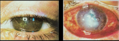
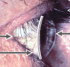
what is this condition associated with? |
herpesvirus (keratitis/corneal scarring) |
|
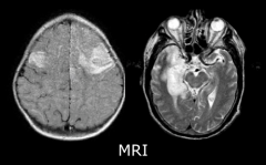
if a patient presented with fever, personality change, and this MRI, what is high on the list? |
herpesvirus encephalitis |
|
if this was seen on a peripheral blood smear, what is the diagnosis? |
CMV (large intranuclear inclusions) |
|
|
infection with this is one of the most common causes of hearing loss in neonates |
CMV |
|
|
if you see a form of encephalitis that is present seasonally or with travel to a certain area what must be on the differential? |
arthropod-bourne encephalitis (ticks, etc.) |
|
|
this is the most important flavivirus encephalitis worldwide and is found widespread throughout Southeast Asia; 1/2 of these infections are neuroinvasive |
Japanese encephalitis |
|
|
this patient presents with a fever that began 2-3 days ago with chills and malaise as well as encephalitis; symptoms include headache, delirium, and a tremor; what is the diagnosis? |
Japanese Encephalitis |
|
|
this type of viral infection presents in two stages; the mild stage includes a fever, headache, nausea, and a characteristic rash on the back and stomach; the more severe stage includes high fever, stiff neck, convulsions, stupor, and eventually death; peak of onset is late summer |
West Nile Virus |
|
|
4 main types of inflammatory reactions to CNS infections: |
|
|
|
what type of inflammatory infiltrate is seen in CNS infections such as acute meningitis, cerebritis, and abcesses? |
neutrophils |
|
|
what type of inflammatory infiltrate is seen in CNS infections such as chronic meningitis and encephalitis? |
mononuclear cells |
|
|
what type of inflammatory infiltrate is seen in CNS infections such as TB, fungal, and parasitic infections? |
granulomatous |
|
|
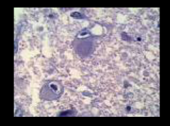
what type of inflammatory infiltrate is seen in viral encephalitis? |
microglial nodules |
|
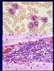
if this type of infiltrate was seen in a CNS infection, what would be the diagnosis? |
neutrophil acute meningitis (bacterial), cerebritis, abcess |
|
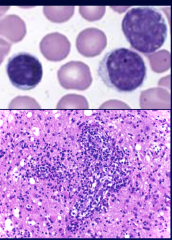
if this type of infiltrate was seen in a CNS infection, what would be the diagnosis? |
mononuclear chronic meningitis, encephalitis |
|
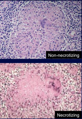
if this type of infiltrate was seen in a CNS infection, what would be the diagnosis? |
granulomatous TB, fungi, parasite |
|
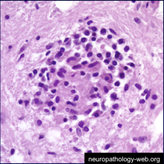
if this type of infiltrate was seen in a CNS infection, what would be the diagnosis? |
microglial nodules viral encephalitis |
|
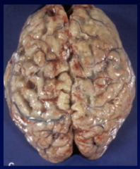
what type of CNS infection would produce this gross appearance? (cloudy meninges) |
acute bacterial meningitis |
|
|
what are 3 complications that can arise out of bacterial meningitis? |
cerebral edema infarcts hydrocephalus |
|
|
this is a complication that can arise specific to meningitis due to N. Meningitisl; meningococcal septicemia, hemorrhagic rash and necrosis |
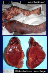
Waterhouse-Friderichsen Syndrome |
|
|
this is described as an area with acute inflammation and edema which develops a necrotic center; characteristic of a brain abscess |
cerebritis |
|
|
after a few days, what forms around the necrotic center of a brain abscess in the brain? |
fibrotic capsule |
|
|
this type of focal infection usually occurs as a complication of meningitis in infants |
subdural empyema |
|
|
this type of focal CNS infection can occur secondary to bone/soft tissue infection and is rare in the brain but can spread rapidly in the spinal cord, forming abscesses that cause cord compression; seen commonly with S. aureus |
epidural empyema |
|
|
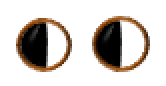
this is when you have a defect within the same location of the visual field in each eye |
homonymous defect |
|
|
this is when you have a defect within different locations of the visual field in each eye |
heteronymous defect |
|
|
this is when you have a defect in one half of the visual field |
hemianopsia |
|
|
this is when you have a defect in one quadrant of the visual field |
quadrantic anopsia |
|
|
this is a defect in the visual field |
anopsia |
|
|
this is the first order neuron in the visual pathway; receives sensory info from the rods and cones |
bipolar neurons (of the retina) |
|
|
this is the second order neuron of the visual pathway; receives input from the bipolar cells and axons form the optic nerve |
ganglion cell (of the retina) |
|
|
this is the third order neuron of the visual pathway; receives input from the optic tracts and relays that information to the primary visual cortex |
lateral geniculate nucleus |
|
|
the optic nerves converge at the ____________ and continue to the LGN as the ____________. |
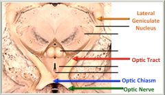
optic chiasm optic tracts |
|
|
this is found in the angle of the optic chasm; explains the visual damage caused by aneurysm |
internal carotid artery |
|
|
what is the fate of those fibers of the optic tract that don't synapse on the LGN? |
bypass, join the brachium of the superior colliculus to influence light reflexes |
|
|
what visual information is carried by the right optic tract? |
the medial visual field of the left eye the lateral visual field of the right eye |
|
|
the LGN is composed of ____________ layers
|
six |
|
|
within the LGN, the ____________ layers are the part of the visual pathway concerned with the location and movement of an object in the visual field; the "where" of the visual field |
magnocellular |
|
|
within the LGN, the ____________ are the part of the visual pathway concerned with the color and form of the object; the "what" of the visual field |
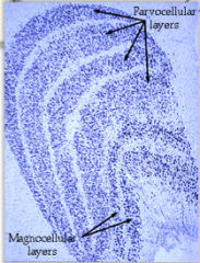
parvicellular |
|
|
from the LGN, tertiary neurons form the ____________ which enters the posterior limb of the internal capsule and forms a triangular area known as ____________ |
optic radiation (geniculocalcarine tract) Wernicke zone |
|
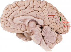
what is the circled area? |
lateral geniculate nucleus |
|
|
the fibers of the optic tract terminate in the walls of the ____________ sulcus |
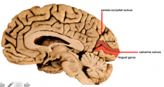
calcarine |
|
|
the dorsal part of the optic radiation projects to the ____________ while the ventral part of the optic radiation (Loop of Meyer) projects to the ____________. |

cuneus gyrus lingual gyrus |
|
|
what is the retinotopic organization of the primary visual cortex? |
|
|
|
after receiving the visual stimulus in the primary visual cortex, further processing and conscious interpretation of visual information is carried out where? |
extrastriate cortical areas |
|
|
from the primary visual cortex, the magnocellul ar stream (the where) projects to the ____________ where it is combined with other outputs to form the spatial visual pathway |
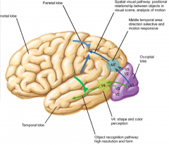
posterior parietal lobe |
|
|
from the primary visual cortex, the parvicellular stream (the what) projects to the ____________ where it is combined with other senses to form the object recognition pathway (eg. looks and sounds like mom) |

inferior temporal lobe |
|
|
what would a lesion in the right optic nerve produce? |
right eye blindness |
|
|
what would a longitudinal transection of the optic chiasm produce (pituitary tumor) |

bitemporal hemianopsia (lateral visual fields are knocked out in both eyes) |
|
|
what would a lesion in the right angle of the optic chiasm produce? (aneurysm of ICA) |

right nasal hemianopsia |
|
|
what would a lesion of the right optic tract produce? (temporal lobe tumor that compresses tract against cerebral crus) |

contralateral homonymous hemianopsia (left visual field knocked out in each eye) |
|
|
what would a complete destruction of the right optic radiation produce? |
contralateral homonymous hemianopsia (left visual field knocked out in each eye) |
|
|
what would a lesion in the ventral optic radiation (loop of Meyer) produce? (temporal/occipital lobe tumor) |

contralateral, homonymous, superior quadrantic anopsia (pie in the sky) |
|
|
what would a lesion in the dorsal optic radiation produce (parietal/occipital lobe tumor) |
contralateral, homonymous, inferior quadrantic anopsia (pie on the ground) |
|
|
what would a lesion in the right visual cortex (both cuneus and lingual gyri) produce? |
left homonymous hemianopsia with macular sparing |
|
|
what are the 3 visual reflexes that govern pupil size/lens curvature? |
pupil constriction (light reflex) pupil dilation accommodation |
|
|
in the light reflex, impulses from the retina pass through the optic tract and bypass the lateral geniculate nucleus, traveling through the ____________ and synapsing on the ____________ |

brachium of superior colliculus pretectal nucleus |
|
|
in the light reflex, impulses from the pretectal nucleus pass to the ____________ which contains preganglionic parasympathetic neurons |
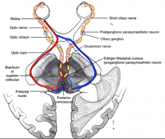
Edinger-Westfall nucleus |
|
|
in the light reflex, preganglionic parasympathetic neurons from the Edinger-Westfall nucleus pass through the ____________ to synapse onto postganglionic parasympathetic neurons in the ____________. |
occulomotor nerve ciliary ganglion |
|
|
in the light reflex, postganglionic parasympathetic nerves from the ciliary ganglion travel through ____________ to the pupil constrictor muscles |
short ciliary nerves |
|
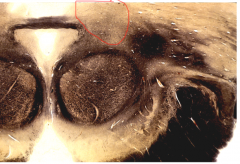
what is the circled area of the rostral midbrain? |
pretectal area |
|
|
would a lesion in the LGN or optic radiation produce a light reflex deficit? |
no - afferents are still intact (because they bypass the LGN) |
|
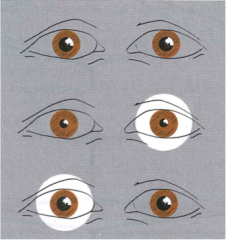
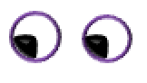
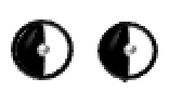
what would produce this effect? (when shined into left eye, both eyes constrict; when shine into right eye, neither eye constricts) |
lesion in right optic nerve |
|
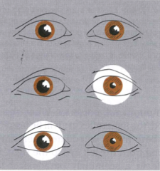
what would produce this effect? (right eye doesn't constrict no matter what) |
damage to right oculomotor nerve |
|
|
where does the pupillary dilator reflex originate from? |
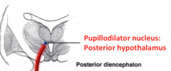
pupillodilator nucleus in posterior hypothalamus |
|
|
from the pupillodilator nucleus, neurons descend through the pupillodilator tract to synapse on the ____________ which contains preganglionic sympathetic neuron |
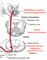
ciliospinal center of C8-T1 (in lateral horn) |
|
|
from the ciliospinal center, pregangionic sympathetic fibers from the pupillodilator reflex pass out of the spinal cord and ascend to synapse on the ____________ which contains postganglionic sympathetic neurons |
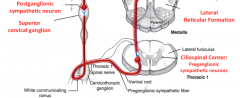
superior cervical ganglion |
|
|
from the superior cervical ganglion, postganglionic sympathetics fibers travel through ____________ nerves to reach the pupil dilator muscle |
nasociliary and long ciliary nerves |
|
|
what is the classic triad of Horner's syndrome? |
miosis (pupil constriction) ptosis anhydrosis |
|
|
what are the three components of the accommodation reflex? |
convergence of eyes pupillary constriction thickening of lens |
|
|
this is the process of maintaining a clear visual image as gaze shifts from distant to a near object |
accomodation |
|
|
in the accommodation reflex, ____________ afferent fibers from the striate cortex project to the ____________ of the midbrain |
corticotectal accommodation center |
|
|
from the accommodation center, fibers pass to the ____________ and the ____________ to constrict the pupil and converge the eyes. |
Edinger-westfall nucleus occulomotor nucleus (to medial rectus) |
|
|
this is a condition where the pupil doesn't constrict the pupil in reaction to increased light but does constrict the pupil in the accommodation reflex; can result from neurosyphilis, MS, or encephalitis |
Argyll Robertson pupil |
|
|
this is the ring around the cornea that connects the cornea and the sclera |
limbus |
|
|
this is the point where the optic nerve contacts the retina; can be visualized in the back of the eye |
optic disk/papilla |
|
|
this condition occurs when the lens calcifies and becomes opaque, can no longer refract light |
cataracts |
|
|
this the structure that produces aqueous humor; also contains the muscles that determine the tension of the suspensory ligaments of the lens (important in accommodation) |
ciliary body |
|
|
this is a condition that results from an imbalance in the ratio of production:drainage of aqueous humor that bathes the eye causing a pressure buildup; on e of most common causes of blindness in the world |
glaucoma |
|
|
lens accommodation and miosis is controlled by ____________ input (sympathetic or parasympathetic?) |
parasympathetic (from the E-W nucleus) |
|
|
pupil dilation (mydriasis) is controlled by ____________ input. (sympathetic or parasympathetic) |
sympathetic (from the superior cervical ganglion)
|
|
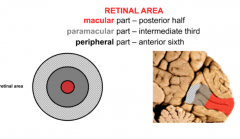
|
this is the area within the retina that contains the highest density of receptors to ganglion cells (1:2); contains only cones |
fovea |
|
|
this is the photopigment with rods that does the signal transduction; changes shape when it absorbs energy resulting in signal cascade |
rhodopsin |
|
|
two types of retinal processing that take place after the excitation of photoreceptors |
spectral (determines color) spatial (location of stimulus) |
|
|
these supporting cells within the retina gather information between neighboring photoreceptors and functions in center surround antagonism |
horizontal cell |
|
|
this a condition where the eye focuses information within the eye; as a result, near objects appear clearly but far objects don't; nearsightedness |
myopia |
|
|
this is a condition where the eye focuses light behind the retina; as a result, near objects appear blurry but far objects don't; farsightedness |
hyperopia |
|
|
there are two general types of eye movements; ____________ movements is when eyes move together in the same direction, whereas ____________ is where eyes move inward or outward |
conjugate vergence |
|
|
what are the 4 different types of conjugate movements |
saccades smooth pursuit optokinetic vestibulo-ocular |
|
|
this conjugate eye movement is voluntary and involves rapid movement of vision to search for a target |
saccades |
|
|
this conjugate eye movement is reflexive and functions to keep an image of a moving target fixed on the retina |
smooth pursuit |
|
|
this conjugate eye movement is reflexive and functions during continuous movement of the target (when you're stationary but objects are moving by quickly) |
optokinetic |
|
|
this conjugate eye movement is reflexive and keeps targets fixed on the retina during head movement |
vestibulo-ocular |
|
|
these eye muscles move the eye horizontally and vertically |
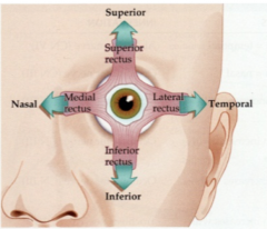
rectus muscles |
|
|
these eye muscles provide torsional eye movements |
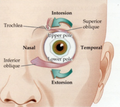
oblique muscles |
|
|
how do you test the superior rectus muscle? |
abduct and elevate |
|
|
how do you test the inferior rectus muscle |
abduct and depress |
|
|
how do you test the superior oblique muscle? |
adduct and depress |
|
|
how do you test the inferior oblique? |
adduct and elevate |
|
|
if the limitation of eye movement isn't restricted to one muscle but rather groups of muscles, what should be on your differential? |
myasthenia gravis |
|
|
the horizontal gaze center is located in the ____________ while the vertical and vergence gaze centers are located in the ____________ |
pons midbrain |
|
|
this is the horizontal gaze center that is found in the pons adjacent to the abducens nucleus |
paramedian pontine reticular formation (PPRF) |
|
|
to gaze to the left, which PPRF is activated? what nuclei does it project to? what muscles does it activate? |
left PPRF projects to left abducens nucleus, right oculomotor nucleus activates left lateral rectus, right medial rectus |
|
|
what would be the outcome of a unilateral lesion in the right PPRF? |
no gaze to the right in either eye |
|
|
in the horizontal gaze pathway, fibers from the abducens nucleus project to the contralateral oculomotor nucleus via the ____________. |
medial longitudinal fasciculus |
|
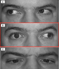
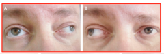
what is this condition? where is the lesion? |
right internuclear opthalmoplegia right eye can't abduct during horizontal gaze to the left, but still functions during convergence |
|
what is this condition? commonly seen in what disease? |
bilateral internuclear opthalmoplegia seen in MS |
|
what is this condition? on what side? |
one and a half syndrome lesion on the left side |
|
|
where is the lesion located in one and a half syndrome? |
unilateral lesion of MLF that also includes adjacent abducens nucleus/PPRF |
|
|
this is the nucleus of the vertical gaze center |
rostral interstitial nucleus of the medial longitudinal fasciculus (riMLF) |
|
|
2 clinical conditions that can cause vertical gaze paralysis |
pineal tumors that compress dorsal midbrain (Parinaud Syndrome) hydrocephalus |
|
|
these are the two nuclei that constitute the vergence gaze center |
nucleus raphe interpositus nucleus reticularis tegmenti pontus |
|
|
the mergence gaze center is controlled by the ____________ eye field |
occipital |
|
|
4 eye fields that constitute the cortical gaze centers |
occipital parietal temporal frontal |
|
|
this eye field, when activated, results in saccades to the contralateral side; projects to the contralateral PPRF |
frontal |
|
|
what would be the result of an irritative lesion (epilepsy) to the right frontal eye field? |

deviation of right towards left |
|
|
this eye field constitutes the visual attention center |

parietal eye field |
|
|
a lesion in what eye field results in a patient neglecting objects in the contralateral side, difficulty making eye movements towards affected side |
parietal eye field |
|
|
what would be the result of an acute lesion (stroke) to the right frontal eye field? |
deviation of eyes towards affected side |
|
|
this eye field is involved in smooth pursuit of the eyes towards the ipsilateral side; also involved in opt-kinetic movements |
temporal eye field |
|
|
what is thought to trigger "attacks" seen in MS |
activation of auto reactive CD4 T cells in PNS (possibly due to EBV or HHV6) |
|
|
these can be seen radiographically and pathologically in patients with MS; represents focal areas of inflammation and injury |
MS plaque |
|
|
T/F: MS only affects white matter |
False - becoming more clear that grey matter is affected |