Reading...
![]()
Play button
![]()
Play button
![]()
Use LEFT and RIGHT arrow keys to navigate between flashcards;
Use UP and DOWN arrow keys to flip the card;
H to show hint;
A reads text to speech;
56 Cards in this Set
- Front
- Back
|
Broadly, what are the three parts of the visual pathway?
|
Optical
Retinocortical Perceptual |
|
|
What is the upper part of the perceptual pathway responsible for?
|
Visuospatial processing
|
|
|
What is the lower part of the perceptual pathway responsible for?
|
Recgnition
|
|
|
What are some of the clinical manifestations of the retinocortical disorders?
|
Blurred vision
Dim vision Scotomas Light spots |
|
|
What is a cause of dim vision?
|
In general, less traffic through the retinocortical system
|
|
|
What is a cause of scotoma?
|
Lesion in a part of the retinocortical pathway
|
|
|
What is a clinical manifestation of the occiipito-temporal part of the perceptual system?
|
Problems with the what: object, color recognition
|
|
|
What is a clinical manifestation of the occiipito-parietal part of the perceptual system?
|
Problems with the where: distributed attention, trouble with spatial relations
|
|
|
How do you eliminate optical problems when thinking about optical disorders?
|
Put pinholes!
It gets rid of media problems, refractive errors, etc. |
|
|
What are two things you should always check if you're evaluating what you think is a retinocortical disorder?
|
Pupil reactions
Visual fields |
|
|
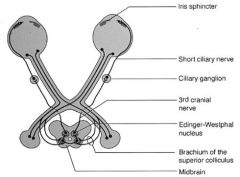
What is the pupillary reflex pathway?
|
|
|
|
What parts of the midbrain are active in the pupillary reflex pathway?
|
Edinger-westphal nucleus
Branchium of the superior colliculus |
|
|
How do you test for a relative afferent pupil defect? Wht do you find?
|
Swinging light test
If a pupil expands when there's light, you've got one of two problems: -Defect in that pupil -Bilateral defect, but the one that expands is worse |
|
|
What test do you use to see where a lesion is in the retinocortical pathway?
|
Visual fields
|
|
|
Where are different places on the retinocortical pathway that you can have lesions?
|
1. Retinal ganglion cells, optic nerve
2. Optic chiasm 3. Optic tracts, optic radiations, visual cortex |
|
|
Where are some different places in the optic nerve that you can get defext?
|
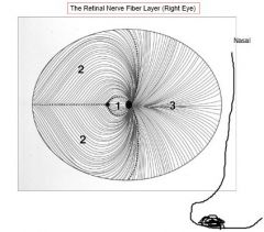
Right eye:
1.Fovea/macula 2.Arcurate 3.Radial (YOU SEE THE TEMPORAL FIELD) |
|
|
What part of the vision is lost in a central scotoma? Where does this lesion take place?
|
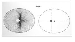
You get a lesion of all of the fibers from the fovea going into the optic nerve, which leads to a blind spot in the center of vision
|
|
|
What part of the vision is lost in a cecocentral scotoma? Where does this lesion take place?
|
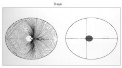
Not only in the fovea, but the other ganglia that extend over are effected
It's a scotoma from the blind spot to the point of vision |
|
|
What part of the vision is lost in an arcuate scotoma? Where does this lesion take place?
|
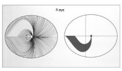
Scimtar with a border along the meridian of sight
|
|
|
What part of the vision is lost in a temporal scotoma? Where does this lesion take place?
|
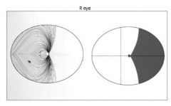
If this is the RIGHT edge, you're losing the NASAL cells, which means that you're having a defect in the TEMPORAL field of sight
|
|
|
What happens after the optic chiasm to the visual information?
|
The fibers are segregated into hemifields: "right world" goes to left brain, vice versa
|
|
|
If you've got a defect in the optic chiasm, what will be the effect on the visual field?
|
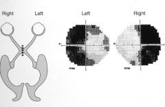
Bitemporal hemianopia: you're losing the sight of the nasal fibers, which are the ones that cross.
This results in an inability to see the TEMPORAL fields |
|
|
If you have a visual field defect in he retrochiasma, what will be the effect on visual fields?
|
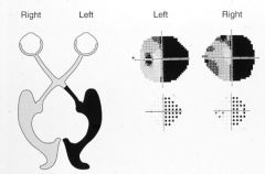
Homonymous hemianopia - this could be a defect anywhere past the chiasm
|
|
|
How do you want to go about evaluating the perceptual disorders of the visual system?
|
See if they can identify "what" (temporal)
See if they can identify the "where"/spatial (parietal lobe) |
|
|
What are the different types of eye movements performed by the ocular motor system?
|
Saccades
Pursuit Vergence Vestibulo-ocular |
|
|
What is the function of a saccade? What is unique about it?
|
Move the eyes from one equidistant target to another
You don't see anything between the two points! |
|
|
What is the function of a pursuit?
|
Keep the eyes on a target when the target moves
|
|
|
What is the function of a vergence?
|
Keep the eyes on a target as the distance from the viewer changes
|
|
|
What is the function of a vestibulo-ocular eye movements?
|
Keep the eyes on the target when the viewer's head moves
|
|
|
What are the two types of saccades?
|
Voluntary
Invountary |
|
|
What are some of the voluntary saccades?
|
Move the eyes to a target seen in a peripheral field
Move the eyes to an unseen target |
|
|
What are some of the involuntary saccades/
|
REM
Fast phases of nystagmus Random |
|
|
In general, where does the control of involuntary horizontal saccades take place?
|
Front of the brain
|
|
|
In general, where does the control of voluntary horizontal saccades take place?
|
Back of the brain
|
|
|
What is the signalling pathway for the control of horizontal saccades?
|
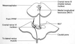
1. Signal comes in from PPRF
2. Synapse in the CNVInucleus 3.2 signals sent out: -Lateral rectus -Interneuron to the CNIII nucleus through the MLF Moving the eyes SIgnalling is ipsilateral once you get to hte pons |
|
|
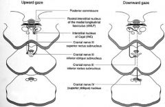
In general, how are vertical saccades controlled?
|
Bihemispheric
One group of pathways for upgaze, another for downgaze |
|
|
What pathways are involved in the control of vertical saccades?
|
|
|
|
What are some of the different disorders of saccade?
|
Absent (gaze palsy)
Reduced amplitude (hypometric) Slow Inaccurate (dysmetric) Intrusive |
|
|
Where is pursuit generated in the brain?
|
Occipito-parietal region
|
|
|
In order to perform pursuit eye movements, what is a precondition?
|
You've got to be able to see first!
|
|
|
What are the pathways involved in the control of horizontal pursuit?
|
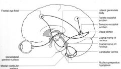
The posterior part of the brain dominates
The final common pathway is the CNVI nucleus Cerebellum is also involved. |
|
|
How do you evaluate pursuit?
|
Have the patient follow your moving light/finger
Is it smooth? Is it complete? Are there oscillations? |
|
|
If a patient is unable to pursue, what will you see?
|
They'll be able to follow your finger for a while, stop, and then have to perform a catchup saccade
|
|
|
What are various causes of problems with pursuit?
|
Lack of sleep
Alcohol intoxication Pursuit is quick to go, as far as eye movements go |
|
|
What are some of the disorders of pursuit?
|
Cogwheel
Absent |
|
|
What are the two types of vergent eye movements?
|
Convergence: move the eyes closer to one another
Divergence (hard to do): move the eyes farther apart from one another |
|
|
Generally, what are the parts of the brain involved in the vergence pathway?
|
Generation in the parieto-occipital regions
Signal travels to the midbrain We don't know, exactly |
|
|
How do you go about evaluating vergence?
|
Measure ocular alignment when the eyes are focusing on a distant target, then when they're focusing on a near target
|
|
|
What are the different disorders of vergent?
|
Too much convergence
Too little convergence |
|
|
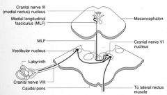
Where is the vestibulo-ocular reflex generated?
|
Labyrinths
|
|
|
Does the vestibulo-ocular reflex reach the cerebrum?
|
No.
|
|
|
What parts of the brain are involved in the control of the vestibulo-ocular reflex?
|
|
|
|
How do you evaluate the vestibulo-ocular reflex?
|
It's hard: if people are awake, they make voluntary eye movements
1. Doll's headmaneuver; move head rapidly, look for slow contraversive conjugate eye movements 2. Cold water calorics: look for ipsiversive slow conjugate eye movements and perhaps contraversive involuntary saccades |
|
|
What are some of the vestibulo-ocular disorders?
|
If the entire thing is underfunctioning, the entire world looks like it's a hand-held camera: "oscillopsia"
Unilateral: nystagmus |
|
|
What are the features of a supranuclear gaze palsy?
|
1. Absent voluntary gaze
2. Intact vestibulo-ocular reflex |
|
|
What are some of the different levels of the visual field pathways?
|
Supranuclear: brain cerebral centers to the PPRF
Intranuclear: MLF Nuclear: PPRF, pons |