![]()
![]()
![]()
Use LEFT and RIGHT arrow keys to navigate between flashcards;
Use UP and DOWN arrow keys to flip the card;
H to show hint;
A reads text to speech;
47 Cards in this Set
- Front
- Back
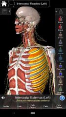
External Intercostal muscle |
Superior Att: inferior border of the rib above. Ex: 4th external Intercostal muscle attached to bottom of rib 4. Inferior Att: superior border of the rib below. Ex: 4th external intercostal muscle attached to top of rib 5. Actions: elevates the rib below. Innervation: Intercostal nerve, Ex: 4th Intercostal nerve.
|
|
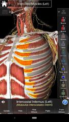
Internal Intercostal muscle |
Superior Att: inferior border of the rib above. Inferior Att: superior border of the rib below. Actions: depresses the rib above. Innervation: Intercostal nerve |
|
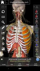
Innermost Intercostal muscle |
Superior Att: inferior border of the rib above. Inferior Att: superior border of the rib below. Actions: depresses the rib above. Innervation: intercostal nerve |
|
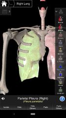
Parietal pluera |
The outer lining of serous membrane enclosing the plueral cavity. |
|
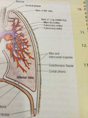
Costal pleura |
Inner surface of the thoracic wall |
|
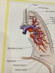
Mediastinal parietal pleura |
Lining the mediastinum medially |
|
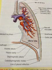
Diaphragmatic parietal pleura |
Covering the superior surface of the diaphragm |
|
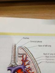
Cervical pleura |
Extends superior to the 1st rib |
|
|
Pleural recesses |
Areas where one parietal pleura contacts another parietal pleura |
|
Visceral pleura |
Delicate serous membrane that covers each lung |
|
|
Pleural cavity |
The space between the parietal pleura and visceral pleura. |
|
|
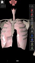
How many lobes does the right lung have? |
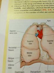
3 lobes; superior, middle, & inferior |
|
|
How many lobes does the left lung have? |
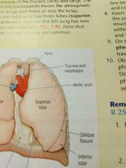
2 lobes; superior & inferior |
|
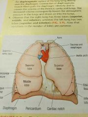
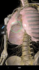
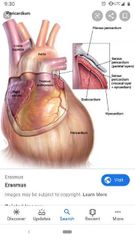
Pericardium |
Between right & left pleural cavities. Occupies the midline between lungs, lies posterior to the sternum, & contains the heart |
|
Phrenic nerve |
Innervates diaphragm, motor and sensory nerves. Important for breathing. Located between mediastinal pleura and pericardium, passes down the heart. Anterior to the root of the lung. |
|
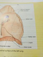
Lingula |
On the left lung, inferior, medial portion of the superior love. Homolog of the middle lobe of right lung. |
|
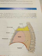
Mediastinum Boundaries |
Superior: superior thoracic aperture. Inferior: diaphragm. Anterior: sternum. Posterior: vertebral bodies of T1-T12. Lateral: mediastinal pleura (left & right). |
|
Pericardium (pericardial sac) |
Encloses the heart. Pierced by the aorta, pulmonary trunk, superior vena cava, the 4 pulmonary veins, & inferior vena cava |
|
Fibrous pericardium (parietal pleura) |
Layer of connective tissue that is non-elastic. Provides support and protection to the heart. |
|
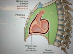
Serous pericardium (visceral/parietal) |
Visceral and Parietal layers. The visceral layer is intimately attached to the heart and the roots of the great vessels. The Parietal layer is attached to the inner lining of the fibrous pericardium. |
|
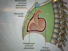
Pericardial cavity |
The space between the Parietal serous pericardium and the visceral serous pericardium. It contains small amounts of serous fluid that acts to reduce surface tension and lubricate. |
|
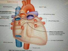
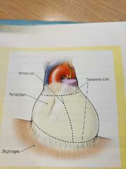
Oblique pericardial sinus |
Posterior to the heart within the pericardial cavity. |
|
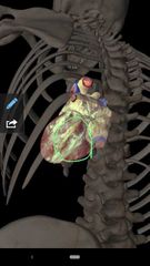
Transverse pericardial sinus |
Posterior to the pulmonary trunk and ascending aorta within the pericardial cavity. |
|
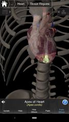
Apex of the heart |
Inferior left side of heart, part of left ventricle. |
|
Base of the heart |
Formed by the left atrium and part of the right atrium and the emergence of the great vessels. |
|
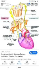
Vagus nerve (CN X) |
Stimulates some muscles in the heart to slow down the heart rate. Descends within the thorax posterior to the root of the lung. |
|
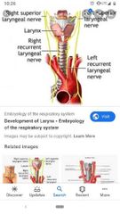
Left Recurrent Laryngeal nerve |
Branches from left vagus nerve inferior to the aortic arch and posterior to the ligamentum arteriosum. |
|
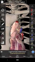
Ligamentum arteriosum |
Between the aortic arch and pulmonary trunk. Connects the left pulmonary artery to the inferior part of the arch of the aorta. |
|
|
Coronary Sulcus |
Courses the circumference of the heart, separating the atria from the ventricles. |
|
|
Anterior interventricular sulcus |
Separates the right & left ventricles. Also, indicates the location of the interventricular septum internally. |
|
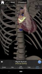
Right auricle |
Extends from the right atrium. |
|
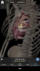
Left auricle |
Extends over the left atrium |
|

Aortic semilunar valve |
A valve between the left ventricle and the aorta |
|
Pulmonary semilunar valve |
A valve between the pulmonary trunk and the right ventricle. |
|
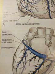
Coronary Sinus |
On the diaphragmatic surface of the heart, located on the coronary sulcus. Opens into the right atrium. |
|
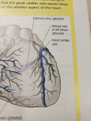
Great cardiac vein |
Runs into the coronary Sinus. Passes deep to the arteries on the anterior aspect of the heart. Returns deoxygenated blood from anterior surfaces of the left ventricle. |
|
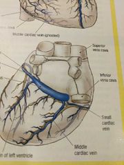
Middle cardiac vein |
Runs along the posterior interventricular artery. Drains into the coronary sinus and drains part of the right and left ventricles. |
|
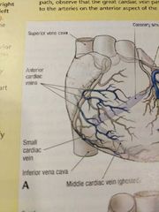
Small Cardiac vein |
Runs along side the marginal artery. Runs into the coronary sinus between right atrium and ventricle. Drains the coronary sinus and the right atrium |
|
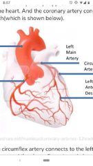
Left Coronary artery |
Arises from the aorta, above the left cusp of the aortic valve. Feeds blood to the left side of the heart. Divides into the LAD branch & circumflex branch. |
|
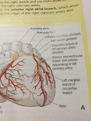
Anterior interventricular artery LAD (left anterior descending artery) |
Major blood supply to the interventricular septum. |
|
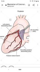
Circumflex artery |
In the coronary sulcus around the left side of the heart. Supplies the left atrium with oxygenated blood. |
|
|
Right coronary artery |
Opens into the right aortic sinus. Begins in the coronary sulcus between the right auricle and the ascending aorta. |
|
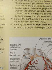
Anterior right atrial branch |
Arises close to origin of the right coronary artery and ascends along the anterior wall of the right atrium toward the superior vena cava. |
|
|
Sinuatrial nodal branch |
Supplies the sinuatrial node |
|
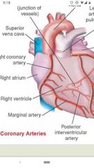
Right marginal branch |
Follows the margin of the heart off of the right coronary artery. Arises near the inferior border of the heart, where it accompanies the small cardiac vein. |
|
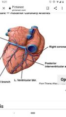
Posterior interventricular branch or PDA (Posterior descending artery) |
Runs off the right coronary artery (right dominant heart) or runs off the left coronary artery (left dominant heart). Accompanies the middle cardiac vein. Supplies oxygenated blood to posterior part of interventricular septum and posterior inferior wall of the left ventricle. |
|
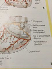
Crux of the heart |
The point where the posterior interventricular sulcus meets the coronary sulcus. The artery to the AV node arises from the right coronary artery at this location. |