Reading...
![]()
Play button
![]()
Play button
![]()
Use LEFT and RIGHT arrow keys to navigate between flashcards;
Use UP and DOWN arrow keys to flip the card;
H to show hint;
A reads text to speech;
28 Cards in this Set
- Front
- Back
|
Functions of the spinal cord
|
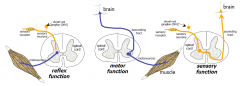
1. sensory
2. motor 3. reflex |
|
|
The 3 types of sensory receptors and their cells?
|
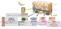
TACTILE
Meissner Pacinian Ruffini Merkel PROPRIOCEPTION muscle spindles joint receptors Golgi tendon organs PAIN (&TEMP) free (nociceptors) |
|
|
TACTILE FIBRES
|
Tactile: all alphabeta fibres or group 2
- medium size (6-12um) - pretty quick (30-70m/s) - all mylenated BUT Differ in their locations and functions |
|
|
TACTILE LOCATIONS AND FUNCTIONS
|
Meissner: glabrous (hairless) skin F: touch and press
Pacinian: Subcut. F: deep press, vibration Ruffini: all skin F: stretch Merkel: All skin F: touch and press |
|
|
PROPRIOCEPTION fibres
|
- pretty quick (70-120m/s)
- all myleinated - v.sensitive to relaxation |
|
|
PROPRIOCEPTION Receptors
|
Muscle spindles: location in muscles and function in muscle legth (propioception)
|
|
|
Grey Matter cells
|
1. Spinal cells
- projection cells (large, MAKE, PNS and CNS, EXCITE) - interneurons (Small, MODIFY, LOTS (7:1), inhbit or Excite) |
|
|
Subdivisions of the spinal cord
|
(1) dorsal horn: somatic sensory
- tactile, pain*, proprioception - tract cells, interneurones (2) intermediate grey matter: autonomic - sympathetics (thoracic/lumbar) - parasympathetics (sacral) (3) ventral horn: somatic motor - α motoneurones (motor end-plate), γ motoneurones (spindles) |
|
|
Degeneration and Regeneration
|
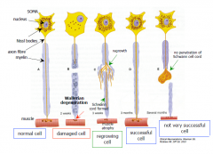
• little CNS regrowth
- NoGo, glial scars, afferents? (may provide nutrition) - some plasticity: sprouts, synapses • PNS better regrowth - different stages after injury - motor/sensory nerve preferences • why so little? - no connection better than wrong one? |
|
|
NoGo
|
protein made by oligodendrocytes, inhibits regeneration
|
|
|
Adult Neurogenesis
|
• no set cell number
• cell birth in adult: STEM CELLS - subventricular zone - subgranular zone hippocampus (memory) • new cells: - survive and work - do not live long - learning and exercise helps survival - stress not good |
|
|
Reflexes
|
|
|
|
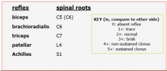
Clinical Testing POPULAR
|
|
|
|
the 4 reflexes?
|
Stretch
Gamma reflex loop Flexor reflex golgi tendon |
|
|
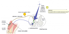
The stretch Reflex
|
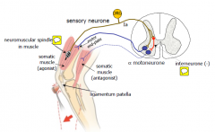
- muscle tone
- movement: reflex contraction (deep tendon) NB: synapse on inhibitory (-) interneurone (antagonists) |
|
|
(2) γ reflex loop
|
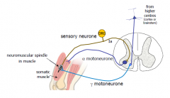
- prompted contraction/tone from higher centres
- tricks into stretch |
|
|
Flexor reflex
|
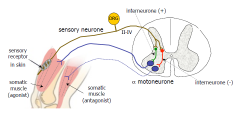
- movement: reflex contraction
- withdrawal of limb (eg pain) - several levels/joints involved (they go up ad sown a few spinal levels so if your hand is affected by a noxious stimulus the shoulder and trunk are also pulled away |
|
|
(4) Golgi tendon reflex
|
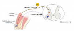
- STOP contraction
- releases tension (eg, heavy loads), too heavy the tendons under too much stress |
|
|
Ramus
|
communication between 2 nerves
|
|
|
Gray and white communicating rami
|
When used without further definition, it almost always refers to a communicating branch between a spinal nerve and the sympathetic trunk. More specifically, it usually refers to one of the following:
Gray ramus communicans: White ramus communicans |
|
|
Gray Communicating Rami
|
umyelinated, postganglionic sympathetic fibres
- found at all levels |
|
|
White Communicating Rami
|
myelinated preganglionic sympathetic fibres
- T1- L3 - lateral and intermediolateral cell column |
|
|
Termination of Conus Medullaris
|
L3 in newborns
Lower border L1 in adults |
|
|
Cauda Equina
|
Motor and sensory roots L2-Co
- subarachnoid below conus medullaris |
|
|
Major Motor and Sensory Nuclei of the Spinal Cord
|
1. ciliospinal center of Budge (C8-T1)- SNS eye
2. Intermediolateral cell cloumn; C8-L3; entire SNS 3. nucleus dorsalis of clark; C8-L3; dorsal spinocerebellar tract 4. Parasympathetic nucleus (S2-4). |
|
|
Myotatic Reflex
|
- afferent; spindle, DRG & Ia fiber
- efferent: ventral horn motor neuron |
|
|
Gamma motor neurons
|
Gamma motoneurons (γ-motoneurons), also called gamma motor neurons, are a type of lower motor neuron used in the process of muscle contraction, and represents about 30% of fibers going to the muscle.[1] Like alpha motor neurons, another type of motor neuron, its cell bodies are located in the anterior horn of the spinal cord
|
|
|
Oligodendrocytes
|
Oligodendrocytes (from Greek, meaning cells with a few branches), or oligodendroglia (Greek, few tree glue),[1] are a type of brain cell. They are a variety of neuroglia. Their main functions are to provide support and to insulate the axons (the long projection of nerve cells) in the central nervous system (the brain and spinal cord) of some vertebrates. (The same function is performed by Schwann cells in the peripheral nervous system.) Oligodendrocytes do this by creating the myelin sheath, which is 80% lipid and 20% protein.[2] A single oligodendrocyte can extend its processes to 50 axons, wrapping approximately 1 μm of myelin sheath around each axon; Schwann cells, on the other hand, can wrap around only 1 axon. Each oligodendrocyte forms one segment of myelin for several adjacent axons
|