Reading...
![]()
Play button
![]()
Play button
![]()
Use LEFT and RIGHT arrow keys to navigate between flashcards;
Use UP and DOWN arrow keys to flip the card;
H to show hint;
A reads text to speech;
49 Cards in this Set
- Front
- Back
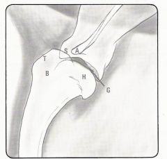
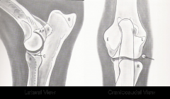
Lateral Shoulder
|
T = Greater Tubercle
B = Bicipital Groove Between H and A = Glenoid cavity |
|
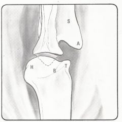
CdCr Shoulder
|
A = Acromonion
T = Greater Tubercle |
|
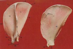
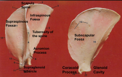
Scapula Anatomy
|
|
|
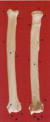
Radius Anatomy
|
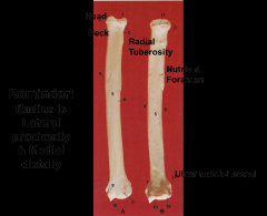
Head radial tubersoity
neck nutrient foramen ulnar notch lateral radius is lateral proximal and medially distally |
|
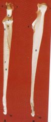
Ulnar Anatomy
|
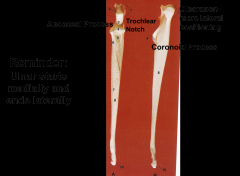
Aconeal process olecranon-more lateral positioning
trochlear notch coronoid process |
|
Elbow Lateral and CrCd
|
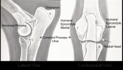
Lateral and CrCd
|
|
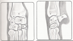
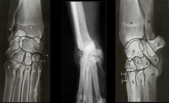
Carpal Dorsopalmar and Lateral
|
Carpal Dorsopalmar and Lateral
|
|
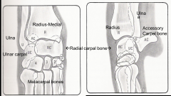
Dpa of carpus
DMPLO carpus DPLMO carpus |
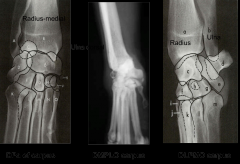
Dpa of carpus
DMPLO carpus DLPMO carpus |
|
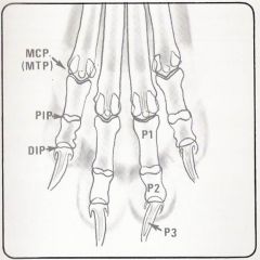
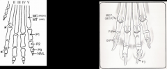
Digit Anatomy
|
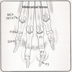
MCP = metacarpal phalangeal joint
PIP = proximal interphalangeal joint DIP = distal interphalangeal joint |
|
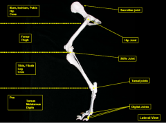
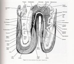
Hind leg anatomy
|
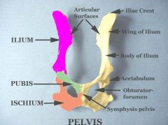
Pelvic Anatomy
|
|
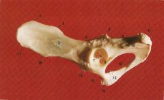
Pelvic Anatomy again
|
Feline Capital Physeal Dysplasia
|
|
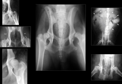
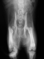
Hip Dysplasia
|
Hip Dysplasia
|
|
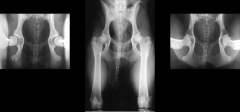
Penn Hip views
From left to right Distraction, Exended and Compression |
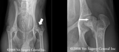
Legge Calves Perthes Disease
|
|
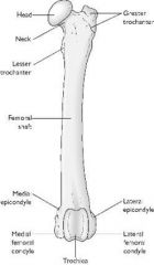
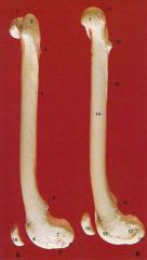
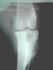
Femur CrCd
|
head is medial
greater trochanter is lateral less trochanter is medial condyoes medial and lateral epicondyles seen caudally trochlear groove = home of the patella |
|
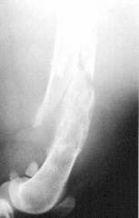
Femur Lateral and Medial
|
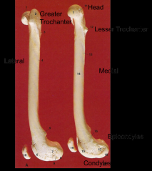
greater trochanter head
lateral lesser trochanter medial epicondyles condyles |
|
|
Panosteitis
|
|
|
DJD
|
|
|
Osteosarcoma
prox tibia/fibula |
|
|
pathological fracture
|
|
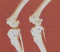
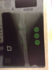
Stifle anatomy
Left- Lateral Aspect Right – Medial Aspect |
Joints:
A Stifle joint B Femoropatellar joint C Femorotibial joint D Proximal Tibiofibular joint 1 Body of femur 2 vPatella 3 Lateral suprecondylar tuberosity 4 Lateral ridge of trochlea 5 Lateral epicondyle 6 Lateral fabella- in origin of gastrocnemius muscle 7 Medial fabella 8 Lateral condyle of femur 9 Tibial tuberosity 10 Cranial border tibial crest 11 Muscular extensor groove 12 lateral condyle of tibia 13 Proximal tibiofibular joint 14 Head of fibula 15 Body of fibula 16 Interosseous space 17 Body of tibia 18 Medial ridge of trochlea 19 Medial supracondylar tuberosity 20 Medial epicondyle 21 Medial fabella 22 Medial condyle of femur 23 Tibial crest 24 Cranial border 25 Medial condyle of tibia 26 Sesamoid of tendon of popliteius muscle |
|
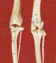
Fibula is lateral
left is cranial, right is caudal |
1. Body of femur
2. patella 3. medial ridge of trochlea 4. lateral ridge of trochlea 5. medial epicondyle 6. lateral epicondyle 7. medial condyle 8. lateral condyle 9. medial condyle of tibia 10 Lateral condyle of tibia 11 Intercondylar area 12 Tibial tuberosity 13 Cranial border of tibial crest 14 Head of fibula 15 Body of fibula 16 Interoseous space 17 Body of tibia 18 Medial and lateral suprecondylar tuberosities 19 Medial and lateral fabella 20 Popliteal surface 21 Sesamoids in tendon of popliteus 22 Intercondylar fossa 23 Caudal intercondylar area 24 Proximal tibiofibular joint |
|
|
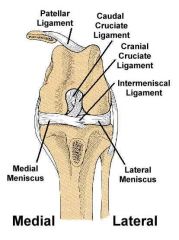
Cruciate ligament
Collateral ligament Meniscus Patellar ligament |
|
|
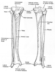
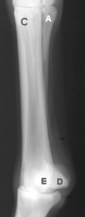
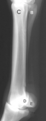
Tibia: condyles, malleolus-medial pointed, intercondylar eminence, tibial tuberosity
Fibula: fibula lateral, head, lateral malleolus |
|
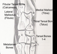
Tarsal Joint Anatomy
|
|
|
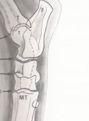
lateral tarsus
|
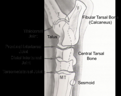
|
|
Metatarsus and Phalanges
|
cool
|
|
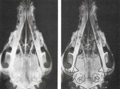
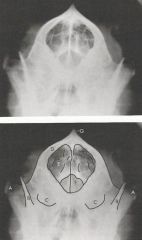
DV Skull radiograph
|
Zygomatic Arch (A)
Mandible (C) Frontal Sinus (E) Ethmoid Turbinates (G) Cranium (I) Nasal Septum (H) Tympanic Bullae (N) Occipital Condyles (O) |
|
|
Zygomatic Arch (A)
Coronoid Process (B) Tympanic Bullae ( C ) Calvarium (D) Cranial Cavity (E) Foramen Magnum (F) Sagittal Crest (G) |
|
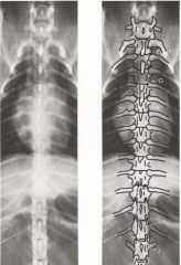
VD Thoracic
|
C7 (A)
T1( B) Spinous process of T1 Head of 2nd rib (D) Tubercle of 2nd rib (E) Intervertebral disc space b/t T3-T4 (G) T13 (I) L1 (J) |
|
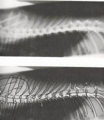
Lateral Thoracic Spine
|
Body of T1 (A)
1st rib (B) Spinous process of T5 (c ) Neural Canal (D) Caudal Articular process of T6 (E) Cranial articular Process of T7 (F) Intervertebral disc space b/t T6-T7 Intervertebral foramen b/t T7-T8 (H) Spinous Process of T 10 (I) Articular process of T 13 (K) |
|
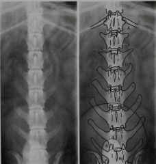
VD Lumbar Spine
|
T13 (A)
L1 (B) Intervertebral disc space b/t L1-L2 (C ) Transverse Process of L4 (F) Spinous Process of L4 (G) |
|
Lateral Lumber Spine
|
T 13 (A)
12th Rib (B) 13th Rib ( C) L1 (D) Neural Canal (E) Caudal articular process of L2 (F) Cranial Articular process of L3 (G) Intervertebral disc space between L2-L3 (H) Transverse process of L4 (I) Spinous process of L4 (J) Intervertebral foramen between L4-L5 (K) Accessory process (L) L7 (M) Sacrum (N) |
|
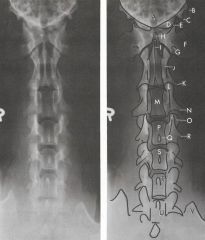
VD Cervical Spine
|
Occipital Condyles (D)
Atlanto-occipital joint (E) Dens (H) Wings of the Atlas C1 (F) Axis C2 (J) Spinous Process of axis C2 (I) Transverse process of axis (K) C3 (M) Cranial articular process of C3 (L) Caudal articular process of C3-C4 articulation (O) C4 (P) C5 (S) C6 (T) C7 (U) Transverse Foramen (G) |
|
|
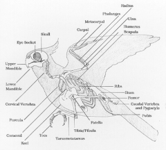
Corocoid
Ulna larger than radius Tibiotarsal Bone Tarsusmetatarsus Cervical vertebrae varies Thoracic vertebrae usually 8 and assoc. with ribs Lumbar and sacrum vertebrae fused=synsacrum Pygostyle Pneumatic bones Keel Zygodactyl |
|
|
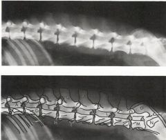
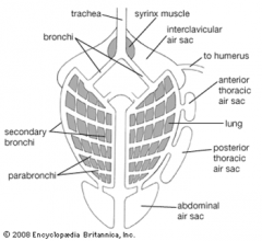
Crop
Proventriculus=stomach Ventriculus=gizzard Lungs and airsacs (8-9) NO Diaphragm Choana Cloaca Kidney Unable to distinquish sex organs on rads |
|
Snake Anatomy
|
0-20% = trachea/esophagus
25% = heart 30-40% = lung 30-50% = liver 50% = stomach 60-80% = reproductive organs 70-85% = kidney No bladder Right sided organs bigger than left sided If you see kidney on radiograph of snake-abnormal |
|
|
good luck memorizing this. you're in trouble.
|
|
|
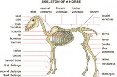
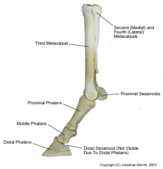
MC 3 = Cannon bone
MC 2 & 4 = Splint bones P1 = Long Pastern P2 = Short Pastern P3 = Coffin or Pedal bone Distal sesamoid = Navicular MCphalangeal joint = Fetlock = ankle Coronary band Radius/ulna and cannon bone = knee |
|
|
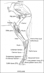
Stifle = knee
Hock = tarsus Point of hock = calcaneus Gaskin = between stifle and hock Lower limb |
|
|
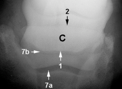
1 = flexor cortex
C = navicular bone 2 = prox. Border of navicular bone 7a/b = distal interphalangeal joint prox. and distal |
|
|
1 = flexor cortex
C = navicular bone 2 = prox. Border of navicular bone 7a/b = distal interphalangeal joint prox. and distal |
|
Metacarpal/tarsal DMPLO
|
A = MC/MT 2
B = MC/MT 4 C = MC/MT 3 D = Medial Sesamoid E = Lateral Sesamoid |
|
Metacarpal/tarsal DLPMO
|
A = MC/MT 2
B = MC/MT 4 C = MC/MT 3 D = Medial Sesamoid E = Lateral Sesamoid |
|
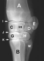
Carpal Medial Oblique - DLPMO
|
A = Radius
B = Cannon bone (MC 3) C = Radial carpal bone D= Ulnar carpal bone E = Accessory carpal bone G = 3rd carpal bone H = 4th carpal bone M = MC 4 |
|
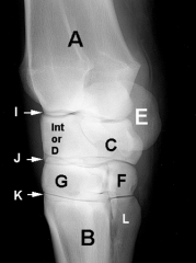
Carpal Lateral Oblique DMPLO
|
A = Radius
B = Cannon bone (MC 3) C = Radial carpal bone D= Ulnar carpal bone E = Accessory carpal bone G = 3rd carpal bone H = 4th carpal bone L = MC 2 (splint 2) |
|
Tarsal Lateral Oblique DMPLO
|
highlights:
Dorsolateral surfaces MT 2 (H) Talus (4-5) |
|
Tarsal Medial Oblique DLPMO
|
highlights:
Dorsomedial surfaces Medial malleolus (2) Calcaneus Mt4 |
|
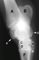
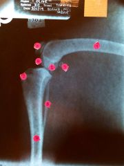
Left Caudocranial stifle
|
1 medial condyle of femur
2 lateral condyle of femur 3 medial condyle of tibia 4 fibula 5 tibia FOV = distal 3rd of femur to proximal 3rds of tibia/fibular Beam = over stifle joint Measure distal end of femur |
|
|
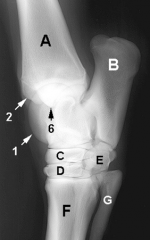
1) medial condyle
2) tibial tuberosity 3) sesamoid bone 4) femur 5) patella 6) lateral condyle 7) tibia 8) fibula |