Reading...
![]()
Play button
![]()
Play button
![]()
Use LEFT and RIGHT arrow keys to navigate between flashcards;
Use UP and DOWN arrow keys to flip the card;
H to show hint;
A reads text to speech;
121 Cards in this Set
- Front
- Back
|
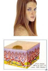
What are the three skin layers?
|
Epidermis
Dermis Subcutaneous |
|
|
What is contained in the Dermis layer of the skin?
|
Blood Vessels
hair follicles sebaceous glands, sweat glands |
|
|
What are the four appendages of skin?
|
hair
Nails sebaceous glands sweat glands |
|
|
What are the two types of hair?
|
Vellus- fine body hair
Terminal- thicker head hair |
|
|
What are the two types of sweat glands?
|
Eccrine-
Apocrine- pits and groin- BO |
|
|
What are the five purposes of the skin listed in lecture?
|
Protect from trauma and infection.
Prevent fluid loss Regulate body temperature Provide sensory information Produce vitamin D |
|
|
What are the 6 HISTORY Factors when evaluating a patient's skin condition?
|
Duration
Relationship of lesion to external factors History of sun exposure Associated symptoms Constitutional Symptoms Review of Systems |
|
|
name a few of the External factors to be considered when evaluting the relationship of skin lesions to external factors
|
• Topical irritants / allergens
• season • travel • temperature • previous treatment • occupation • hobbies • pregnancy |
|
|
name a few of the Associated symptoms to be considered when evaluting the relationship of skin lesions to associated symptoms
|
pruritis, dryness, rashes/sores, color
changes, changes in hair or nails |
|
|
What are the constitutional symptoms evaluated in an individual with a skin disorder?
|
URI
Wt loss Mailaise Fatigue * All have to be evaluated to be chronic or acute |
|
|
During the physical examination what is tengential lighting good for?
|
Tangential lighting is good for contours
Ophthalmoscope “trick” for illuminated magnification |
|
|
Along with Adequate lighting, adequate ___ is also important?
|
Adequate exposure is important
• Be sure to check axillae, buttocks, back of thighs, between fingers & toes |
|
|
What are the 8 aspects of Inspection for skin disorders?
|
Symmetry
Color Hair Nails Moisture Temperature Texture Mobility and Turgor |
|
|
What should be evaluated when inspecting the color of a skin lesion?
|
Color
• Increased v. decreased pigmentation • Pallor v. cyanosis Central v. peripheral cyanosis • Central cyanosis consistent with pulmonary or cardiac disease; peripheral cyanosis seen with cooler temps or anxiety • Jaundice - also check conjunctivae |
|
|
What should be evaluated when inspecting the Hair around a skin lesion?
|
Hair – distribution, quantity
|
|
|
What should be evaluated when inspecting the nails proximal to a skin lesion?
|
Nails color lesions 12
– color, lesions, capillary refill |
|
|
How do we examine Mobility and Turgor?
|
Mobility & turgor – pinch section
of skin on the forearm…should return to place immediately • Avoid back of hand…too loose • Pinch skin on the thigh in the elderly |
|
|
What are the three skin lesion Distribution terms?
|
Localized
Regional Generalized/disseminated |
|
|
What is a localized skin lesion?
|
Lesion appears in one small area
|
|
|
What is a regional skin lesion?
|
Lesions appear in specific region of the body
|
|
|
What is a generalized/disseminated skin lesion?
|
Lesions appear widely distributed or in
multiple areas simultaneously |
|
|
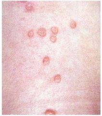
Describe Round/discoid shaped lesions
and associated conditions |
Round / discoid
• Coin shaped • No central clearing • Seen with eczema; • Umbilicated lesions seen with Molluscum contagiosum (photo) |
|
|
Describe Oval shaped lesions and associated conditions
|
Oval
• Ovoid • Seen with Pityriasis rosea |
|
|
Describe annular shaped lesions and associated conditions
|
Round; active
margins with central clearing Seen with tinea (fungal) infections • Example – Tinea corporis |
|
|
Describe zosteriform shaped lesions and associated conditions
|
Zosteriform
(dermatomal) – following a nerve segment Example: • Herpes Zoster |
|
|
Describe Iris / Target shaped lesions and associated conditions
|
Pink macules with purple central papules
-Erythema multiforme |
|
|
What are the three Arrangements of lesions listed in the lecture
|
Linear- Contact dermatitis
Serpiginous- Cutaneous larva migrans Morbilliform- measles. |
|
|
Describe Morbilliform shape/arrangement of lesions
|
Measles-like
• Erythematous maculopapular lesions that become confluent on the face and body |
|
|
What are the five terms used to describe borders of skin lesions?
|
Distinct
Indistinct Active Irregular Raised Borders |
|
|
Define Distinct Borders of a skin lesion
|
Well-demarcated or
defined; able to draw a line around the area with confidence |
|
|
Describe indistinct boders of a skin lesion
|
Poorly defined;
borders merge with normal skin |
|
|
Describe active boders of a skin lesion and example
|
Active – margin of lesion shows greater
activity than the center Example – tinea infections |
|
|
Describe irregular boders of a skin lesion and example
|
Irregular – notched margins; not smooth
Example – malignant melanoma |
|
|
Describe Raised boders of a skin lesion and example
|
Raised borders – center of lesion is
depressed compared to the edge Example – basal cell 27 carcinoma |
|
|
What are the six color terms when defining a skin lesion?
|
Flesh – same tone as surrounding skin
Erythematous – variable shades of red • Pink, salmon, coppery, reddish-blue Violaceous – light violet Tan-brown Black or blue-black White |
|
|
What are the six things being observed when palpating a skin lesion?
|
Consistency
Mobility Blanchable Tenderness Depth of lesion Deviation in temperature |
|
|
What are the ABCDs of Malignant Melanoma
|
A – asymmetry
B – borders (irregular) C – color (variegated) D – diameter > 6mm E – elevation |
|
|
What is a primary skin lesion?
|
• Arise from previously normal skin
• Key to accurate diagnosis |
|
|
What is a secondary skin lesion?
|
• Arise from changes in primary lesions
• Usually due to scratching and/or infection |
|
|
What are the three types of primary lesions (basic)
|
Circumscribed, flat, non-palpable
Superficial elevations by free fluid Palpable, elevated solid masses |
|
|
What are two types of Circumscribed, flat, nonpalpable primary skin lesions?
|
Macule
Patch |
|
|
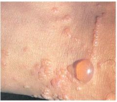
What are three types of Superficial elevations by free fluid in primary skin lesions?
|
Vessicle
Bulla Pustule |
|
|
What are five types of Palpable elevated solid masses in primary skin lesions?
|
Papule
Plaque Nodule Tumor Wheal |
|
|
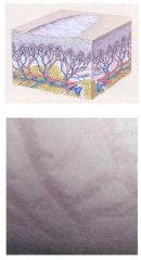
Macule Definition and example
|
Definition:
• Flat, non-palpable • Circumscribed color change • <1 cm in diameter • Variable color Examples: • Measles • Freckles • Petechiae |
|
|
Patch Definition and example
|
Definition:
• Flat, non palpable • Irregular shape • > 1 cm in diameter Examples: • Mongolian spots • Café au lait spots • Port wine stain • Vitiligo |
|
|
Papule Definition and example
|
Definition:
• Up to 1 cm • Palpable, firm • Circumscribed • Colors - Flesh colored, red, brown • May be confluent and form plaques Examples: • Molluscum contagiosum • Warts • Nevi |
|
|
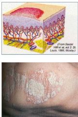
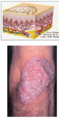
Plaque Definition and example
|
Definition:
• Elevated, firm, rough • > 1 cm • Well-circumscribed Examples: • Psoriasis (photo) • Eczema |
|
|
Nodule Definition and Example
|
Definition:
• > 0.5 cm • Deeper and firmer than a papule • Usually round Example: • Lipomas • Skin cancers Malignant melanoma, basal cell or squamous cell carcinomas |
|
|
Tumor Definition and Example
|
Definition:
• A large nodule • Deeper in the dermis • >2 cm Examples: • Hemangioma • Benign tumor |
|
|
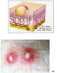
Wheal Definition and example
|
Definition:
• Irregular, transient, superficial edema Examples: • Mosquito bites • Hives (Urticaria) See photo • Allergic reaction |
|
|
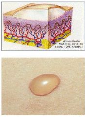
Vessicle Definition and example
|
Definition:
• Well-circumscribed • Up to 1.0 cm • Filled with serous fluid Example: • Herpes simplex “Dew drops on rose petals |
|
|
Bulla Definition and example
|
Definition:
• Well-circumscribed • Greater than 1.0 cm • Filled with serous fluid Examples: • 2nd degree burns • Blisters |
|
|
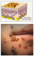
Pustule Definition and example
|
Definition:
• Elevated, superficial; well-circumscribed • Epidermal • Filled with pus Examples: • Acne • Impetigo • Fire ant bites (photo) |
|
|
What are the three types of secondary skin lesions (basic)
|
Loss of skin surface
Material on the skin surface Misc. |
|
|
What are the types of secondary lesions from loss of skin surface?
|
Erosion, Ulcer, Fissure
|
|
|
What are the types of secondary lesions considered "miscellaneous"
|
Lichenification
Excoriation Atropy Scar Burrow |
|
|
What are the types of secondary lesions considered Material on the skin surface
|
Crust
Scale |
|
|
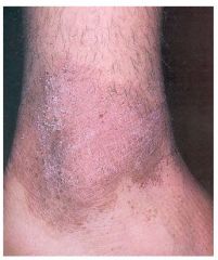
Skin erosion definition and example.
|
Definition:
• Loss of a superficial epidermis • Surface is moist, but doesn’t bleed • Heal without scarring Examples: • Ruptured varicella vesicles (pock marks) • Eczema |
|
|
Skin ulcer definition and example.
|
Definition:
• Deeper loss of epidermis and dermis • Heals with scarring Examples: • Stasis ulcer (photo) • Decubitus |
|
|
Skin fissure definition and example.
|
Definition:
• Linear crack, or break, from the epidermis to the dermis Examples: • Eczema (photo) • Tinea pedis • Angular cheilitis |
|
|
Skin Excoriation definition and example
|
Definition:
• Abrasion or scratch mark • May be linear or rounded • Usually due to scratching Examples: • Scabies • Atopic dermatitis • Dry skin |
|
|
Skin Crust definition and example
|
Definition:
• Dried residue of serum, pus, or blood Examples: • Impetigo (photo) • Tinea capitis • Kerion Raised boggy secondarily infected fungal lesion of hair |
|
|
Skin Scale Definition and example
|
Definition:
• A thin flake of exfoliated epidermis Example: • Dandruff • Psoriasis (photo) • Seborrheic dermatitis |
|
|
Skin Lichenification definition and example
|
Definition:
• Thickening and roughening of the skin • Increased visibility of skin markings Examples: • Atopic dermatitis • Chronic dermatitis |
|
|
Skin Atrophy Definition and example
|
Definition:
• Thinning of skin with loss of normal skin markings • Skin looks shinier and more translucent Examples: • Stretch marks / striae (photo) • Topical steroid use |
|
|
what are the four categories of Skin diseases?
|
Papulosquamous
Nodular Vesiculobullous Maculopapular |
|
|
What are the papulosquamous lesions, what are examples?
|
Papules, plaques and scales
Examples: • Psoriasis • Lichen planus Pityriasis rosea |
|
|
What are the 5 P's of this certain skin disease?
|
Lichen planus
The Five P’s Pruritic Polygonal Purple Planar Papules |
|
|
What are the nodular lesions?
|
Benign and malignant epidermal and dermal
nodules |
|
|
What are examples of benign and malignmant nodular lesions?
|
Examples of benign lesions:
• Nevi (photo) • Cherry angiomas • Epidermoid cysts Malignant: Squamous cell carcinoma • Isolated keratotic, eroded papule or nodule • Located in a sun exposed area Basal cell carcinoma • “Pearly” nodules in sun exposed areas • Associated with central ulcerations and telangiectases |
|
|
What are vesiculobullous lesions?
|
Vesicles and bullae
Examples: • Impetigo • Herpes • Pemphigus |
|
|
What is Pemphigus
|
Autoimmune disease
affecting the skin and mucous membranes Associated with vesicles and bullae that can rupture and weep Can be fatal |
|
|
What are maculopapular lesions, what are examples?
|
Macules and papules
Examples: • Viral exanthems Generalized, erythematous maculopapular rash • Drug eruptions |
|
|
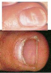
What is the definition and cause of clubbed nails?
|
Definition:
• Rounded, bulbous nail base. Feels spongy. Causes: • Chronic hypoxia • Congenital heart disease • Lung cancer |
|
|
What are Beau's lines?
|
Definition:
• Transverse depressions secondary to trauma or acute or severe illness Lines grow out with the nail |
|
|
What are the lines in the nails secondary to trama that grow out with the nail?
|
Beau's lines
|
|
|
Define Paronychia, what is a cause?
|
Definition:
• Acute or chronic inflammation of the proximal & lateral nail folds • Nail folds swollen, reddened, & tender Cause: • Frequent immersion in water |
|
|
What is onychocryptosis, what is a cause?
|
Definition:
• INGROWN TOENAIL • Usually involving the large toe. Nail grows into the dermis. Cause: • Improperly cutting nails • Tight shoes |
|
|
Define Terry's nails. What is a cause?
|
Definition:
• Mostly white with a distal band of reddish brown Cause: • Aging • Chronic disease such as diabetes cirrhosis, heart failure |
|
|
Define Leukonychia, what is a cause?
|
Definition:
• Trauma to nails causing areas of white discoloration Cause: • Trauma • Repeated manicuring |
|
|
Define Koilonychia
|
"spoon nail"
|
|
|
Define Onycholysis- what is a cause?
|
Definition:
• Painless separation of the nail plate from the nail bed Causes: • Most common cause: trauma to long finger nails • Other causes: psoriasis, contact dermatitis |
|
|
What is Onychomycosis? What can be a cause?
|
Definition:
• Fungal infection of nail bed, plate or matrix Cause: • Occlusive footwear, dissemination of fungal infections, and locker room exposure |
|
|
What can be some causes of Nail pitting?
|
e.g., psoriasis, arthritis, SLE,
alopecia areata |
|
|
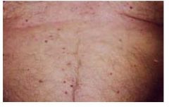
Define Petechiae
|
Deep red/purple-red
lesions < 0.5 cm Round, irregular Non-blanchable Variable distribution Represent blood outside of vessel • Seen with infections and bleeding 57 disorders |
|
|
Define Purpura
|
Deep red/purple-red
lesions > 0.5 cm Same descriptors as petechiae, just larger. |
|
|
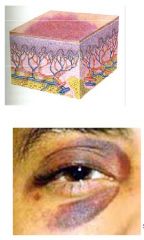
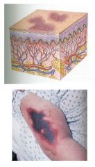
Define Ecchymosis
|
Purple lesions of
variable size • Fade to green, yellow, brown Round/oval, irregular borders Represent blood outside of vessels due to trauma or bleeding disorder |
|
|
Define Spider Angioma and causes
|
Fiery red lesions
• Small in size…up to 2 cm Central body with surrounding erythema and radiating legs Blanch with pressure Seen on face, neck, arms & upper trunk Seen with liver disease, pregnancy; may be normal |
|
|
Define Cherry Angiomas
|
Bright –red papules,
1-3 mm size, red, flat or raised, nonpulsatile, seen on the trunk; don’t blanch associated with aging |
|
|
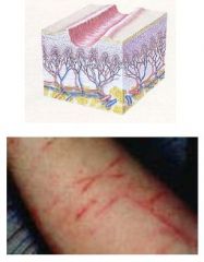
Define Telangectasias and causes
|
Fine, irregular red
lines secondary to dilation of capillaries Blanch Seen with basal cell carcinomas, sundamaged skin, rosacea |
|
|
Define Hemangioma
|
Red, irregular lesion
secondary to dilation of dermal capillaries Starts as macular patch, can progress to plaque or nodule Example: “Strawberry hemangioma” |
|
|
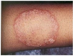
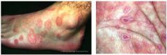
Annular Tinea Corporis
|
|
|
Atrophy
|
|
|
Beau's lines
|
|
|
Bulla
|
|
|
Cherry Angioma
|
|
|
Crust
|
|
|
Ecchymosis
|
|
|
Excoriation
|
|
|
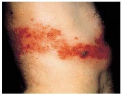
Fissure
|
|
|
Hemangioma
|
|
|
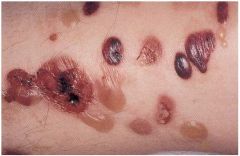
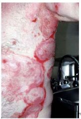
Hemorrhagic Bullae
|
|
|
Iris-Target lesions, Pink Macules with purple central papules Erythema Multiforme
|
|
|
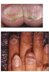
Koilonychia
|
|
|
Lichenification
|
|
|
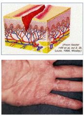
Linear Arrangement Contact Dermatitis
|
|
|
Macule
|
|
|
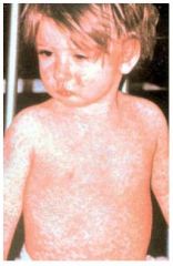
Morbilliform Arrangment of Erythematous maculopapular lesions from Measles
|
|
|
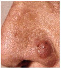
Nodular lesion- Basal Cell carcinoma
|
|

|
Nodule
|
|
|
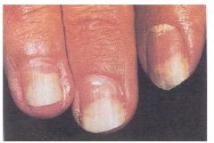
Onycholysis
|
|
|
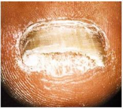
Onychomycosis
|
|
|
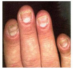
paronychea
|
|
|
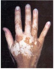
patch lesion Vitiligo
|
|
|
Pemphigus
|
|
|
Plaque
|
|
|
Purpura
|
|
|
Pustule
|
|
|
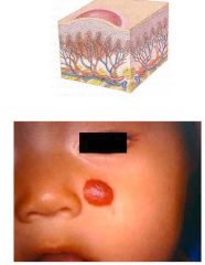
Round-Discoid umbilicated Molluscum Contagiosum
|
|
|
Scale
|
|
|
Vesiculobullous lesion- Herpes Zoster
|