![]()
![]()
![]()
Use LEFT and RIGHT arrow keys to navigate between flashcards;
Use UP and DOWN arrow keys to flip the card;
H to show hint;
A reads text to speech;
70 Cards in this Set
- Front
- Back
|
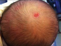
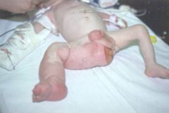
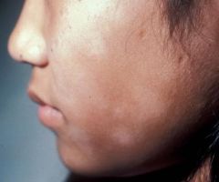
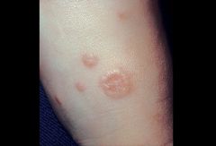
congenital absence of skin in a small area, after it heals a bald spot is left if occurs in multiple places on the scalp, then associated with _________ if occurs as a midline defect, then look for _________ and get a ________ study |
aplasia cutis Trisomy 13 spinal dysraphia and underlying skull defects (especially with hair collar sign - distorted/dense hair surrounding the lesion)....get an MRI |
|
|
aplasia cutis |
|
|
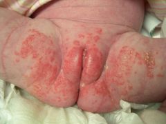
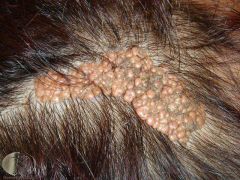
tiny, pinhead-sized, white papules or epidermal inclusion cysts, seen in newborns most often on the face
|
Milia |
|
|
Milia |
|
|
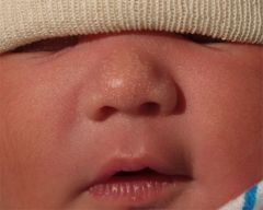
Pinpoint white-yellow papules appear on the nose and central face. Caused by maternal androgen exposure. |
Sebaceous Hyperplasia |
|
|
Sebaceous Hyperplasia |
|
|
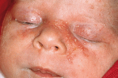
inflammatory pustules on the cheeks and forehead without comedones; resolves spontaneously within first few weeks |
Neonatal acne |
|
|
Neonatal acne |
|
|
inflammatory pustules with open and close comedones over the face, in an infant of 3-4 months of age; caused by androgenic stimulation of sebaceous glands |
Infantile acne |
|
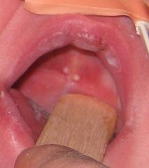
small, benign whitish-yellow masses on either side of the raphe on the hard palate of a newborn |
Epstein pearls |
|
very superficial vesicles that are easily ruptured; occurs due to obstruction of sweat glands and is also called “prickly heat rash.” |
Miliaria rubra |
|
|
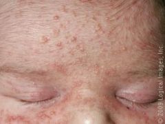
erythematous macules with raised central lesions (papules or vesicles filled with EOSINOPHILS) Usually seen at birth or by DOL 2. Usually disappears by DOL 7. |
Erythema toxicum |
|
|
Erythema toxicum |
|
|
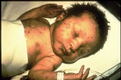
present at birth, pustules that transform into scaly hyperpigmented macules of uniform size (no associated erythema), more common in African-American babies hyperpigmented macules may persist for months NEUTROPHILS on Tzanck smear |
Transient neonatal pustular melanosis |
|
|
Transient neonatal pustular melanosis |
|
|
Neonatal candidiasis |
|
|
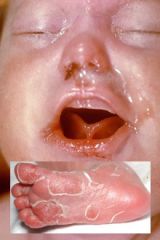
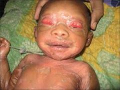
Congenital Rubella |
|
|
Congenital Syphilis |
|
|
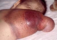
Red lesion that is elevated and enlarges, proliferating rapidly during first 6 months then eventually self-involute (usually starts at 2 yo and disappear by 5-10yo) Only need treatment if found in certain areas: eyes, beard, ear/nose/lips, midline lumbosacral region (get MRI!) Treatment involves propranolol, steroids |
Hemangioma |
|
|
PHACES Syndrome |
* Posterior fossa malformation (DANDY WALKER) * Hemangioma. Often in the distribution of the Facial Nerve. Look for a large segmental hemangioma on the FACE. Segmental refers to what looks like a nerve distribution (usually V1). This can be associated with STROKES. * Arterial cerebrovascular anomaly * Cardiac anomalies: Especially COARCTATION OF THE AORTA * Eye anomalies: MICROPHTHALMIA, STRABISMUS * Sternal defect |
|
|
PHACES Syndrome |
|
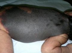
large, congenital vascular tumors (not true hemangiomas but can cause a severe CONSUMPTIVE COAGULOPATHY and death) |
Kasabach-Merritt syndrome |
|
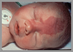
Salmon colored lesion often called a stork bite or salmon patch; blanch with pressure |
Nevus Simplex |
|
|
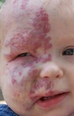
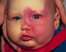
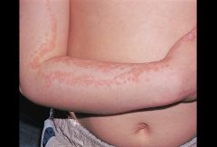
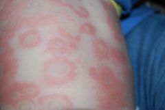
Capillary malformations that start as pink/flat lesions that become dark red-purple. They then progress to being thick/raised in adulthood. Present at birth and are PERMANENT. They are benign if noted in isolation. If noted on the face, they can be associated with glaucoma (increased intraocular pressure that can present as a red eye). |
Nevus Flammeus (Port-Wine Stain) |
|
what should you be concerned about this presentation? |
port-wine stains on the face may be associated with glaucoma |
|
port-wine stains involving the ophthalmic branch of the trigeminal nerve (V1) are associated with? |
Sturge-Weber syndrome port-wine stain trigeminal nerve distribution + INTRACRANIAL VASCULAR MALFORMATION (look for with MRI) also glaucoma, seizures, cognitive defects |
|
port-wine stains + hemihypertrophy on the lower extremities |
Klippel-Trenaunay syndrome *remember hemihypertrophy can also be related to Neuroblastomas, Beckwith-Wiedemann Syndrome, Russell-Silver Syndrome, Proteus Syndrome |
|
|
Sebaceous Nevus |
|
"cape-like" "coat sleeve" "bathing trunk" "garment type" |
Giant congenital melanocytic nevus 5-15% lifetime risk of developing melanoma Get MRI if overlying spine |
|
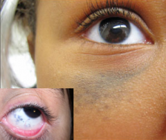
unilateral, irregularly speckled areas of blush, should receive yearly ophtho checkups to look for melanoma |
Nevus of Ota |
|
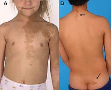
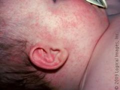
Syndromes associated with Cafe-Au-Lait spots |
Neurofibromatosis type I: >6 REGULAR café-au-lait macules. If prepubertal,these are> 5 mm, if postpubertal, > 15 mm. ---Freckle-like in axilla or inguinal areas McCune-Albright syndrome (in photo): IRREGULAR café-au-lait macules, PRECOCIOUS PUBERTY, BONE PROBLEMS (fractures, cranial deformities) |
|
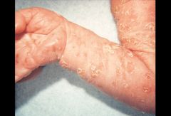
"fish-scale" disease often seen in atopic dermatitis patients Treatment with ammonium lactate or alpha-hydroxyacid-containing agents (ex: urea-containing cream) |
Ichythyosis Vulgaris |
|
noted at birth with thin transparent film (similar to cellophane) eyelashes missing, eyelids seem inverted |
Lamellar ichthyosis (AKA collodion baby) |
|
covering is hard (“armor-like”) and horny, movement is restricted, poor prognosis |
Harlequin ichthyosis |
|
|
Autosomal-dominant disorder: *Cafe au last spots *Axillary/Inguinal freckling *Fibromatosis *Eye-Lisch nodules *Skeletal bowing/Scoliosis *Positive family history (first-degree relative) *Optic nerve glioma |
"CAFE SPOT" Neurofibromatosis Type 1 |
|
|
Autosomal-dominant disorder: *acoustic nerve tumors (AKA neuromas or schwannomas) - can cause tinnitus or even hearing loss *spinal cord tumors (ependymomas) *meningiomas |
Neurofibromatosis Type 2 |
|
|
*NEUTROPENIA, poor neutrophil chemotaxis, and platelet dysfunction (Neutrophils containing giant lysosomal granules) *OCULOCUTANEOUS ALBINISM and frequent SKIN and LUNG INFECTIONS with Staphylococcus and Streptococcus. (They can have recurrent pneumonias.) |
Chediak-Higashi Syndrome |
|
|
Autosomal-dominant disorder with: *facial angiofibromas *ash-leaf spots *shagreen patch *periungual fibromas *periventricular or cortical TUBERS: Usually associated with INFANTILE SPASMS or seizures *cardiac rhabdomyomas: Look for a kid with arrhythmias! *renal angiomyolipoma |
Tuberous Sclerosis |
|
|
x-linked dominant disorder (Lethal in males!) Inflammatory vesicular phase Verrucous phase Hyperpigmentation phase noted along the lines of Blaschko, and finally a phase in which the hyperpigmentation disappears. Can leave atrophy or hypopigmentation behind. also a/w cicatricial alopecia, delayed tooth eruption (peg-shaped teeth) |
Incontinentia Pigmenti |
|
|
X-linked recessive (similar to incontinentia pigmenti but can occur in males) HYPOHIDROSIS, decreased sweating, which can lead to hyperthermia; HYPOTRICHOSIS, sparse hair, so no eyebrows/lashes; DELAYED TOOTH ERUPTION;and DEFORMED/PEG TEETH. |
Hypohidrotic Ectodermal Dysplasia |
|
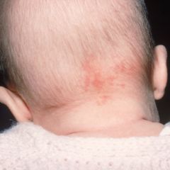
rash that looks like eczema, but is linear or papular and can follow the Lines of Blaschko |
Lichen Striatus |
|
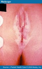
chronic, inflammatory, dry, white, and somewhat scaly rash that is usually found in the genital area. |
Lichen Sclerosus |
|
|
rashes that spare the inguinal folds |
contact dermatitis, eczema |
|
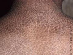
many tiny skin-colored/pink follicular papules |
keratosis pilaris |
|
redness and fissuring of the weight-bearing part of the plantar surface of the foot spares the interdigital skin area! |
juvenile plantar dermatosis |
|
hypopigmented dry white patches, on the cheeks and extensor extremities |
pityriasis alba |
|
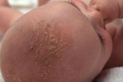
erythema with greasy yellowish scales, usually on scalp in infants treat with selenium sulfide or ketoconazole |
seborrheic dermatitis (cradle cap!) |
|
|
pruritic erythematous lesions that spare the folds in older kids found in the antecubital fossa, popliteal fossa, back of neck, ankles, wrists, back of hands/feet |
atopic dermatitis, eczema |
|
coin-shaped eczematous lesions usually on the extensor surfaces of extremities. Lesions are uniform, without any central clearing. |
nummular eczema |
|
|
round or oval patches of hair loss rarely associated with autoimmune diseases like thyroiditis |
alopecia areata |
|
Vesicles + Crusted Lesions.
**high index of suspicion for a rash “not improving with steroids and/or antibiotics. |
eczema herpeticum |
|
|
form of acute hair shedding that occurs diffusely. Instead of patches, you see “thinning” of the hair often related to a psychological or medical stressor |
telogen effluvium |
|
|
loss of hair in an irregular pattern (not a nice circle) and hair of differing lengths |
trichotillomania |
|
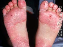
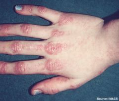
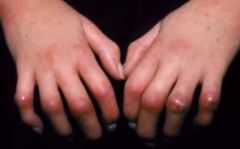
well-defined erythematous skin lesions with silvery scales sometimes results in punctate bleeding when scales are removed (this is called the Auspitz sign) infants may have in diaper area and goes into the inguinal folds. |
psoriasis |
|
periorbital heliotropic rash with proximal muscle weakness gottron papules - flat-topped reddish-violet skin over the knuckles diagnose by biopsy CK level will be high |
dermatomyositis |
|
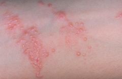
vesicular rash, may be linear will not spread once the affected area is washed with soap/water (fluid from vesicles CANNOT spread the rash) |
Poison Ivy (Rhus) reaction Type IV hypersensitivity rash, Allergic contact dermatitis |
|
|
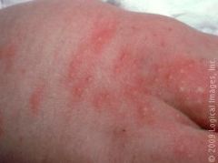
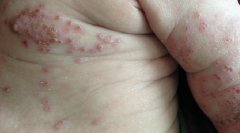
rash due to hypersensitivities to insect bites of bedbugs/fleas/mosquitoes that results in edema, erythema, and pruritis, some lesions may be umbilicated recurrent crops that wax and wane every few weeks or months |
Papular urticaria |
|
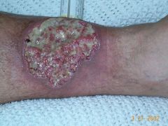
deep, bluish, necrotic and boggy-looking ulcers |
Pyoderma Gangrenosum associated with Crohn's disease |
|
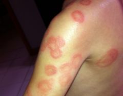
chronic skin condition with annular (circular) lesions, can by slightly pruritic (can look like ringworm without scaling) |
Granuloma Annulare |
|
|
severe blistering! may be confluent some lesions may look like a target or bullseye with the center dark/dusky +Nikolsky sign +at least two mucous membranes involved associated with aromatic seizure meds, PCNs, NSAIDs, sulfa drugs |
Stevens-Johnson Syndrome Toxic Epidermal Necrolysis (if >30% BSA) |
|
target lesions with dusky centers only 0-1 mucous membranes involved |
Erythema Multiforme (major if patient appears toxic) |
|
thickened tightened skin with a waxy appearance |
Scleroderma |
|
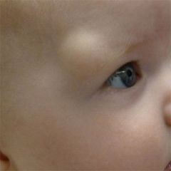
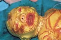
saclike growths presents at birth may contain hair or teeth slow growing and can get infected so should be removed |
Dermoid cyst (aka Epidermoid cyst) |
|
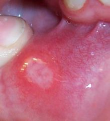
painful lesions in the oral mucosa with a grayish-white base and rim of erythema can occur in isolation or be associated with Behcet's or Schwachman-Diamond Syndrome |
Aphthous Ulcers |
|
erythema, very painful, warm nodules on the shins causes: sarcoidosis, Ulcerative colitis, OCPs/sulfas/PCNs |
erythema nodosum |
|
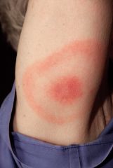
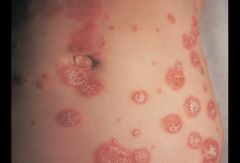
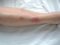
Bullseye lesion 1-2 weeks after tick bite Treatment: doxy if >8yo (PCN or amox if not) If has carditis, recurrent arthritis, or neuritis then treat with IV PCN or ceftriaxone |
Lyme Disease (Borrelia Burgdorferi) |
|
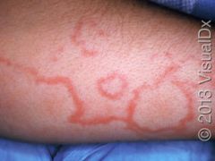
transient, erythematous, macular light-colored rash that is serpentiginous, margins progress with clear centers associated with Rheumatic fever |
Erythema Marginatum |
|
|
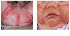
slapped cheek rash with diffuse lacy or reticular rash on extremities |
Erythema infectiosum (Parvovirus B19, Fifth Disease) |
|
scaly and extremely erythematous rash in the premolar and perianal area that can desquamate |
Zinc deficiency |
|
|
Rash of zinc deficiency + alopecia + diarrhea + FTT |
Acrodermatitis Enteropathica |