Reading...
![]()
Play button
![]()
Play button
![]()
Use LEFT and RIGHT arrow keys to navigate between flashcards;
Use UP and DOWN arrow keys to flip the card;
H to show hint;
A reads text to speech;
119 Cards in this Set
- Front
- Back
|
Describe these planes:
sagittal coronal frontal median transverse axial |
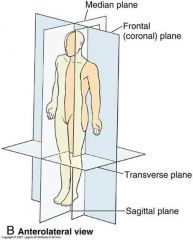
sagittal--divides into left and right sides
coronal--aka frontal, divides into front and back frontal--aka coronal, front and back median--middle sagittal plane transverse-- aka axial divides into top and bottom axial--aka transverse, top and bottom |
|
|
What are the 4 types of tissues?
|
Epithelial
Connective Muscle Nerve |
|
|
What are the main functions of epithelial tissue?
|
Protection
Absorption (O2 in lungs) Secretion (sweat, mucous) Excretion (kidneys filtering) |
|
|
What is the difference between epithelial tissue and connective tissue? How does this affect their function?
|
Epithelial is tightly packed to form a boundary. Connective is surrounded by extracellular matrix which allows it to do things like bear weight, withstand tension
|
|
|
What are the characteristics of epithelial tissue?
|
avascular
prolific basement mem with free face nourishment thru diffusion divides rapidly (quick repair) tightly packed |
|
|
What are the proper CT?
|
loose
adipose dense reticular |
|
|
What are the Specialzied CT?
|
bones
cartilage blood |
|
|
What are the 3 types of cartilage?
|
Hyaline--strong and flexible (nose, costal, joints)
Elastic--rigid but elastic (outer ear) Fibrocartilage--very tough, slightly compressible (intervertebral disks, pubic symphysis) |
|
|
What are the 3 types of muscle tissue?
|
skeletal
cardiac smooth |
|
|
What does sesmoidal mean?
|
round bone usually impedded in a tendon.
patella |
|
|
what's the difference between epiphysis and diaphysis in bone?
|
epiphysis is the knobby parts on the end and diaphysis is the long middle part on long bones
|
|
|
In long bone where is the yellow and red marrow?
|
Yellow marrow in the diaphysis
red marrow in the epiphyses |
|
|
what is wolf's law?
|
bone will grow when weight bearing and will diminish when not in use
|
|
|
Describe a fibrous joint and why this joint is characteristic of a fibrous joint.
|
skull sutures (non-moving, no joint cavity)
tibiofibular joint (bones further apart but still dense CT (ligaments flex allows for slight movement) (syndesmoses) teeth (dentoalveolar--gomphoses) |
|
|
name some cartilagenous joints and why they are characteristic of a cartilagenous joint.
|
epiphysial cartilage is a synchondroses because it joins 2 bones with hyaline cartilage with little movement
pubic symphyses has fibrocartilage joining 2 bones and has very slight movement |
|
|
Name some synovial joints and why they are characteristic of synovial joints.
|
elbow--has synovial fluid, joins two bones, lots of movement
glenohumaral--joins two bones, synovial fluid, lots of movement |
|
|
How does CT combine with muscle tissue?
|
CT or fascia that is surrounding the muscle will extend beyond fibers and form tendon to attach to bone and provide a means for nutrition and innervation to the muscle.
|
|
|
name the ligaments of the clavicle
|
costaclavicular ligament (main stabilizer)
anterior sternoclavicular lig articular capsule and disk interclavicular lig posterior sternoclavicular lig |
|
|
what is the coracoclavicular ligament and what 2 ligs does it contain?
|
coracoclavicular lig attaches the clavicle to the coracoid process on the scapula
contains trapezoid lig (more lateral) and conoid lig (more medial) |
|
|
which muscles are innervated by the anterior rami branch?
|
ALL muscles except intrinsic back muscles
|
|
|
which muscles are innervated by the anterior rami branch?
|
ALL muscles except intrinsic back muscles
|
|
|
What is the O, I and nerve of trapezius?
|
Originates on occipital bone, nuchal ligament, and C7-T12 spinous processes
Inserts into the lateral clavicle, acromion, and spine of the scapula Innervated by the accessory nerve (CN XI) |
|
|
what is the O, I and nerve of the levator muscle?
|
Origin is C1-4
Inserts into medial border of scapula above spine Innervated by dorsal scapular nerve (C5) |
|
|
which is superior, the rhombus major or minor?
|
minor
has origin C7-T1 major is lower with origin of T2-T5 both insert on medial border of scapula |
|
|
what nerves serves the rhombus major and minor?
|
dorsal scapular nerve C5
|
|
|
What is the O, I and nerve of the latissimus dorsi muscle?
|
Origin is fascia of thoracolumbar transverse processes T6-sacrum, iliac crest
Insertion is intertubercular groove of humerous and often inferior angle of scapula as well nerve is thoracodorsal nerve C6-8 nerve is |
|
|
Describe the serratus anterior muscles.
|
origin is ribs 1-8/9
insertion is medial border of scapula innervated by long thoracic nerve protracts and rotates scapula |
|
|
what is scapularhumoral rhythm?
|
once humerus is raised above 30 degrees, scapula must rotate 1-2 degrees for every degree of humerus raising
|
|
|
Where do most vertebral herniations occur?
|
Cervical or Lumbar vertebrae.
Nucleus pulposus herniates outside of anulous fibrosus into the cauda equina or compression of spinal cord |
|
|
What are the intrinsic back muscles from superficial to deep?
How are they innervated? |
Splenius
(capitus and cervicus) erector spinae transversospinal small segmental all are innervated by the posterior rami of spinal nerves |
|
|
What are the 3 Intermediate Deep muscles of the back?
|
Erector spinae
Iliocostalis longissimus Spinalis |
|
|
What are the 3 transversospinalis muscles?
|
semispinalis
multifidus rotatores |
|
|
What makes up the pectoral girdle?
|
bones =clavicle and scapula
joints = sternoclavicular joint, acromioclavicular joint (AC), scapulothoracic "joint", glenohumeral joint |
|
|
what's the only bony articulation between the pectoral girdle and axial skeleton?
|
sternoclavicular joint
|
|
|
what's the prevelance and reason for more middle 1/3 clavicle fractures?
|
80% due to less ligamental tissue support and the superficiality of the bone
|
|
|
what's a common injury in MVA's with regard to pectoral girdle?
|
broken clavicle
|
|
|
Describe the muscle "pulls" that cause the clavicle to displace.
|
SCM elevates medial end of clavicle
weight of arm pulls lateral end down b/c the trap can't handle it pec draws the humerus medially |
|
|
What's the treatment for a broken clavicle?
|
Use of several types of slings (figure 8)
Sometimes surgery is displacement is severe |
|
|
What type of joint is the sternoclavicular joint?
Name all the ligaments involved. |
synovial joint with articular disc (fibrocartilage)
interclavicular lig ant. and post. sternoclavicular ligs costalclavicular ligs |
|
|
What are the 3 ligs of the AC joint?
|
AC lig
Coracoclavicular (trapezoid lig and conoid lig) |
|
|
Which muscles are innervated by anterior rami?
Posterior rami? |
anterior rami innervate every muscle except the intrinsic back muscles which are innervated by the posterior rami
|
|

Name these arteries
|
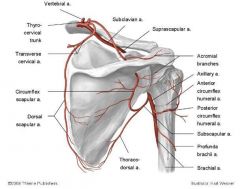
|
|
|
What are the rotator cuff muscles?
|
supraspinitus
infraspinitus subscapularis teres minor |
|
|
What nerve innervates the serratus anterior muscles?
|
long thoracic nerve (c5-7)
|
|
|
What is the scapulohumural rhythm?
|
After the humerus is abducted beyond 30 degrees the scapula must rotate 1 degree for every 2 degrees the humerus goes up.
120 and 60 |
|
|
What are the intermediate back muscles good for?
What are they? |
proprioception and likely some respiration
Serratus posterior superior (angled a bit) Serratus posterior inferior (pretty horizontal across) |
|
|
Spinal cord as it exits divides into 2 things. What are they and what are they associated with in terms of nerve signals
|
ventral and dorsal roots
ventral innervates all muscles except intrinsic back muscles and is associated with efferent (outgoing) motor signals dorsal root innervates the intrinsic back muscles and is associated with incoming afferent nerve signals |
|
|
Where do dorsal sensory neurons get their signals?
|
Somatic division = skin, tendons, joints, skeletal muscles
Visceral Division: smooth, muscle, cardiac, organs, glands |
|
|
Where do motor neurons get their signals from?
|
Somatic Division= skin, tendons, joints, skeletal muscles
ANS = SNS, PSNS |
|
|
What are the 3 types of ganglia in the PNS?
|
Sensory
Autonomic Enteric |
|
|
What are the names of the parasympathetic ganglia associated with cranial nerves?
|
cilliary
pterygopalatine submandibular Otic COPS |
|
|
List 4 PNS ganglia.
|
DRG
Paravertebral aka sympathetic chain gang Prevertebral Enteric |
|
|
What type of neuron cell bodies are associated with DRG?
|
sensory--somatic and visceral
|
|
|
What type of cell bodies are associated with paravertebral ganglia?
|
postgangliotic visceral motor
this bad boy is mostly associated with SNS |
|
|
What are white and grey rami communicans?
|
little bridges connecting sympathetic nerves throughout the paravertebral ganglia.
|
|
|
What type of neuron cell bodies are in the prevertebral ganglia?
|
postganglionic visceral motor
associated with plexuses via the abdominal aorta |
|
|
What are the plexuses associated with the prevertebral ganglia?
|
celiac ganglion
superior and inferior mesenteric ganglion aorticorenal ganglion |
|
|
What is the enteric ganglia?
|
Sort of its only little nervous system. It can be modulated by the SNS and PNS but it sort of does it's own thing.
associated with GI tract plexuses |
|
|
What are plexuses?
|
Different spinal nerves mingle and eventually give rise to a named peripheral nerve that has axons from all these minglers.
|
|
|
What are the 3 major somatic plexuses?
|
Cervical C1-5 short (scalp and neck)
Brachial C5-T1 (longer upper limbs Lumbosacral L1-S4 (down in the sacral region, lower limb) |
|
|
Have you done a dermatome quiz?
|
Hope so...
|
|
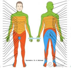
name these dermatomes
|
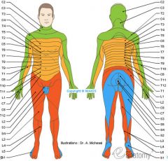
good luck!
|
|
|
Why is a myotome significant?
|
A myotome is the muscle mass that is innervated by a single spinal nerve.
Muscles have nerves from more than one spinal nerve but some spinal nerves are more primary than others in certain muscles. Weakness in a certain muscle can direct you to knowing if a spinal nerve could be injured or affected. |
|
|
How could you tell the difference between a spinal nerve involvement vs. a peripheral nerve involvement in a muscle?
|
testing the myotomes and muscle strengths of those regions associated with spinal versus peripheral nerves could tell you.
|
|
|
Name some visceral plexuses in the thorax.
|
esophogeal
cardiac pulmonary |
|
|
What type of nerve is associated with visceromotor fibers?
|
splanchnic nerves
splanchnic = guts |
|
|
What is the difference in the sizes of the pre and post ganglionic neurons between SNS and PSNS?
|
SNS
pre is short and post is long PSNS pre is long and post is short. The post are in the walls of the organ innervated. |
|
|
What is referred pain?
|
internal organs have nerves that carry sensory info. These nerves follow the SNS and any pain signals can be interpreted by CNS as somatic structure pain
|
|
|
What is the myenteric plexus?
submucosal plexus? |
Both in enteric nervous system
interconnected |
|
|
Deltoid
|
origin:
lateral 1/3 of the clavicle acromion and spine of the scapula insertion: deltoid tuberosity of the humerus innervation: axillary nerve (C5,C6) |
|
|
What are the parts of the deltoid muscle?
Function? |
CAS
clavicular (long parallel fibers) acromial (multipennate; more powerful) spinous (long parallel fibers) Almost every motion of upper limb except first 15 degrees of abduction |
|
|
what are rotator cuffs muscles?
|
Supraspinatus
Infraspinatus Teres minor—teres means round Subscapularis—Deep=sub |
|
|
You have spinal cord damage at C7. Which muscles will still work?
|
The shoulder muscles will still move arm but dexterity of hands and fingers will be lost due to trouble with radial, ulnar, muscolocutaneous, median nerve losses
|
|
|
cervical vertebrae nerve functions
|
C1-C6
Neck flexors C1-T1 Neck extensors C3, C4, C5 Supply diaphragm (mostly C4) C5, C6 Shoulder movement, raise arm (deltoid); flexion of elbow (biceps); C6 externally rotates the arm (supinates) C6, C7 Extends elbow and wrist (triceps and wrist extensors); pronates wrist C7, T1 Flexes wrist C7, T1 Supply small muscles of the hand |
|
|
what does damage to C6 mess with?
|
all the muscles of the shoulder
All the muscles of the arm except ulnar |
|
|
what are shoulder muscles innervated by axillary
|
deltoid
teres minor |
|
|
name the shoulder muscles from C5-C6
|
supraspinatus
infraspinatus deltoid subscapularis teres major pectoralis major (clavicular part) subclavius serratus anterior |
|
|
Where are the rotator cuff muscles located?
|
supraspinatus--superior
subscapularis--anterior infraspin--posterior teres minor--poster |
|
|
If the glenohumeral joint dislocates, what nerve could be damaged?
|
axillary
|
|
|
what do bursae do?
|
lie between tendons and muscles to reduce friction as they pass over bones.
|
|
|
what is supraspinatus tendinitis and calcific supraspinitus tendinitis?
|
inflammation of the tendon from repetitive motions or calcium deposits in the tendon that can lead to spontaneous rupture
|
|
|
What nerves innervate the subscapularis muscle and what is its function?
|
upper and lower subscapular nerves from C5,6
medially rotates humerus stabilize head of humerus in glenoid cavity |
|
|
What is the suprascapular and spinoglenoid notches used for?
|
foramen for the suprascapular nerve (C5-6) to go innervate the infrascapularis muscle
|
|
|
Which nerve being damaged would result in winged scapula?
|
long thoracic nerve (serratus anterior muscle failure to hold scapula in proper position)
|
|
|
Where does the axillary artery come from?
|
It is an extension from the subclavian artery.
It begins at lateral margin of 1st rib and it ends at the inferior border of teres major (there it becomes brachial artery) |
|
|
What branches off the axillary artery?
|
1st part: superior thoracic artery
2nd : thora-coac-romial a. lateral thoracic a 3rd: subscapular a ant. circumflex hum a post. circumflex hum a |
|
|
Describe the veins of the upper limb
|
basilic vein--->axillary vein-->subclavian vein-->vena cava
cephalic vein dumps from arm to axillary vein portion. |
|
|
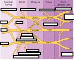
What are the stages of the brachial plexus?
|
Randy Travis Drinks Cold Beer
Roots Trunks Divisions Cords Branches (terminal named nerves) |
|
|
What divisions form the lateral cord of b.p.?
|
anterior divisions of upper and middle trunks
called lateral because it is lateral to axillary artery |
|
|
What divisions make the medial cord of b.p.?
|
anterior division of lower trunk
called medial because it is medial relative to the axillary artery |
|
|
What divisions make up the posterior cord?
|
union on all three posterior divisions from the upper, middle and lower trunks
called posterior cord because it lies posterior to axillary artery |
|
|
What are the 3 major anterior nerves of the arm?
|
musculocutaneous
ulnar median |
|
|
What are the 2 posterior named nerves of the arm?
|
radial and axillary
|
|
|
Where does the musculocutaneous nerve come from in the b.p?
|
lateral cord divides into lateral median and musculocutaneous
C5,6,7) |
|
|
Where does the lateral median come from in the b.p.?
|
lateral cord (C5,6, 7)
|
|
|
Where does the median nerve come from in the b.p.?
|
lateral cord
|
|
|
Where does the ulnar nerve come from in the b.p.?
|
medial cord
(C8, T1) |
|
|
Where does the median root of median nerve come from in b.p.?
|
median cord (C8, T1)
|
|
|
Where does the radial nerve come from in the b.p.?
|
posterior cord
(C5-T1) |
|
|
What does the posterior cord of b.p. branch into named nerve-wise?
|
radial nerve and axillary nerve
|
|
Fill in
|
|
|
|
What comes from the C5 root of the bp?
|
Dorsal scapular nerve
Runs dorsal (shark fin) along the medial border of the scapula |
|
|
What comes from C5-7 roots of bp?
|
Long Thoracic nerve
under pec innervates the serratus anterior winged scapula |
|
|
What nerves come from the just the superior trunk of bp?
|
suparscapular nerve
(suprascapular muscle and goes through spinoglenoid notch to innervate the infraspinatus muscle too) subclavian nerve subclavian muscle (little guy under clavicle) |
|
|
What nerve comes from the lateral cord before it becomes the musculo and 1/2 median in bp?
|
lateral pectoral nerve--this is the one that actually looks median in the picture yokley gave us
(the median pectoral nerve comes from the median cord) |
|
|
What nerves arise from the medial cord of the bp before it goes to med. half of Median and Ulnar nerves?
|
medial pectoral (this is the one that looks lateral in the picture yokely gave us)
medial cutaneous of arm AND medial cutaneous of forearm |
|
|
What nerves come from the posterior cord of bp before it becomes Radial / axillary nerve?
|
upper subscapular nerve
thoracodorsal nerve lower subscapular nerve axillary |
|
|
What happens with a rupture of transverse ligament of atlas:
and what 2 conditions might you see this with? |
Dens is free
Atlantoaxial joint can dislocate resulting in spinal cord damage, particularly during flexion e.g. rheumatoid conditions, Down’s syndrome |
|
|
how are prokaryotes different than eukaryotes in how they store DNA?
|
No nucleus so they hold DNA in protein complex in nucleoid
|
|
|
What is a chromatin?
|
groups of the histones that DNA wraps around
|
|
|
what's the difference between euchromatin and heterochromatin?
|
euchromatin is active and being used to make proteins
heterochromatin is hibernating or inhibited transcription |
|
|
what is semiconservative replication.
|
Double strand is separated and each one makes a daughter molecule, but in those daughter molecules each one has a newly synthesized strand and a parent strand
|
|
|
How is the starting point for replication different between prokaryotes and eukaryotes?
|
pro's, replication begins at a single unique site
euk's begin at multiple sites, usually AT rich regions |
|
|
What protein binds to dsDNA to melt it?
|
Dna A
|
|
|
What is DNA helicase?
|
the unwinder
|
|
|
Type I DNA Topoisomerase
|
removes supercoils by cutting one strand
|
|
|
Which direction does DNA polymerase read DNA sequence?
|
reads it in 3'-->5' (needs RNA primer) so it can make it in 5'-->3' direction
|
|
|
what is primase?
|
the RNA polymerase that puts the primers on the lagging strand (on the Okazaki fragments)
|
|
|
What's the name of the guy that replicates the DNA on the lagging strand?
|
DNA polymerase III
|