Reading...
![]()
Play button
![]()
Play button
![]()
Use LEFT and RIGHT arrow keys to navigate between flashcards;
Use UP and DOWN arrow keys to flip the card;
H to show hint;
A reads text to speech;
18 Cards in this Set
- Front
- Back
|
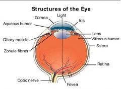
Anatomy of the eye
• Dura mater - is continuous w/ this eye structure - covers this nerve • Blood vessels are found at this location of the eye • Structure that replaces the sclera at the anterior pole • Fluids found in specific regions of the eye • These structures fxn in accommodation of the lens |
Anatomy of the eye
• Dura mater - is continuous with sclera - covers the optic nerve • found at the surface of the retina • Cornea • Fluids - - Aqueous humor in the anterior & posterior chambers - Vitrous humor everywhere else • ciliary muscle and zonule fibers |
|
|
The optic disk only contains these parts of these cell types
|
contains only the axons of retinal ganglion cells
|
|
|
Def of Fovea and Macula
|
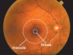
The Macula is a region (~5mm) of high visual acuity near the center of the retina. The Fovea (~1.5mm) is the central spot of the retina w/ the highest visual acuity, highest number of photoreceptors (cones). It is surrounded by the macula.
|
|
|
Name for the location of the back of the eyeball
|
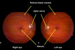
Ocular fundus
*Notice the relative location of structures btwn the L & R eyes |
|
|
Refractive power of an optical system:
• Def of refraction • Def of focal distance and its dependent variables (2) • The reciprocal value of focal distance and its units • The structure responsible for the main refractive power of the eye |
Refractive power of an optical system:
• the interaction btwn light and its environment that causes light to change its direction • the distance btwn the midline of the lens where converging light rays meet. It depends on the lens material and lens curvature • Refractive power, measured in diopters (D) • The cornea |
|
|
Far Vision:
• With parallel light rays coming from a far distance, this eye structure serves as the main dependent for refractive power • the state of the ciliary muscles and suspensory ligaments during far vision |
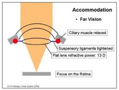
Far Vision:
• Cornea • Ciliary muscles - relaxed, Suspensory ligaments - contracted |
|
|
Near Vision:
• The state of the ciliary muscles and suspensory ligaments • Describe the elasticity of the lens |
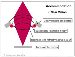
Near Vision:
• Ciliary muscles - contract, Suspensory ligaments - relax • The lens becomes more rounded/convex in shape, increasing its refractive index (13D --> 26D on average) |
|
|
Refractive plasticity:
• Def • Condition where the lens loses its elasticity |
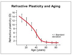
Refractive plasticity:
• the variability of the refractive power of the lens btwn far and near vision • Presbyopia |
|
|
Visual Acuity:
• def • How it can be measured • relationship btwn visual acuity and retinal photoreceptors • what it is dependent on (2) • Place in the visual field where it is lowest |
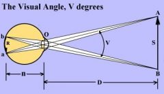
Visual Acuity:
• the ability t distinguish btwn two nearby points • finding the visual angle btwn the pupil and the object • As photoreceptor density increases, so does visual acuity • it is dependent on the number of photoreceptors and the proper fxning of the optical apparatus (lens and cornea) • Lowest in the periphery |
|
|
Pupil diameter and the ANS
• Sympathetic NS - how it influences the pupil - where the post-ganglionic cell bodies that iNN the eye are located - the eye muscle it iNN • Parasympathetic NS - how it influences the pupil - where its pre-ganglionic fibers originate and synapse - the eye muscle the post-ganglionic fibers iNN |
Pupil diameter and the ANS
• Sympathetic NS - causes dilation - superior cervical ganglion - Dilator pupillae • Parasympathetic NS - causes constriction - EW nucleus, synapse in the ciliary ganglion - Constrictor pupillae |
|
|
Def of Emmetropia
|
Normal sightedness
|
|
|
Myopia:
• another term • Where the image is produced, relative to the retina • state of ciliary muscle and lens |
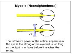
Myopia:
• "near sightedness" • image is produced in front of the retina • ciliary muscle is contracted, lens is rounded |
|
|
Hyperopia:
• another term • Where the image is produced, relative to the retina • state of ciliary muscle and lens |
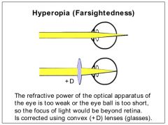
Hyperopia:
• "Far-sightedness" • Image is produced beyond the retina • ciliary muscle is relaxed, lens is flat |
|
|
Interpretation of distance equivalents in a Neurological Examination of Visual acuity
|
numerator = the distance a patient can read the number
denominator = the distance someone with normal vision can read that same number |
|
|
Papilledema:
• Another term • What it can indicate • causes dilation of these veins • How the disk looks through a opthalm-\oscope |
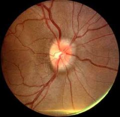
Papilledema:
• Optic disk edema • can indicate high ICP • increased pressure compromises venous drainage, leading to dilation of retinal veins • it appears pushed forward and white instead of pink |
|
|
Detached retina - which areas separate
|
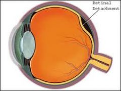
The retina separates from the retinal pigment epithelium
|
|
|
The leading cause of vision loss in individuals 60yrs of age in the US and many European countries
|
Age-related Macular degeneration
|
|
|
Diabetic retinopathy:
• Cause of retinal defects • vision loss is most noticeable for the patient when this area begins to be compromised |
Diabetic retinopathy:
• blood supply dysfxn due to permeability of the basal membrane of capillary endothelial cells and blood vessel damage • is most noticeable when the macula is involved |