Reading...
![]()
Play button
![]()
Play button
![]()
Use LEFT and RIGHT arrow keys to navigate between flashcards;
Use UP and DOWN arrow keys to flip the card;
H to show hint;
A reads text to speech;
62 Cards in this Set
- Front
- Back
|
Describe the 2 broad groups of fungi
|
Yeast: Single cells; round; reproduce by budding or fission
Mold: Germinate to branching fibers (hyphae); ends of hyphae have round forms that are NOT yeast but conidia, or spores Dimorphic species are coded as molds |
|
|
Name the 5 phyla of the kingdom Fungus: (and their corresponding sexual spores)
|
Ascomycetes (ascospores)
Basidiomycetes (basidiospores) Zygomycetes (zygospores) Chytridiomycetes or chytrids (oospores) (not a human pathogen) Fungi Imperfecti (asexual) |
|
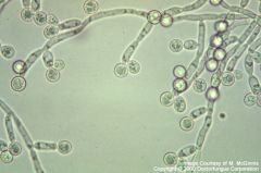
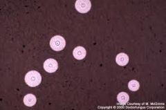
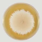
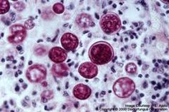
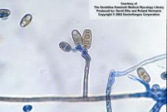
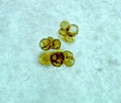
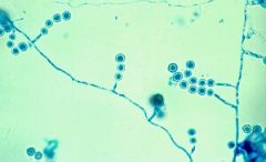
Name the fungus
|
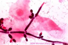
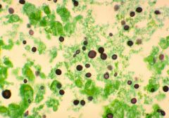
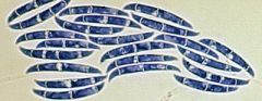
Candida albicans.
On cornmeal agar, chlamydospores are diagnostic for C albicans (see on tips). Also can see blastoconidia arranged in dense clusters on pseudohyphae On chromagar: C alb colonies are yellow-green to blue-green |
|
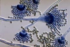
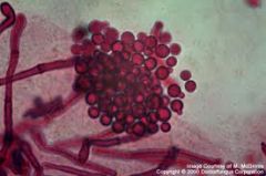
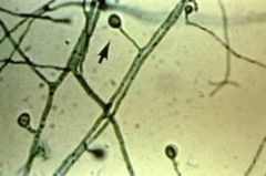
Name the fungus
|
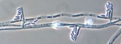
Candida.
Forms filamentous extensions (FEET or SPIDERS) on blood agar |
|
|
You see a small creamy colony on culture. Next step?
|
Think yeast. Unicellular. Plate to cornmeal agar for initial categorization based on morphology. Some biochemical tests can help.
Growth on PFA & Mycosel: think candida --> germ tube test, + is albicans Gr on PFA not mycosel: possible crypto, do urease test. |
|
|
You see a large fuzzy colony on culture. Next step?
|
Major considerations:
-Hyaline septate molds -Pigmented/dematiaceous septate molds -Aseptate molds -Dimorphic fungi (will present as a mold. R/O with yeast conversion) Consider rate of growth, type of hyphae, pigmentation, and type of sporulation (instant pattern recognition) |
|
|
Rapid growers
|
ZYGOMYCETES ('lid lifters")
yeasts C immitis Aspergillus spp |
|
|
Slow growers
|
Dimorphic fungi
Dematiaceous fungi (>1 wk) |
|
|
How to ID a dimorph?
|
Must demonstrate capacity for conversion
Grow as mold at 25-30C Grow as yeast 37C (TISSUE FORM) Some need coaxing (H caps - BHI; B dermatitidis - BHI; C immitis never converts) Exoantigan test largely replaced conversion tests. Performed on culture. |
|
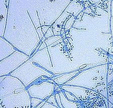
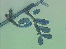
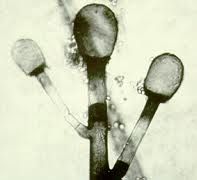
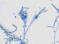
Name the fungus
|
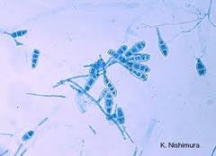
Aspergillus fumigatus
|
|
|
Aspergillus fumigatus
-Rapid grower -Cx: Blue-green with white apron -Morph: Conidia terminate in swollen vesicle A fumigatus: single row of phialides (vs penicillium - branches; vs A terreus - double row) A niger: heavy pigment A flavus: lollipop. circumferential phialides |
|
|
Aspergillus niger
- Cx: black on one side, yellow on other vs dematiaceous fungi - black both sides |
|
|
Aspergillus terreus
- cinnamon colonies - Double row of phialides (vs A. fumigatus - single row) |
|
|
Aspergillus flavus
Circumferential phialides LOLLIPOP |
|
|
Aspergillus
Penicillium Geotricum Trichophyton Microsporum Epidermophyton Rhizopus |
|
|
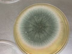
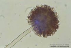
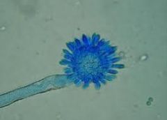
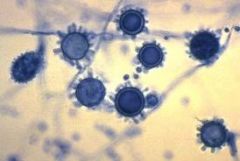
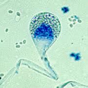
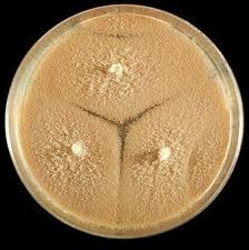
Aspergillus: Major diseases & identifying characteristics
|
Usually A. fumigatus
-ABPA (allx bronchopulm asp - colonizes airway and elicits allx response. Dx serology IgE) -IBPA (invasive bp asp - invades. immunocompromised host) -Aspergilloma (non-invasive. upper lobes) ID: - Tissue: hyaline septate mold (i.e. clear with hyphae), 45 degree branching (not specific) - Cx: Rapid growth, colonies with distinct margin & white apron, FRUITING HEADS |
|
|
What grows on Mycosel?
|
Dimorph or dermatophyte (slow growers)
Mycosel contains cycloheximide that inhibits contaminants, as well as Zygomycetes, Aspergillus, and Crypto. |
|
|
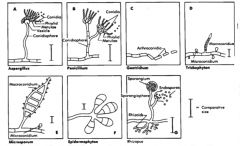
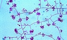
dermatophyte chart
|
|
|
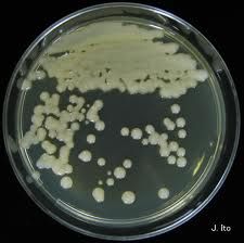
Candida
|
|
|
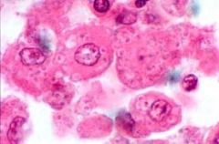
Cryptococcus
May mimic broad/narrow based budding ** Characteristic variation in size! ** 3-15 um |
|
|
Cryptococcus
India ink preparation Lack of staining = CAPSULE |
|
|
Malassezia
Causes tinea versicolor & seborrheic dermatitis Lipophilic SPAGHETTI & MEATBALL hyphae BOTTLE SHAPED YEAST ~wobbles |
|
|
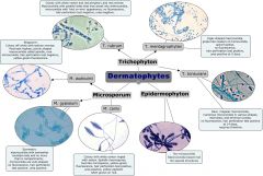
Microsporum
(this is M. gypseum) Pear-shaped macroconidia M.gypseum: flat tan cx. M.canis: fluffy; yellow reverse cx. Tinea capitis. |
|
|
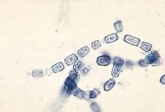
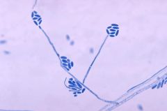
Trichophyton mentagrophytes
causes athletes foot colonies: white fuzzy on front, brownish on back BIRDS ON A FENCE (characteristic microconidia; can have cigar-shaped macroconidia) (more jax-like than T rubrum) Dermatophyte (with microsporon, epidermophyton) |
|
|
BIRDS ON A FENCE
Trichophyton rubrum #1 cause jock itch, athlete's foot, onychomycosis (dermatophyte) (reddish colonies) |
|
|
Microsporum canis
Dermatophyte (along with trichophyton & epidermophyton) Spindle-shaped macroconidia with pointed ends & transverse septa Causes SCALP infection (tinea capitis) Microsporon do NOT infect nails |
|
|
Microsporum gypseum
Dermatophyte Oval-shaped macroconidia with BLUNT ends (vs M canis) and transverse septa |
|
|
Epidermophyton floccosum
Dermatophyte Club-shaped macroconidia with transverse septa; NO microconidia |
|
|
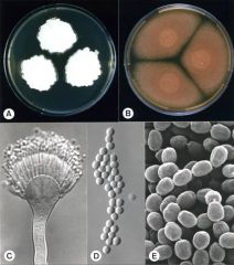
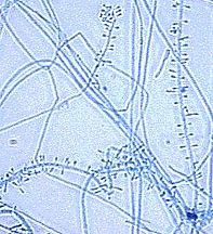
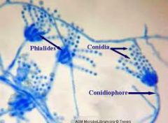
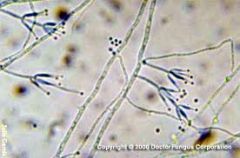
Morphology of aspergillus vs. penicillium
|
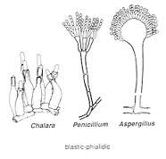
Aspergillus: Toilet brush: Has a circle, then spikes
Penicillium: Paintbrush: Bristles emanate directly from surface |
|
|
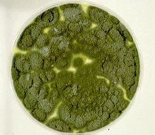
Penicillium
"paintbrush" hyaline mold (~aspergillus, fusarium) GREEN COLONIES common bread mold. ubiquitous |
|
|
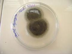
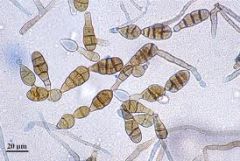
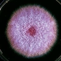
Fusarium
PINKISH Hyaline mold (~aspergillus, penicillium) Unique to hyaline filamentous molds in that produces micro & macro conidia (micro are small and ~acremonium; macro are canoes) May cause opportunistic infection Important plant pathogen (the banana killer!) |
|
|
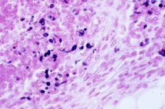
Histoplasmosis of the lung
2-5um yeasts, narrow-based budding, seen in tissue within histiocytes OH & MS river valleys GUANO |
|
Dimorphic fungus
Slow grower |
Histoplasmosis
-Grows as yeast in tissue; mold at <30C Hyaline septate mold with lollipop-like smooth microconidia & spiked macroconidia (in more mature colonies. Early may mimic birds on a fence) Grows within histiocytes in tissue, 2-5um, narrow based budding. necrotizing granulomas. |
|
|
Blastomyces dermatitidis
8-12um BROAD BASED BUDDING MS & OH river valley; dogs. lung --> skin & bones |
|
|
Blastomyces
LOLLIPOP CONIDIA |
|
|
DDx for LOLLIPOP CONIDIA in fungal culture:
|
Blastomyces dermatitidis
Histoplasma capsulatum (older colonies develop spiked macroconidia) Paracoccidioides Scedosporium & Chrysosporium Sepedonium (~histopl) |
|
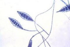
|
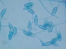
Sporothrix schenkii
2-5um elongated cigar-shaped yeasts with NARROW budding Purulent background Rose thorn puncture --> subcutaneous mycosis |
|
|
Sporothrix schenckii
Dimorphic fungus, NOT spread by inhalation (rose thorn puncture) Mold form: hyphae with DAISY conidia |
|
Dimorphic fungus; more rapid growth (7d)
|
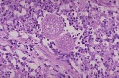
Coccidioides immitis
Granulomas with large 50-200um spherules with thick walls; enclose tiny 2-5um endospores California - Valley fever DDx: Rhinosporidium (nasal lesion), P wickerhami (olecranon bursitis). Endospores may look like histopl but DO NOT BUD |
|
|
Coccidioides immitis
Barrel-shaped arthroconidia Rapid growth (7d) vs other dimorphs Inhaled |
|
|
What blood group is more susceptible to infection with coccidoides? What are other risk factors?
|
Blood group B
Third trimester pregnancy Immunosuppression |
|

Dimorph; v slow growth (10-30d)
|
Paracoccidioides braziliensis
Tissue: diagnostic 10-15um yeast with circumferential budding = MARINER'S WHEEL Cultures: identical to blastomyces (lollipop conidia) Central & S America. Inhaled spores --> dissemination to reticuloendothelial system, bowel, liver. |
|
- hyaline mold
- slow growth (unlike other hyalines) |
Acremonium
- plant & soil; may be opportunisic infection - source of cephalosporins Filamentous hyphae with simple 1-celled conidia |
|
|
Name the hyaline molds
|
Aspergillus
Penicillium Fusarium Acremonium dermatophytes ... |
|
|
Name the dematiaceous molds
|
Alternaria
Bipolaris Cladosporium Exophiala Fonsecaea Wangiella Phialophora (pigmented, slow-growing) |
|
|
Name the dermatophytes
|
Trichophyton
Microsporon Epidermophyton |
|
|
Name the zygomycetes (aseptate molds)
|
Mucor
Rhizomucor Rhizopus Cunninghamella Absidia |
|
|
Alternaria
Dematiaceous mold Large conidia with ALTERNATING transverse & longitudinal crosswalls |
|
|
Bipolaris
(a dematiaceous mold) Bipolar = can form germ tubes from both poles Colonies: white --> black. Rapid. Woolly. Knobby (GENICULATE) zigzag conidia on microscopy |
|
|
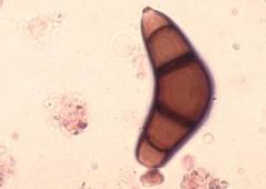
Curvularia
(a dematiaceous mold) Woolly gray black colonies. rapid. Geniculate (knobby) conidia with 4 cells (3rd cell larger & curved) Cause of sinusitis, keratitis, pulm infxn, dissemination |
|
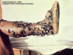
Name this skin condition & the most common pathogen associated with it
|
Chromoblastomycosis
- Chronic skin infection of the legs - Tropical regions Most common cause is: Fonsecae (a dematiaceous mold). KOH skin scrapings show COPPER PENNY bodies |
|
|
Absidia
(zygomycete) Woolly, rapid. Hershey's kiss columella; Pear-shaped sporangia Rudimentary rhizoids Growth 25-42C. Can be human pathogen, causing pulmonary, rhinocerebral, or disseminated infection. |
|
|
Rhizomucor
Zygomycete (woolly, fast) short sporangiophores; tufts; branched. Some rhizoids. Growth up to 55C |
|
|
Yeast colonies growing on culture; germ tube test is positive. Possibilities?
|
Candida albicans
Candida dublinensis (Must VITEK if sterile site) |
|
|
Hyaline molds characterized by production of conidia in chains
|
Penicillium sp
Paecilomyes sp Scopulariopsis sp |
|
|
Hyaline molds producing conidia in clusters
|
Acremonium sp
Fusarium sp Gliocladium sp Trichoderma sp |
|
|
Paecilomyces
Long tapered phialides Oval irregular conidia DDX: PENICILLIUM (but never green) -blunt phialides |
|
|
Scopulariopsis
radial rugae chains of conidia soil; can cause onychomycosis |
|
|
Penicillium
Characteristic green |
|
|
Fusarium
Characteristic pink color Woolly |
|
Not growing on mycosel
|
White yeast not growing on mycosel: Think cryptococcus. Candida albicans grows on mycosel. Do germ tube test & urease.
|
|
|
Name some candida non-albicans
|
C. glabrata: UTIs. Slower growth (72h). Budding yeast, NO pseudohyphae
C. krusei: resistant to ketoconazole. matchstick blastoconidia C. dubliniensis: res to fluconazole. Very ~albicans - must ID on VITEK |