Reading...
![]()
Play button
![]()
Play button
![]()
Use LEFT and RIGHT arrow keys to navigate between flashcards;
Use UP and DOWN arrow keys to flip the card;
H to show hint;
A reads text to speech;
83 Cards in this Set
- Front
- Back
|
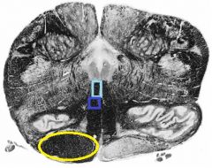
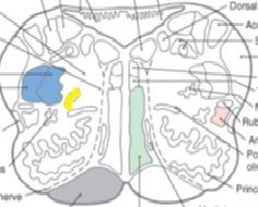
Where do fibers within the fasciculus gracilis and cuneatus begin to synapse in?
|
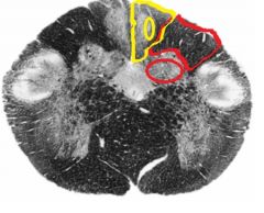
Yellow indicates the fasciculus gracilis and its nucleus gracilis (where it synapses). Red indicates fasciculus cuneatus and its nucleus cuneatus (where it synapses).
|
|
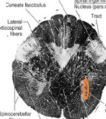
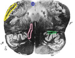
Name the orange area. (hint: name circle and outlined space)
|
Spinal trigeminal nucleus (circle) and tract (outlined space)
|
|
|
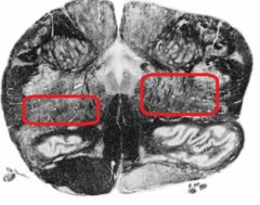
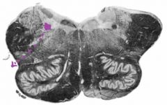
What is the black box labeling?
|
Trigeminal tubercle
|
|
|
what type of afferents does the spinal trigeminal nucleus receive? what do these afferents convey?
|
GSA; pain, temperature and crude touch from the face and external ear.
|
|
|
what surface marking does the spinal trigeminal nucleus and tract give rise to?
|
the tuberculum cinereum or trigeminal tubercle.
|
|
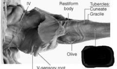
what is the black box labeling?
|
trigeminal tubercle
|
|
|
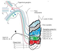
what are the three subnuclei of the spinal nucleus?
|
pars oralis, pars interpolaris, and pars caudalis
|
|
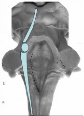
For each number (1-3) give the corresponding subnuclei.
|
1. Pars oralis, 2. Pars interpolaris, and 3. Pars caudalis.
|
|
|
what is the Pars oralis responsible for?
|
tactile input (crude touch)
|
|
|
what is the pars interpolaris responsible for?
|
dental pain
|
|
|
what is the pars caudalis responsible for?
|
pain and temperature input
|
|
where do the nociceptive afferents from around or encircling the mouth synapse in?
|
rostral part of the pars caudalis
|
|
|
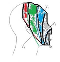
which subnuclei of the spinal nucleus has somatotopic organization? what kind of input does it receive?
|
pars caudalis; V1-3
|
|
|
describe the dorsal- ventral organization of the particular input for the pars caudalis.
|
Ventral - V1, Dorsal -V3
|
|
|
where do the sensory fibers from the external ear travelling in CN 7, 9, and 10 synapse in?
|
dorsal most part of caudal subnucleus.
|
|
|
In what nucleus do GSE neurons originate in?
|
spinal accessory nucleus.
|
|
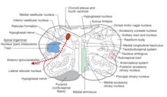
Identify the orange circle.
|
spinal accessory nucleus
|
|
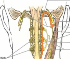
This is the pathway for which CN?
|
spinal accessory
|
|
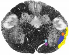
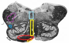
Label all the colored areas with their appropriate tract name.
|
light blue: medial longitundinal fasciculus
purple: tectospinal tract orange: rubrospinal tract dark blue: anterolateral system yellow: anterior and posterior spinocerebellar tracts |
|
Identify where the dorsal column nuclei axonal projections decussate. What are they called?
|
internal arcuate fibers
|
|
|
After the neurons in the dorsal column nuclei send axonal projections that arc across the tegmentum enter the medial lemniscus, where do they ascend to?
|
thalamus
|
|

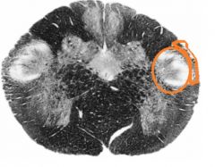
name the orange region and describe its function.
|
accessory cuneate nucleus: receives proprioceptive information from the upper extremity that is destined for the cerebellum.
|
|
|
where do the neurons from the accessory cuneate nucleus project to? Forming what tract?
|
cerebellum as the cuneocerebellar tract
|
|
|
Via what area does the cuneocerebellar tract enter the cerebellum?
|
via inferior cerebellar peduncle (restiform body).
|
|
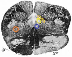
Name the colored areas in this upper medulla cross section.
|
blue: Hypoglossal nucleus
yellow: dorsal motor nucleus goldenrod: solitary nucleus orange: nucleus ambiguus grey: inferior olivary complex |
|
name the colored regions in this cross section of the medulla.
|
dark blue: tectospinal tract
light blue: medial longitudinal fasciculus yellow: pyramidal tracts |
|
|
what are the major vessels that supply the lower medulla?
|
anterior spinal artery, vertbral arteries and posterior spinal artery.
|
|
|
what does the anterior spinal artery supply?
|
anteromedial areas of lower medulla
|
|
|
what does the posterior spinal artery supply?
|
dorsal areas
|
|
|
what do the vertebral arteries supply?
|
lateral areas
|
|
|
At more rostral areas, which area supplies lateral areas?
|
Posterior inferior cerebellar artery
|
|
|
where are the motor CN nuclei usually located? sensory nuclei?
|
ventromedially; anterolateral
|
|
|
the inferior olive is a derivative of what?
|
the alar plate
|
|
|
what cranial nerve is associated with the level of the obex?
|
CN 12- hypoglossal
|
|
|
what are the unique characteristics of the upper medulla?
|
area postrema, restiform body, central tegmental tract, and ventral trigeminothalamic tract
|
|
|
what is the function of the area postrema?
|
chemosensitive trigger zone for emesis stimulated by blood-borne chemicals
|
|
|
what disappears with the appearance of the restiform body?
|
posterior spinocerebellar tract
|
|
|
what does the restiform body provide?
|
a major afferent pathway into the cerebellum from spinal cord and medulla.
|
|
|
at the level of the obex, which tracts begin to form?
|
central tegmental tract and ventral trigeminothalamic tract.
|
|
|
what is the function of the central tegmental tract?
|
local communication pathway within the brainstem.
|
|
|
where is the central tegmental tract far more pronounced?
|
rostral medulla and above
|
|
|
what types of fibers does the central tegmental tract contain?
|
fibers descending from the parvocellar red nucleus, ascending (taste) fibers originating in the gustatory nucleus (rostral solitary nucleus).
|
|
|
where does the ventral trigeminaothalamic tract begin to form?
|
lateral to the medial lemniscus
|
|
|
what does the ventral trigeminothalamic tract carry?
|
pain and temperature from the face.
|
|
name the colored regions on this cross section of the upper medulla.
|
blue: area prostrema
yellow: restiform body pink: ventral trigeminothalamic tract green: central tegmental tract |
|
|
what are the structures retained from the level of sensory decussation?
|
hypoglossal nucleus and nerve, nucleus ambiguus, dorsal motor nucleus of vagus, solitary nucleus and tract, nucleus gracilis and cuneatus, accessory cuneate nucleus, inferior olive and spinal trigeminal nucleus and tract.
|
|
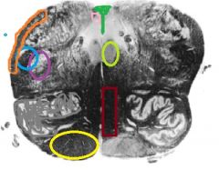
Identify all of the colored areas retained from level of sensory decussation.
|
Yellow: pyramid
Grey: inferior olive Purple: spinal trigeminal nucleus blue: spinal trigeminal tract orange: restiform body green: obex pink: area postrema lime green: hypoglossal nucleus maroon: medial lemniscus |
|
|
what are the prominent features of the midolivary level?
|
inferior olivary complex, hypoglossal nerve and vagus nerve
|
|
|
where does the hypoglossal nerve exit?
|
preolivary sulcus
|
|
|
where does the vagus nerve exit?
|
the postolivary sulcus
|
|
|
what is the principal olivary nucleus involved in?
|
control of planned or skilled, voluntary movements;
|
|
|
what are the two accessory olives called?
|
medial accessory and dorsal accessory olive
|
|
|
how do the output fibers from the inferior olive project to the cerebellum?
|
through the contralateral inferior cerebellar peduncle (olivocerebellar tract).
|
|
|
where is the hypoglossal nucleus found?
|
deep to the hypoglossal trigone.
|
|
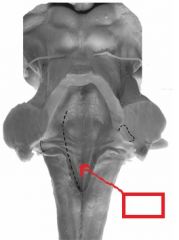
what is the labelled area? what can be found deep to this area?
|
hypoglossal trigone; hypoglossal nucleus
|
|
name the yellow area.
|
nucleus ambiguus
|
|
|
what cranial nerves are associated with the nucleus ambiguus?
|
9, 10, and 11
|
|
|
where does the dorsal motor nucleus of the vagus lie deep to?
|
vagal trigone
|
|
|
where is the vagal trigone?
|
lateral to the hypoglossal trigone
|
|
|
where do the fibers of the dorsal motor nucleus of the vagus synapse? what are the associated structures?
|
postganglionic parasympathetic neurons. associated with visceral structures of thorax and abdomen.
|
|
|
the dorsal motor nucleus of vagus receives input from another nucleus, what is it? what does this input help the DMN take part in?
|
solitary; efferent component of the baroreceptor reflex.
|
|
|
The dorsal motor nucleus of vagus also plays a role in what protective action? (hint: think blood borne chemicals)
|
emesis
|
|
name the red region and its exit pathway.
|
Dorsal motor nucleus and it exits in the postolivary sulcus.
|
|
name the purple region and its exit pathway.
|
solitary nucleus and it exits via the postolivary sulcus.
|
|
|
what are the two general zones of the solitary nucleus? what type of afferents are present?
|
1) rostral and (lateral) gustatory zone. SVA from the oral cavity and pharynx via CN 7.9.10 and 2) a more caudal cardiorespiratory zone.
|
|
|
where do the neurons from the gustatory nucleus project to? via which tract?
|
thalamus (ventral posteromedial nucleus); central tegmental tract
|
|
|
what two CNs contribute to the cardiorespiratory zone of the solitary nucleus?
|
11 and 10
|
|
|
which reflex is most notably taken by the caudal solitary nucleus?
|
baroreceptor
|
|
|
neurons from the cardiorespiratory zone project to which nuclei?
|
dorsal motor neurons, nucleus ambiguus, and the intermediolateral column.
|
|
|
the vestibular nuclei receive input from what?
|
the labyrinth of the inner ear
|
|
|
what is the vestibular nuclei involved in?
|
body equilibrium and control of eye movement.
|
|
|
Damage to the vestibular nuclei will result in what?
|
vertigo, nausea and nystagmus
|
|
|
what nuclei is the arcuate nuclei continuous with?
|
pontine
|
|
|
what do arcuate nuclei send axons into the cerebellum as?
|
ventral external arcuate fibers and striae medullares
|
|
Name all the colored areas.
|
purple: spinal nucleus and tract
dark blue: anterolateral system orange: arcuate nuclei red: pyramidal tract yellow: medial lemniscus light blue: tectospinal tract and medial longitundinal fasciculus |
|
|
what is no longer present at the midolivary level? (hint: vestibular nuclei adopt their position)
|
nucleus gracilis and cuneatus
|
|
|
three things appear in the rostral medulla, what are they?
|
1) dorsal and ventral cochlear nuclei, 2) inferior salivatory nucleus, and 3) nucleus prepositous
|
|
|
what nucleus receives influences from hypothalamus and olfactory system?
|
inferior salivatory nucleus
|
|
|
In the rostral medulla, what two tracts are now part of the restiform body?
|
posterior spinocerebellar tract and cuneocerebellar tract
|
|
|
what are two functions of the reticular formation?
|
consciousness and sleep
|
|
|
what is the blood supply to the upper medulla?
|
vertebral arteries, anterior spinal artery and posterior inferior cerebellar artery.
|
|
|
what are two distinct long tract features that relate to the medulla?
|
in the caudal medulla, both the dorsal column-medial lemniscal system and corticospinal tract decussate.
|
|
|
Ventrally, what is the caudal boundary of the lower medulla? Dorsally, what is the caudal boundary?
|
Ventral- pyramidal decussation
dorsal- obex |