Reading...
![]()
Play button
![]()
Play button
![]()
Use LEFT and RIGHT arrow keys to navigate between flashcards;
Use UP and DOWN arrow keys to flip the card;
H to show hint;
A reads text to speech;
23 Cards in this Set
- Front
- Back
|
Describe Chabertia ovina.
|
1. Strongyloidea
2. Non-equine strongyle - ruminant (sheep/goats) 3. Large mouthed bowel worm 4. Prepatent period ~ 6 weeks 5. 13-20 mm long 6. Characteristic large buccal capsule: plug-feeder |
|
|
What are some clinical signs of Chabertia ovina?
|
1. Usually subclinical
2. Large numbers of plug-feeding adults can cause mucosal ulcerations: local hemorrhage, protein loss into the gut, diarrhea with blood and mucus, weight loss, possible anemia, and decreased wool quality. |
|
|
What is the diagnosis for C. ovina?
|
1. Detection of typical strongyle-type egg
2. Coproculture and ID of L3 needed for definitive diagnosis. |
|
|
What is the treatment and control for C. ovina?
|
Same as other related parasites.
|
|
|
What is Oesophagostomum spp?
|
1."Nodular worm" - reflects lesions produced in large intestinal wall
2. Adults only 3/4 inch long 3. Eggs = strongyle type |
|
|
What is the life cycle of Oesophagostomum?
|
Adults in large intestine are surface feeders; Eggs in feces ( L1 --> L3 in environment)
L3 is ingested --> mucosal encystment --> migrate to colon in submucosa --> emerge to become adults Prepatent period is 6-7 weeks |
|
|
What is the pathology - primary infection of Oesophagostomum?
|
Relatively harmless
Enteritis if large number larvae emerge from mucosal cysts = diarrhea Adult feeding activities: inflammation +/- thickening of mucosa |
|
|
What is the pathology - re-infections of Oesophagostomum?
|
Produce lesions, possible clinical signs
Localized immune-mediated reaction around larvae in mucosa: formation of nodules or "mini-abscesses" |
|
|
What are the lesions or nodules of Oesophagostomum?
|
Larvae persist in nodule for < 3 months
Nodule contents eventually caseate and calcify: larvae either leave or become trapped and die |
|
|
What are the clinical signs of Oesophagostomum?
|
Nodules interfere with function of large intestine
Most common signs are anorexia, weight loss, diarrhea, poor wool quality, etc...Rarely do nodules rupture |
|
|
What is the diagnosis of Oesophagostomum?
|
Detect typical strongylid eggs on fecal exam
No eggs during pre-patent period when larval emergence can cause diarrhea etc... Necropsy: detection of nodules |
|
|
What is Stephanurus dentatus?
|
Swine kidney worm
Adults: stout, 1-1.5 inches long, cream colored; males have poorly developed bursa and thick-walled "squarish" buccal capsule Eggs = typical strongyle type and will be in urine sediment |
|
|
What is the life cycle of Stephanurus dentatus? (Prepatent period = 9-16 months)
|
Direct cycle: earthworms can serve as paratenic hosts
Infection acquired by: ingestion of L3; skin penetration by L3; ingestion of L3 in earthworms that can serve as paratenic hosts L3 migrate from gut to liver (2-9 months) and can cause extensive liver damage --> exit liver capsule --> retro-peritoneally to kidney wall, ureters (mature to adults in cysts); Cysts communicate with ureter --> eggs passed in urine |
|
|
What kind of lesions occur from Stephanurus dentatus and what are the signs?
|
Skin lesions
Liver fibrosis, cirrhosis, abscesses, adhesion Vague signs: unthriftiness, emaciation, death Adults in renal tissues = nonpathogenic |
|
|
How do you diagnosis Stephanurus dentatus?
|
Clinical signs develop in hogs in endemic area (mainly SE US)
Detects eggs in URINE sediment (pigs be 2 years old) Detect worms at necropsy |
|
|
What is Syngamus trachea?
|
1. "Gapeworm" or "Y-worm"
2. Adults in tracheal lumen 3. Fairly common in game birds 4. Bright red = suck blood 5. Males are small (2-6 mm); females are larger ( <20 mm) 6. Male and female in permanent copulation = Y form |
|
|
Explain the structure of Syngamus trachea eggs.
|
1. Atypical strongylid eggs
2. Bipolar opercula 3. Shaped like a hybrid of Nematodirus and trichuris eggs |
|
|
What is the life cycle of Syngamus trachea?
|
1. Adults in trachea produce eggs: eggs coughed up and swallowed; passed in feces in non-embryonated stage
2. Infective L3 develops within egg in 1-2 weeks 3. Infection acquired by ingestion of L3: liver-lung-tracheal migration --> gut mucosa --> hepatic portal vein --> liver --> vena cava --> heart --> lungs --> trachea Mature and copulate in trachea: attach to tracheal mucosa and suck blood (prepatent period ~ 17-20 days) |
|
|
What are the signs and lesions of Syngamus trachea?
|
Larval migration: hemorrhage, edema, possible pneumonia in heavy infections
Adults in trachea: obstructive tracheitis - males deeply embedded in mucosa cause nodules; catarrhal tracheitis with excess mucus; spasmodic dyspnea: head shaking, neck extension, gaping, open-mouth breathing |
|
|
How can you diagnosis Syngamus trachea?
|
Definitive diagnosis based on: detect eggs in feces (differentiate from smaller, brownish Capillaria eggs also with polar plugs) and detect adults attached to lower trachea surrounded by blood streaked mucus
|
|
|
How can you control the spread of Syngamus trachea?
|
1. Avoid keeping birds on same ground for long periods of time
2. Keep pens dry: L3 and paratenic hosts survive in moist areas 3. Minimize cross-transmission among birds |
|
|
What are clinical signs of Cyathostoma variegatum?
|
Severe respiratory distress: dyspnea, head shaking
|
|
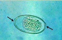
What is this?
|
Syngamus trachea egg
|