Reading...
![]()
Play button
![]()
Play button
![]()
Use LEFT and RIGHT arrow keys to navigate between flashcards;
Use UP and DOWN arrow keys to flip the card;
H to show hint;
A reads text to speech;
25 Cards in this Set
- Front
- Back
- 3rd side (hint)
|
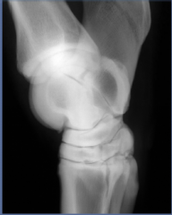
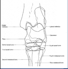
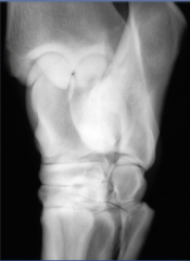
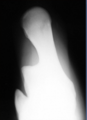
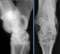
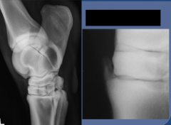
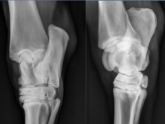
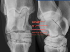
Tarsus
- . . . - . . . . . |
Tarsus
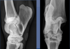
- compound joint with three joint sacs . tibiotarsus and proximal intertarsal . distal intertarsal . tarsometatarsal - standards views . lateromedial . dorsoplantar . DLPMO . PLDMO or DMPLO .skyline |
|
|
|
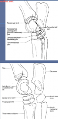
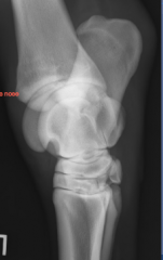
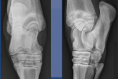
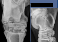
Lateral view
tarsocrural joint = tarsotibial joint |
|
|
|
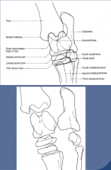
Dorsoplantar
superimposed bony structure |
|
|
|
Oblique Views:
- - |
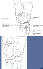
DLPMO
highlights: - - - - - DMPLO (PLDMO) highlights: - - - - |
DLPMO
highlights: - Medial Malleolus - Medial trochlear ridge - Dorsomedial aspect of distal hock - plantarolateral aspect of hock - see tarsal canal if well positioned DMPLO (PLDMO) highlights: - lateral trochlear ridge -dorsolateral aspect of distal hock - plantaromedial aspect of hock - best view for seeing OCD of distal intermediate ridge of tibia |
|
|
DLPMO
dorsomedial border - palmarolateral border |
|
|
|
*gonzo's nose
dorsolateral border - palmaromedial border |
|
|
|
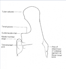
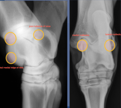
skyline view
highlights: sustenaculum tali tarsal groove (DDF and tendon sheath) tuber calcanei proximal aspect of medial trochlear ridge |
|
|
|
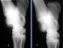
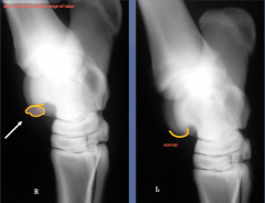
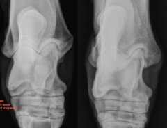
Normal Variations
Medial Trochlear Ridge - normal variability, mistaken for OCD/fracture lesions different between right and left |
|
|
|
Osteochondrosis/Osteochondritis Dissecans
Lesion Locations: |
Lesion Locations:
distal intermediate ridge of the tibia lateral trochlear ridge of the tallus medial trochlear ridge of the talus medial malleolus (less common stie: lateral malleolus, talus, calcaneus) - chronic distention of the tibiotarsal joint may lead to secondary degenerative joint disease >5 mos of age to evaluate |
|
|
|
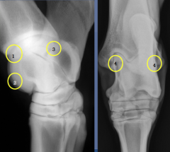
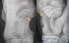
OC/OCD
|
1-distal intermediate ridge of tibia
2- distal medial ridge of tibia 3- proximal tubercle of talus 4-medial and lateral malleolus |
|
|
OCD
distal intermediate ridge of tibia (DIRT) |
contour changes, bone alignment,opacity
change from convex to flattened, irregular shape, sclerotic opaque bone |
|
|
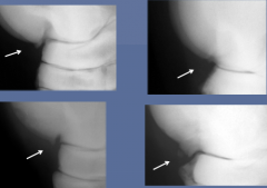
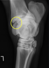
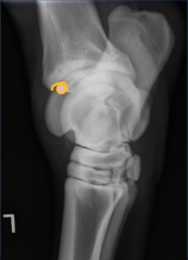
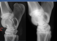
OCD of lateral trochlear ridge of talus
vs normal |
|
|
|
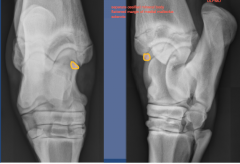
Medial Malleolus
very uncommon DP view and DLPMO seperate ossficied mineral body, flattened margin of medial malleolus, sclerotic |
|
|
|
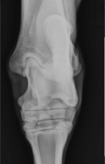
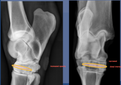
Degenerative joint disease
|
- distal intertarsal and tarsometatarsal joints most common (low motion joints - can cause pain, ankylosis)
- proximal intertarsal joint, tarsocrural joint, talocalcaneal joint (high motion jts, significant, long term lameness and pain of jts) Radiographic findings: |
Radiographic findings:
- osteophytes - periosteal new bone - narrowed joint space - sclerosis and/or lysis (erosive change) of subchondral bone |
|
|
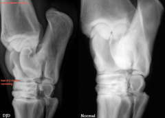
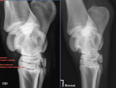
DJD
joint space narrowing - distal intertarsal joint increased opacity, |
|
|
|
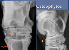
DJD
Osteophytes DP view and lateral view proximal 3rd tarsal bone distal central tarsal bone |
|
|
|

DJD
Sclerosis and Erosions DLPMO view sclerosis and erosion, distal central, proximal 3rd tarsal no joint space definition variable in appearance, focally surrounding DJD or entire central and 3rd tarsal bone lose cortex/medullary distinction all white - increase opacity margin - areas of lysis/erosion, degenerative change central and 3rd tarsal bone irregular margin |
|
|
|
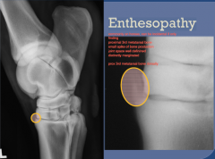
DJD
Enthesopathy commonly on horses can be incidental proximal 3rd metatarsal bone, small spike of bone production joint space well defined, distinctly marginated |
|
|
|
Erosive changes
- horses are usually persistently lame Osteophytes - dorsal and lateral aspects of the distal intertarsal joint margin of bone, lameness, pain |
|
|
|
may or may not be significant
enthesophytes on dorsal metatarsal bone joint space narrowing (distal intertarsal joint, ankylosis = fused) |
|
|
|
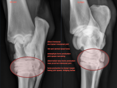
DPLMO view and DLPMO view
distal intertarsal and tarsal metatarsal joint 3rd and central tarsal bone osteophyte and joint space narrowing distomedial talus bone production near proximal intertarsal joint bone production to dorsal margin losing joint space, bridging bones |
|
|
|
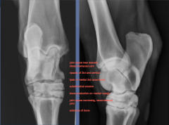
joint space loss laterally
distal intertarsal joint opacity of 3rd and central tarsal bone lysis of medial 3rd tarsal bone subchondral erosion joint space narrowing, tarsometatarsal joint sclerosis of bone |
|
|
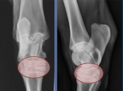
DP view
|
DJD
lateral view osteophyte erosive changes no distinct joint space sclerosis of 3rd and central tarsal bone |
DLPMO
|
|
|
DMPLO view
DJD |
|
|
|
Additional tarsus disease
|
collateral ligament injury (jt instability)
tibia stress fractures (articular margin, osteoarthritis) tarsal bone fractures (difficult to see, take other views off normal views, not displaced, repeat radiographs 7 to 10 days, osteoclast bone aborption) osteomyelitis of the calcaneus (anywhere in tarsus, point of most abuse, predisposed) tarsal sheath tenosynovitis |
|