Reading...
![]()
Play button
![]()
Play button
![]()
Use LEFT and RIGHT arrow keys to navigate between flashcards;
Use UP and DOWN arrow keys to flip the card;
H to show hint;
A reads text to speech;
84 Cards in this Set
- Front
- Back
|
Joint Category, Synarthrosis is ___ & and consists of what time of joints?
|
Interosseus connective tissue
Fibrous & Cartilaginous joints |
|
|
Synarthrosis is:
|
1. Connective tissue used to connect bony components
2. Grouped into two divisions according to type of connective tissue used in the union of bone to bone. 3. Slight to no movement allowed |
|
|
Fibrous Joints consist of:
|
Suture
Gomphosis Syndesmosis |
|
|
Suture Joint is:
|
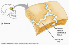
a joint in which two bony components are united by a thin layer of dense fibrous tissue.
**Edges generally interlock or overlap one another. |
|
|
Gomphosis Joint is:
|
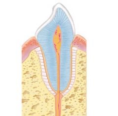
A joint in which the surfaces of bony components are adapted to each other like a peg in a hole
Ex. A tooth – between mandible and maxilla. |
|
|
Syndesmosis Joint is:
|
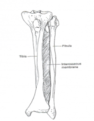
A type of fibrous joint in which two bony components are joined directly by a ligament, a fibrous cord or aponeurotic membrane
|
|
|
Tri-Malleolar Fracture or Severa ankle Sprain can:
|
cause separation of tibia and fibula
**May require surgery to stabilize. |
|
|
Cartilaginous Joints consists of:
|
1. Symphysis
2. Synchondrosis **Sometimes called “Amphiarthrotic Joints" |
|
|
Symphysis Joint is:
|
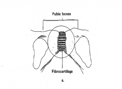
two bony segments are covered by thin lamina of hyaline cartilage and directly joined by fibrocartilage in the form of disks or plate
**Allow limited to moderate amounts of movement |
|
|
Cartilaginous Joints:
|
exist near the midline of the body
|
|
|
Sychondrosis Joints are:
|
1. A type of joint in which the material used for connecting the two components is hyaline growth cartilage. (primary cartilaginous joint)
2. Permits growth while also providing stability & small amount of mobility. |
|
|
Synostoses is:
|
When bone growth is complete, some of these joints ossify and convert to bony unions
|
|
|
Diarthroses:
|
In a synovial joint the ends of bones are free to move in relation to one another because no cartilaginous tissue directly connects adjacent bony surfaces
The bony components are indirectly connected to one another by means of a joint capsule that encloses the joint. |
|
|
Synovial Joints have:
|
A joint capsule that is composed of two layers.
A joint cavity that is enclosed by the joint capsule. Synovial tissue that lines the inner surface of the capsule. Synovial fluid that forms a film over the joint surfaces “Synovial Joint” Hyaline cartilage that covers the surfaces of the enclosed contiguous bones. |
|
|
Stratum Synovium is:
|

Inner layer, highly vascularized but poorly innervated. Insensitive to pain.
Synthesizes hyaluronate of synovial fluid. |
|
|
Subsynovial Tissue:
|

lies outside the intima as loose network of highly vascularized fibrous connective tissue.
Adheres to outer capsule and supports intima. |
|
|
Stratum Fibrosum:
|
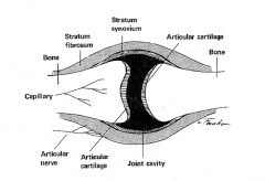
Collagen & elastin account -90% of dry weight
Water-70% of wet weight. |
|
|
Whats the LAYER between Stratum fibrosum and stratum synovium?
|

Subsynovial Tissue
|
|

Know this for the Test!!! :)
|
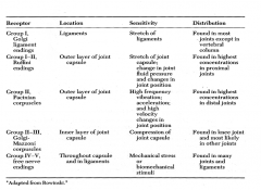
**Do not need to know the Groups for the Receptors!!
|
|
|
Synovial Joint Features consist of:
|
1. A joint capsule that is formed of fibrous tissue.
2. A joint cavity that is enclosed by the joint capsule. 3. A synovial membrane that lines the inner surface of the capsule. 4.Synovial fluid that forms film over the joint surfaces. 5. Hyaline cartilage covering joint surfaces. 6. Ligaments & muscles surrounding. 7. Reduces friction between bony components. 8. Fluid provides nourishment for hyaline cartilage. |
|
|
Synovial Fluid is:
|
Similar to blood plasma, but contains hyaluronic acid and lubricin
Hyaluronate responsible for viscosity of fluid. Hyaluronate reduces friction between synovial folds of capsule and joint surface. Lubricin responsible for cartilage-on cartilage lubrication. |
|
|
In Synovial Fluid, Hyaluronate does what?
|
1. Responsible for viscosity of fluid.
2. Reduces friction between synovial folds of capsule and joint surface. |
|
|
In Synovial Fluid, Lubricin is responsible for:
|
cartilage-on cartilage lubrication
|
|
|
Thixotropic is when:
|
1. Viscosity of fluid varies inversely w/ joint velocity or rate of shear.
2. When bony components moving rapidly viscosity decreased, slowly viscosity increased 3. High temperatures decrease, low temperatures increase viscosity. |
|
|
Boundary Lubrication of Synovial Joints is when:
|
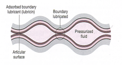
Each surface is coated or impregnated with a thin layer of molecules that keeps the opposing surfaces from touching.
**Forms Gels |
|
|
Fluid Lubrication of Synovial Joints is when:
|
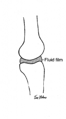
Film of fluid, maintained under pressure, holds two joint surfaces apart
|
|
|
Hydrostatic or Weeping Lubrication is when:
|

Load bearing surfaces are held apart by film of lubricant that is maintained under pressure
Calcified cartilage keeps fluid from being forced into subchondral bone. When load is removed, fluid flows back into articular cartilage. |
|
|
Weeping Lubrication compression causes:
|
Compression causes cartilage to deform & to “weep” fluid, which forms fluid film over the articular surfaces
Calcified cartilage keeps fluid from being forced into subchondral bone. When load is removed, fluid flows back into articular cartilage. |
|
|
Hydrodynamic Lubrication is a:
|
Wedge of fluid is created when non-parallel opposing surfaces slide on one another
**Lifting pressure generated in wedge of fluid & by fluids viscosity keeps joint surfaces apart. |
|
|
Squeeze Film Lubrication is when pressure is:
|
Created in fluid film by movement of articular surfaces that are perpendicular to one another. Good for High Loads short duration!
**As opposing surfaces move closer together, they squeeze fluid film our of area of impending contact. |
|
|
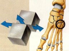
What are the 3 types of Synovial Joints?
|
1. Uniaxial Diarthrodial
2. Biaxial Diarthrodial 3. Triaxial |
|
|
Uniaxial Diarthrodial, of the 3 types of Snyovial Joints:
|
Allows motion in one plane around one axes
Axis located near or in the center of joint (1° of Freedom) Ex. Hinge Joints, Pivot Joints |
|
|
Biaxial Diarthrodial, of the 3 types of Snyovial Joints:
|
Allows motion within two planes around two axes
Ex. Saddle Joints |
|
|
Triaxial, of the 3 types of Snyovial Joints:
|
Allows motion within three planes around three axes
Ex. Plane Joints, Ball-and- Socket Joints |
|
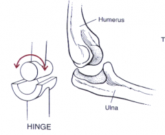
Uniaxial Hinge Joints are:
|
Diarthrodial joints
Permits motion around a single axis. Ex. Similar to door hinge, IP joints of fingers |
|
Pivot (Trochoid) Joints are:
|
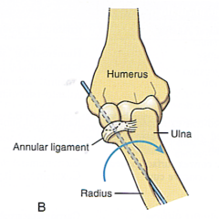
1st Component: Shaped like a ring
2nd Component: Shaped so that it can rotate within the ring Ex. AA joint, radiohumeral joint |
|
|
Biaxial Diarthrodial Joint where:
|
the bony components are free to move in two planes of movement (2° of freedom)
|
|
|
There are two types of Biaxial Joints, what are they?
|
1. Saddle
2. Condyloid |
|
|
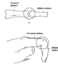
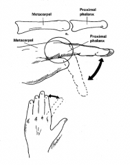
Condyloid Joints are:
|
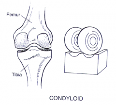
shaped so that the concave surface of one bony component is allowed to slide over the convex surface of another component in two directions
Ex. MCP joint of fingers |
|
This is a type of what joint?
|
Condyloid Joint (Biaxial)
|
|
|
Saddle Joint is
|
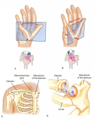
each joint surface is both convex in one plane and concave in another (Fit like a rider on a saddle)
Ex. CMC joint of thumb, sternoclavicular joint |
|
|
Triaxial or Multiaxial Diarthrodial Joints are:
|
Joints in which bony components are allowed to move in three planes around three axes (3° of freedom)
Ex. Plane joints, ball-&–socket joints |
|
Plant Joints:
|
Permit gliding between two or more bones.
Adjacent surfaces may glide or rotate w/ regard to one another in any plane. Ex. Carpal joints |
|
|
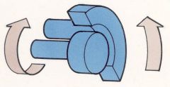
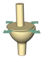
Ball & Socket Joints are:
|
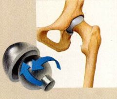
Formed by a ball-like convex surface being fitted into a concave socket
Ex. Hip & shoulder joints. |
|
|
Kinematic Chains can be:
|
Opened or Closed
**In the human body each limb could be thought of as a portion of a rigid chain |
|
|
Open Kinetic Chain is:
|
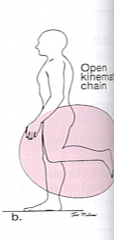
When the ends of limbs or parts of the body are free to move without causing motion at another joint.
Motion does not occur in a predictable pattern. |
|
|
Characteristics of Open Kinetic Chain Examples are:
|
1. Distal end of extremity free in space/not in contact w/ fixed object
2. Movement pattern named by rotary stress in joint. 3. Joint movements occur in isolation. 4. Muscle recruitment & movements are isolated. 5. Joint axis stable during movement. 6. Proximal portion of joint stable – distal segment mobile. 7. Movement pattern often non functional 8. Movement causes shear forces in joint. |
|
|
Closed Kinetic Chain occurs:
|
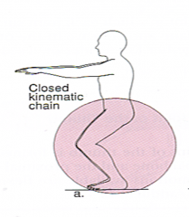
When the ends of limbs meet a fixed resistance such as in weight bearing, like squating
Motion will occur in a predictable pattern. |
|
|
Characteristics of Closed Kinetic Chain Examples are:
|
1. Distal end of segment fixed to something.
2. Movement pattern is characterized by linear stress in joint. 3. Multiple joint movements occur simultaneously/Both segments move together 4. Co-contraction of muscle around joint. 5. Movement is not inhibited. 6. Loading is physiological & more functional. 7. Movement causes compression, enhancing stability |
|
|
According to Steindler, a true closed kinematic chain exist only when?
|
During isometric exercise, since by definition, neither the proximal nor distal joint can move in a closed system
|
|
|
There are 3 Arthrokinematics, What are they?
|
1. Roll
2. Spin 3. Slide |
|
|
Arthrokinematics is:
|
Movement of the joint surfaces
Movements include roll, sliding and spin. |
|
|
The "Roll" in Arthrokinematics refers to:
|
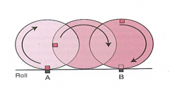
The rolling of one joint surface on another, as in a tire rolling on the road
Ex. In the Knee the femoral condyles roll on the fixed tibia |
|
|
"Sliding" in Arthrokinematics refers to:
|
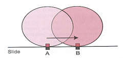
A pure translatory motion, which refers to the gliding of one component over other, like wheels sliding on ice
Ex. In the shoulder the humerus slides inferior during abduction. |
|
|
"Spin" in Arthrokinematics refers to:
|
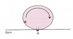
A pure rotary motion
Rotation of a movable component Ex. At the elbow, the head of the radius spins on the capitulum of the humerus during supination or pronation. |
|
|
In Arthrokinematics, what does Ovoid and Sellar mean:
|
Ovoid: One surface convex and one concave.
Sellar: Each surface has both convex and concave. |
|
|
Convex on Concave =
|
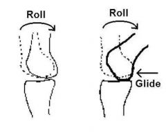
Opposite: So roll has to go in the opposite direction of slide
**Important for Immobilization |
|
|
Concave on Convex =
|
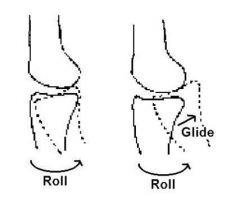
Same, So the Roll and slide go in the same direction
**Important for Immoblization (Slide/Glide meaning the same thing) |
|
|
Osteokinematics refers to:
|
Movement of the bones rather than the movement of the articular surfaces
**Extent of the anatomic range is determined by a number of factors, including the shape of the joint surfaces, the joint capsule, ligaments, muscle bulk, & surrounding musculotendinous & bony structures. |
|
|
Name 3 Osteokinematics:
|
1. Hypermobile
2. Hypomobile 3. Contracture |
|
|
In Osteokinematics, Hypermobile is:
|
When ROM exceeds the normal limit.
May be caused by a failure to limit motion by either the bony or soft tissues and can result in instability or laxity. |
|
|
In Osteokinematics, Hypomobile is when:
|
ROM is less than what would normally be permitted by a normal structure
May be caused by bony or cartilaginous blocks to motion or by the inability of the capsule or ligaments to elongate sufficiently to allow normal ROM. |
|
|
In Osteokinematics, Contracture is:
|
The shortening of soft tissue structures around a joint
**Can be permanent |
|
|
Joint Structure are 4 parts, what are they:
|
1. Ligaments and Tendons
2. Bursae 3. Cartilage 4. Bone |
|
|
Ligaments and Tendons in joint structure are what:
|
Fibroblasts is 20% : tissue volume
80% : extracellular components Collagen fibers: Tendons = parallel arrangement Ligaments =varied arrangement |
|
|
Bursae in joint structure is what:
|
Flat sacs of Synovial Membrane in which the inner sides of the sacs are separated by a fluid film to decrease friction
Located where moving structures are in tight approximation |
|
|
Bursae are located between:
|
Bones and tendon
Bone and skin Muscle and bone Ligament and bone |
|
|
There are three types are Bursae, what are they?
|
Subcutaneous
Subtendinous Submuscular |
|
|
Subcutaneous Bursae is:
|
Generally found between bone and skin.
|
|
|
Subtendinous Bursae lies:
|
Between tendon and bone.
|
|
|
Submuscular Bursae lies:
|
Between muscle and bone
|
|
|
Cartilage in Joint structure is what?
|
Large extracellular matrix & small cellular component
Components different from that of tendons and ligaments **Contains Chondrocytes, Type II collagen & small amounts of Type IX & XI collagens |
|
|
There are 3 types are Cartilage what are they?
|
1. White Fibrocartilage
2. Yellow Fibrocartilage 3. Hyaline Cartilage |
|
|
White Fibrocartilage contains:
|
Type I cartilage
Forms the bonding cement in joints that permit little motion Found in the intervetebral disk. ( Don’t a lot of motion exists) |
|
|
Yellow Fibrocartilage is found:
|
In ears & epiglottis
Has a higher ratio of elastin to collagen fibers **Ligamentum of flavum sometime called this |
|
|
Hyaline Cartilage forms:
|
A thin covering on the ends of many of the bones in adults
|
|
|
Bones Cellular Components consists of:
|
Fibroblasts
Fibrocytes Osteoblasts Osteocytes Osteoclasts Osteoprogenitor cells **Bones have hardest connective tissue in the body |
|
|
Osteoblasts:
|
lay down bone
|
|
|
Osteoclasts:
|
responsible for bone resorption
|
|
|
Muscles Functions have:
|
Active & Passive Insufficiency
|
|
|
Active Insufficiency in muscles function cannot be:
|
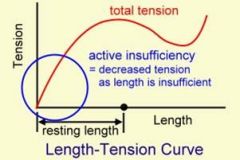
Shorten anymore to a point at , has to be a two joint muscles no more sliding of myofilaments can occur
They also have diminished ability of a bi-articulate muscle to produce or to maintain active tension |
|
|
Passive Insufficieny in muscle functions do not have:
|
enough length to get to full ROM (tightness is occurring & unable to move farther; occurs in two joint muscles)
**Occurs when an inactive, potentially antagonistic muscle is of insufficient length to permit completion of full ROM available at the joints crossed by the passive muscle. |
|
|
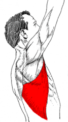
Spurt Muscles have:
|
Proximal attachment that is far from the joint axis & distal attachment that is close to the joint axis
Ex: Biceps, Triceps *These muscles have relatively large rotary component |
|
|
Shunt Muscles have:
|
Proximal attachment that is close to the joint axis & distal attachment far away from the joint axis
Provide a large compression force since they have a large translatory component. Ex: Brachioradialis |