![]()
![]()
![]()
Use LEFT and RIGHT arrow keys to navigate between flashcards;
Use UP and DOWN arrow keys to flip the card;
H to show hint;
A reads text to speech;
69 Cards in this Set
- Front
- Back
|
Developmental Anatomy |
Changes throughout lifespan |
|
|
Gross Amatomy |
Visible to naked eye |
|
|
Microscopic Amatomy |
Too small to be seen |
|
|
Principal of Complementarity |
What a structure can do depends if it’s specific form.
Muscle from anterior elbow will flex elbow |
|
|
Levels of Structural Organization |
1. Chemical level 2. Cellular level 3. Tissue Level 4. Organ Level 5. Organ System Level 6. Organismal Level |
|
|
Chemical Level |
Atoms combined to make molecules |
|
|
Cellular Level |
-Cells are made up of molecules - Smallest thing to carry out life processes - Made up if molecules |
|
|
Tissue Level |
- Made up of similar types of cells that are: - Similar in structure and perform common related function |
|
|
Organ Level |
Made up of different types of tissue. |
|
|
What makes up the organ system? |
- Smooth muscle tissue - Connective Tissue - Epithelial tissue - Blood vessel |
|
|
Organ system level |
Made up of different types of tissues. |
|
|
Organismal Level |
The human organism is made up of many organ systems. |
|
|
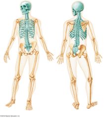
Axial and Appendicular Skeleton - Axial: Axis is around which the body moves (center) -Appendicular: Appendages of the body |
|
|
Anatomical position |
- Standard position of reference for describing anatomical positions - Feet, palms, eyes forward - Erect position - Arms to side - Feet shoulder width apart |
|
|
Anatomical Planes |
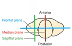
- Transverse plane |
|
|
Superior/Cranial and Inferior Caudal |
- Describes structures relative to each other on vertical axis of body. - S/C: upward surfaces (towards the head) -I/C: downward surfaces away from head |
|
|
Anterior/Ventral and Posterior/Dorsal |
-Describes relativity based on "front and back" A/V: front surface P/D: back surface Ex. Sternum anterior to heart |
|
|
Medial/Lateral |
- Relativity of structures in terms of midline of body - M: Towards midline L: Away from midline |
|
|
Proximal/Distal |
1. Used to describe location of extremes (upper and lower limb) P: Closer to midline D: Farther from midline Ex. Elbow distal to shoulder 2. Start of the extremity vs end of extremity Ex. Digestive system |
|
|
Superficial/Deep |
- Describe depth of structure Superficial- Close to surface of body Deep - Far from surface more internal |
|
|
Flexion |
- Motion that decreases joint angle |
|
|
Dorsiflexion and Plantarflexion |
D: Toes toward legs P: Toes away from leg |
|
|
Adduction and Abduction |
- Move towards/away the midline of body. |
|
|
Lateral Flexion |
Moving away from midline of body |
|
|
Wrist Deviation |
Lateral flexion that moves hand towards radius (lateral) or ulna (medial) Radial Deviation and Ulna Deviation |
|
|
Rotation (axial twist) |
Turning about the axis of the body |
|
|
Internal/Medial and External/Lateral |
Turning limb around limb access |
|
|
Anatomical Movements Specific to Forearms |
Supination and Pronation S: Palm up P: Palm down |
|
|
Anatomical Movements Specific to Foot |
Inversion and Eversion I: Soul of feet inwards E: Soul of feet outwards |
|
|
4 Types of Tissue in Body |
-Epithelial Tissue - Protect - Connective Tissue - Support - Muscle Tissue - Moves -Nervous Tissue -Control |
|
|
Types of Epithelial Tissue |
- Covering/Lining Epithelium- forms outer layer of skim and internal structures (skin, organ wall) - Glandular Epithelium - form glands of body |
|
|
Functions of Epithelium |
-Physical Protection -Absorption -Sensation -Secretion |
|
|
Physical Protection with Epithelium |
- Stops harmful substances from entering body or organs Ex. Placed between lumen of urinary bladder and connective tissue |
|
|
Absorption with Epithelial Tissue |
-Can form selective barrier where certain things can or can not pass Ex. Fluids in Kidneys |
|
|
Sensation of Epithelial Tissue |
Hair like extensions that can detect changes in temperature, chemical composition. |
|
|
Secretion of Epithelial Tissue |
Occurs in glands; group of one or more epithelial cells that make or secrete particular substance |
|
|
Types of Glands |
- Endocrine: Secretes products directly into bloodstream (thyroid) -Exocrine: Secrete products into ducts (sweat, earwax, saliva) which are in specific areas (localized impact) |
|
|
Structure of Exocrine Glands |
- Ring shaped with a lumen of duct that take product to specific location - Can have multiple layers |
|
|
Why is basement membrane thicker in exocrine cells than epithelial? |
- Exocrine does not want product to leave gland to nearby tissue but go through ducts. |
|
|
Basement Membrane |
Basal Lamina: Adhesive sheet beneath basal surface; joins epithelial tissue to connective tissue Reticular lamina -Both act as filter |
|
|
Apical Surface (Free Surface) |
Free surface exposed to exterior environment or cavity of organ -Can have microvilli or cilia |
|
|
Characteristics of Structure of Epithelial Tissue |
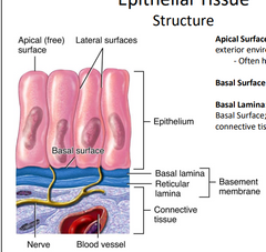
- Fit closely together to form continuous sheets - Sit upon and are supported by connective tissue - Avascular: blood vessels do not go directly to them -Highly regenerative |
|
|
Basal Surface |
Anchored lower surface |
|
|
How do Epithelial Tissues get nutrients? |
Nearby blood vessels when they are anchored to connective tissue |
|
|
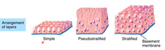
Arrangement of Layers of Epithelial Tissue |
|
|
|
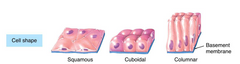
Cell Shape of Epithelial Tissue |
|
|
|
Single layer of ET means it is easier for _________ |
Substances to pass through |
|
|
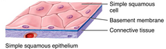
Simple Squamous Epithelium |
- Thin and specialized for moving molecules across - Placed wherever absorption or secretion occurs |
|
|
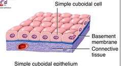
Simple Cuboidal Epithelium |
- Absorption of fluids |
|
|
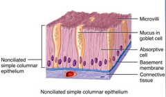
Noncilitilated Simple Columnar Epithelium |
- Microvilli located on surface - Contains mucus for slip surface -Absorption and secretion |
|
|
Ciliated Simple Columnar Epithelium |
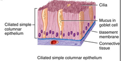
- Covered with cilia for movement - Absorption and secretion |
|
|
Non-Keratinized Stratified squamous epithelium |
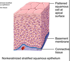
- Where protection is needed - Top layers are dead - Tissue grows from Basal Layer to top |
|
|
Stratified Cuboidal Epithelium |
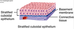
- Protection but not as much -Esophagus, lining of sweat glands and salivary glands |
|
|
Stratified Columnar Epithelium |
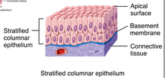
- Protection but not as well as squamous -Pharynx |
|
|
Pseudostratified Ciliated Columnar Epithelium |
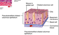
- Appears to be stratified but all tissue is attached to basement membrane -Respiratory movement and found in trachea |
|
|
Relaxed Transitional Epithelium |
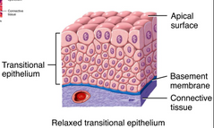
- Stratified layer of cells that change shape based on location and loads applied -Allows stretchy structures to expand from within - Ex. Located in bladder; change shape when full/empty |
|
|
Role of Connective Tissue |
- Supports body structures - Develops from mesenchyme |
|
|
Types of Connective Tissue |
- Connective Tissue Proper (loose and dense) - Cartilage -Bone -Blood |
|
|
Function of Connective Tissue |
- Support (Ex. Bone: framework and supports body) - Binding (Ex. Tendons binding muscles to bone) -Storage (Ex. Storage of fats) - Transport (Blood; carries waste, nutrients around the body) - Protection (Skeleton protects organs) - Immune Protection (Connective tissue stores white blood cells |
|
|
Three Structural Elements of CT |
- Cells (Adipocytes, Macrophages, Fibroblasts, Mast cells Plasma Cells, Macrophages, Plasma Cells, Eosinophils, Neutrophils) - Ground Substance (Non-living tissues; thick, fluid, gelatinous) - Fibers (Elastic Fibers, Collagen Fibers, Reticular Fibers |
|
|
Connective Tissue Paper- Loose |
- Areolar: Cells found in areolar (least specialized - Adipose - primary cell type is lipid - Reticular - Found in "hollow" organs as supported framework (contains reticular fibers act as sponges ) |
|
|
Areolar Connective Tissue |
Fibroblasts: Produce protein fibers Collagen Fibers: Strong, flexible and resist stretch Elastic Fibers: Reticular fiber Reticular Fibers: Tough but flexible branching Ground substance |
|
|
Connective Tissue Proper - Dense |
- Regular: Collagen fibers, running parallel, density packed (provides strength in one direction) - Irregular: Mostly collagen fibers, irregular/ mesh pattern, provides strength in multiple directions) - Elastic: Collagen and elastic fibers allows for stretch and recoil |
|
|
Types of Membranes |
- Cutaneous Membrane (skin) - epidermis (epithelial) and dermis (connective) - Mucous: Epithelial + loose connective tissue + goblet cells (mostly columnar in shape) - Serous: Simple epithelia + connective tissue; provides thin, watery fluid to allow easy motion over surfaces (heart, lungs don't stick) - Synovial: Synovial cells + loose connective tissue (not technically epithelial membrane), lines joint cavities and secretes synovial fluid |
|
|
Where do cartilage cells (chondrocytes) come from? |
- Developed from chondroblasts: collagen + elastic fibers + ground substance |
|
|
What are the three different types of Cartilage? |
Hyaline Cartilage- most common, high fine collagen fibers, precursor to skeleton in fetus Elastic cartilage- collagen + elastic fibers Fibrocartilage- Toughest type of cartilage, chondrocytes and collagen fibers arranged in parallel rows, resists compression, "shock" absorption |
|
|
What fibers are in bone? |
- Collagen |
|
|
Importance of Blood |
- Transport all over body |
|
|
mmm |
hhhh |