![]()
![]()
![]()
Use LEFT and RIGHT arrow keys to navigate between flashcards;
Use UP and DOWN arrow keys to flip the card;
H to show hint;
A reads text to speech;
83 Cards in this Set
- Front
- Back
|
What are BAP and CAP? |
Supportive media (allow growth of all except the most fastidious organisms) |
|
|
How does CAP differ from BAP? |
The RBCs of CAP have been lysed |
|
|
Two examples of bacterial genera that grow on CAP but not BAP |
Neisseria
Haemophilus |
|
|
How is BAP useful for bacterial identification? |
Certain bacteria exhibit hemolytic properties on BAP |
|
|
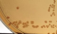
What are the three hemolysis patterns, and how do they appear on BAP? |
α - partial hemolysis (greenish zone around isolated colonies)
β - total hemolysis (clear zone around isolated colonies)
γ - no hemolysis |
|
|
Selective medium (Definition) |
A medium that contains inhibitory reagents that restrict growth of some or most organisms and allow selective growth of desired organisms |
|
|
Differential medium (Definition) |
A medium that contains a factor or factors that reveal specific metabolic or cultural characteristics of specific bacterial isolates |
|
|
Characteristics of Colistin-Nalidixic Acid (CNA) agar |
Selective for Gram-positive bacteria
Differential for hemolytic properties |
|
|
Characteristics of MacConkey (Mac) agar |
Selective for Gram-negative bacteria
Differential for lactose fermentation |
|
|
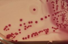
Describe the appearance of lactose-fermenting bacteria on a MacConkey agar |
Light pink to reddish colonies surrounded by a zone of precipitated bile |
|
|
What test is used to further differentiate Gram-positive cocci? |
Catalase test |
|
|
Which Gram-positive cocci are catalase-positive? |
Staphylococci |
|
|
Which Gram-positive cocci are catalase-negative? |
Streptococci and Enterococci |
|
|
Describe the appearance of a positive catalase test |
Bubbling (effervescence) is observable |
|
|
Describe the appearance of a negative catalase test |
No bubbles |
|
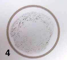
Is this catalase test positive or negative? |
Negative |
|
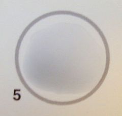
Is this catalase test positive or negative? |
Positive |
|
|
Which test is used for further identification of the staphylococci? |
Coagulase test (latex agglutination test) |
|
|
Which of the staphylococci is coagulase-positive? |
Staphylococcus aureus |
|
|
Which of the staphylococci are coagulase-negative? |
Staphylococcus epidermidis
Staphylococcus saprophyticus |
|
|
Describe the appearance of a positive coagulase latex agglutination test |
Aggregation of the black latex suspension with loss of the black background |
|
|
Describe the appearance of a negative coagulase latex agglutination test |
Little or no agglutination without loss of the black background |
|
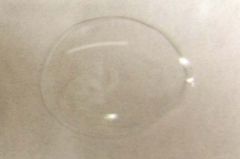
Is this coagulase latex agglutination test positive or negative? |
Positive |
|
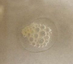
Is this coagulase latex agglutination test positive or negative? |
Negative |
|
|
How can S. aureus be distinguished from the coagulase-negative staphylococci without a coagulase test? |
S. aureus is β-hemolytic
S. epidermidis and S. saprophyticus are γ-hemolytic |
|
|
How are the coagulase-negative staphylococci further differentiated? |
Novobiocin susceptibility test |
|
|
Which coagulase-negative staphylococcus is novobiocin susceptible? |
S. epidermidis |
|
|
Which coagulase-negative staphylococcus is novobiocin resistant? |
S. saprophyticus |
|
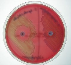
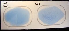
Based on this novobiocin susceptibility test, identify the bacteria on the left and right halves of the agar |
Left - S. epidermidis
Right - S. saprophyticus |
|
|
Which test is used for further identification of the streptocococci? |
Hemolysis on blood agar |
|
|
Which streptococci are β-hemolytic? |
Group A streptococci
Group B streptococci |
|
|
Which streptococci are α-hemolytic? |
Group D streptococci
Viridans streptococci
Streptococcus pneumoniae |
|
|
Which streptococci are γ-hemolytic? |
Group B streptococci
Group D streptococci
Enterococcus faecalis (not actually a streptococcus) |
|
What type of streptococci are on this agar? |
γ-hemolytic streptococci |
|
What type of streptococci are on this agar? |
α-hemolytic streptococci |
|
What type of streptococci are on this agar? |
β-hemolytic streptococci |
|
|
How are the β-hemolytic streptococci further differentiated? |
Latex agglutination test for Group A or Group B |
|
|
Describe the appearance of a positive reaction in the latex agglutination test for Group A or Group B |
Agglutination of the blue latex with loss of the blue background |
|
|
Describe the appearance of a negative reaction in the latex agglutination test for Group A or Group B |
Uniform blue, milky appearance |
|
The Group A latex reagent was applied to the circle on the left, and the Group B latex reagent was applied to the circle on the right. Identify the bacteria being tested. |
Group B streptococci |
|
|
How are the γ-hemolytic streptococci further differentiated? |
Group B latex agglutination test followed by bile-esculin slant and the BHI-6.5% NaCl test |
|
|
Which streptococci return a positive bile-esculin test? |
Group D streptococci
Enterococcus faecalis |
|
|
Describe the appearance of a positive bile-esculin test |
Blackening of the bile-esculin agar |
|
|
Describe the appearance of a negative bile-esculin test |
Yellow/amber agar |
|
Which of these bile-esculin slants is positive? |
Right |
|
|
How are Group D streptococci and Enterococcus faecalis differentiated? |
BHI-6.5% NaCl test |
|
|
Which bacterium is capable of growing in BHI-6.5% NaCl? |
Enterococcus faecalis |
|
|
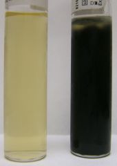
Describe the appearance of a positive BHI-6.5% NaCl test |
The culture is turbid (cloudy) |
|
|
Describe the appearance of a negative BHI-6.5% NaCl test |
The culture is clear |
|
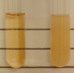
Which of these BHI-6.5% NaCl tests is positive? |
Right |
|
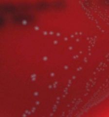
Are these bacteria lactose fermenters? |
Yes |
|
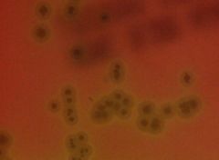
Are these bacteria lactose fermenters? |
No |
|
|
What does "IMViC" stand for? |
Indole production
Methyl red
Voges-Proskauer
Citrate utilization |
|
|
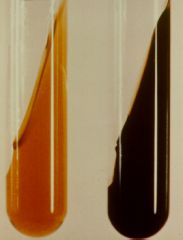
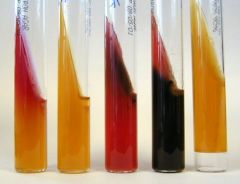
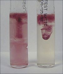
Describe the appearance of a positive indole production test |
Culture medium is pink to red |
|
|
Describe the appearance of a negative indole production test |
Culture medium is lime-green |
|
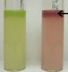
Which of these two indole production tests is positive? |
Right |
|
|
Describe the appearance of a positive methyl red test |
Culture is red |
|
|
Describe the appearance of a negative methyl red test |
Culture is yellow |
|
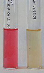
Which of these methyl red tests is positive? |
Left |
|
|
Describe the appearance of a positive Voges-Proskauer test |
Top layer of culture is red, rest is yellow |
|
|
Describe the appearance of a negative Voges-Proskauer test |
Entire culture is yellow |
|
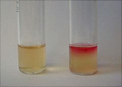
Which of the following Voges-Proskauer tests is positive? |
Right |
|
|
Describe the appearance of a positive citrate utilization test |
Slanted region is blue, the rest is green |
|
|
Describe the appearance of a negative citrate utilization test |
Green |
|
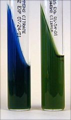
Which of these citrate utilization tests is positive? |
Left |
|
|
What does "TSI" stand for? |
Triple sugar iron test |
|
|
What are the potential results of a TSI test (describe the appearance of each)? |
K/A - red slant, yellow butt
A/A - yellow throughout
K/K - red throughout |
|
|
Describe the appearance of H₂S production in a TSI test |
Blackening of the butt and/or slant |
|
|
Describe the appearance of gas (CO₂) in a TSI test |
Gas bubbles in butt, medium sometimes split |
|
Identify each TSI reaction from left to right |
K/A → A/A → K/K → K/A with H₂S → A/A with gas (CO₂) |
|
|
What does the SIM Deeps test determine? |
Whether an organism can produce H₂S |
|
|
Describe the appearance of a positive SIM Deeps test |
Culture medium is black |
|
|
Describe the appearance of a negative SIM Deeps test |
Culture medium is yellowish |
|
Which of these two SIM Deeps tests is positive for production of H₂S? |
Right |
|
|
Describe the appearance of a positive urease test |
Color change to magenta |
|
|
Describe the appearance of a negative urease test? |
No color change (remains light orange) |
|
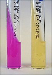
Which of these urease tests is positive? |
Left |
|
|
Describe the appearance of a positive motility test |
The edges of the inoculation site appear fuzzy |
|
|
Describe the appearance of a negative motility test |
The edges of the inoculation site appear sharp |
|
Which of these motility tests is positive? |
Left |
|
|
Describe the appearance of a positive oxidase test |
Dark purple smear |
|
|
Describe the appearance of a negative oxidase test |
Colorless smear |
|
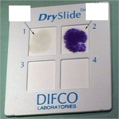
Which of the slides is oxidase positive? |
Top right |