Reading...
![]()
Play button
![]()
Play button
![]()
Use LEFT and RIGHT arrow keys to navigate between flashcards;
Use UP and DOWN arrow keys to flip the card;
H to show hint;
A reads text to speech;
37 Cards in this Set
- Front
- Back
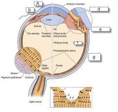
Identify the labels
|
Look
|
|
|
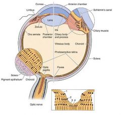
3 tunics of the eye and their contents:
|
Fibrous tunic
--cornea (transparent) --sclera (white)-Dense regular CT limbus is between Vascular tunic --choroid --cilliary body --iris Neural tunic --retina |
|
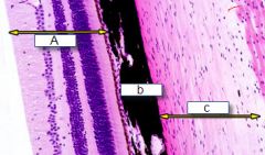
Identify the labels
|

A-retina
b-choroid c-sclera |
|
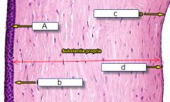
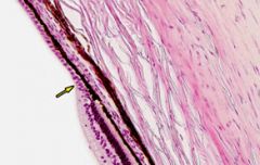
What is this? Identify the labels
|

Cornea of the eye
MN: EBSDEin "ebstein" A-epithelium-stratified sqamous B-bowman's membrane C-endothelial cells D-descemet's membrane |
|
|
How does the cornea get oxygen?
|
It is avascular, so it must receive nutrients from diffusion through the aqueous humor
|
|
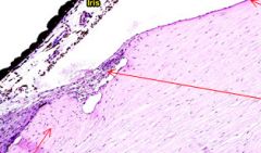
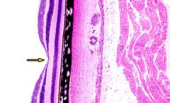
Where is this? What is that hole?
|
Corneosclera junction : limbus
SChlemm's canal-drains aqueous humor |
|
|
What does the choroid of the eye contain?
|
vascular layer that includes capillaries and connective tissue, the innermost layer that separates it from the retina is the Brusch's membrane. It also contains melanocytes,
|
|
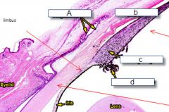
What is this?
identify the labels |
Limbus area again
A-conjunctiva b-sclera (covered in bulbar fascia) C-cilliary body d-cilliary processes |
|
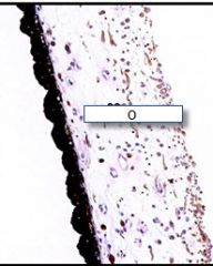
What is this?
|
The iris, it contains melanocytes and muscle tissue
|
|
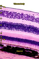
Name each layer 1-10
|

1: pigment epithelium
2: photoreceptors 3: outer limiting membrane 4: outer nuclear layer 5: outer plexiform layer 6: inner nuclear layer 7: inner plexiform layer 8: ganglion 9 optic nerve 10: inner limiting membrane |
|

What is this? What is the arrow pointing at?
|
Optic disk, central retinal artery
|
|
What is this?
|
ora serrata is the serrated junction between the retina and the ciliary body. This junction marks the transition from the simple non-photosensitive area of the retina to the complex, multi-layered photosensitive region. I
|
|
What is this?
|
Macula lutea/fovea centralis- only cones, no rods, best visual acuity in the eye
|
|
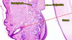
What is this?
|
Eye lid- tarsal glands secrete an oily substance, the arrow is pointing to an eye lash
|
|
|
Which two layers detach in retinal detachment?
|
The pigment epithelium (1) from the photoreceptor layer (2)
|
|
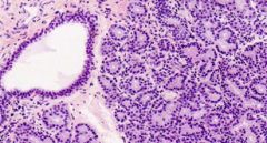
What is this?
|
Lacrimal gland,
similar to parotid gland, has tubular alveolar serous cells that secrete the tears, located in the upper lateral region of the orbit |
|
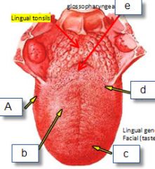
Identify the labels of what kind of papillae is found at each point.
|

A-foliate
b-fungiform c-filiform d-vallate e-foramen cecum and circus terminales |
|

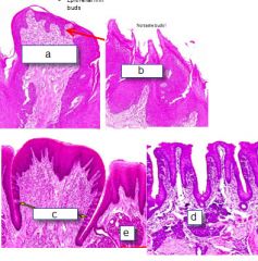
Name each structure:
|

A-fungiform
B-filiform C-Vallate D-foliate E-von ebner gland |
|
Answer each with the letter that matches the characteristic:
1-has no taste buds 2-is present on the entire surface of the tongue 3-covered in strat squam epithelium 4-non keratinized strat squam epith 5-located on the posterior and lateral border of the tongue 6has von ebner glands opening on it |

1,2,3 =B
4=A 5=D 6=C |
|
|
What does a von Ebner gland secrete?
|
serous solution that cleans out the groove so new things can be tasted
|
|
|
What is the structure of a taste bud?
|
oval in shape, with spindle cells inside of it, that have microvilli that project out through a taste pore and into the surrounding environments
|
|
|
Where are taste receptors located?
|
on the microvili of the taste bud cells
|
|

What is this? Identify the labeled structures and their function:
|
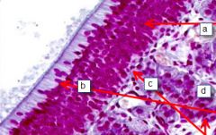
A-olfactory cells-bipolar neurons, allow the sense of smell
b=sustentacular cells-tall columnar cells with a microvilli border, nucelli are found superficial to the nuclei of the olfactory cells c-basal cells-lie against the basal lamina, replace the basal and sustentacular cells D- Bowman's glands-secretes odor binding protein |
|
|
Which concha has olfactory epithelium?
|
Only the superior concha.
|
|

Identify the labels:
|
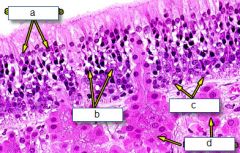
A-sustentacular cells
b-olfactory cells c-basal cells d-bowman's glands |
|
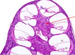
What is this?
What is the red arrow pointing at? |
Cochlea; spiral ganglion
|
|

What is this?
Identify the labels? |
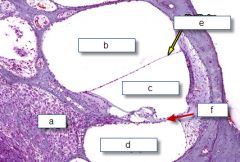
Cochlea
A-spiral ganglion b-scala vestibuli c-scala media d-scala tympani e-vestibular membrane f-basilar membrane |
|
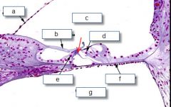
What is this?
Identify the labels? |

A-vestibular membrane
b-tectorial membrane c-scala media d-outer hair cell e-inner hair cell f-basilar membrane g-scala tympani |
|

1. Which label points to the place that connects to the oval window to the helicotrema?
2. Which label points to the one space that is full of endolymph? 3. Which extends from the round window to the helicotrema? |

1. B
2. C 3. D |
|
|
What nerve runs down the center of the cochlea?
|
Cochlear nerve.
|
|
|
What type of cell is found between the inner and outer haif cells and has between it the inner tunnel?
|
Pillar cell
|
|
|
What is a flap of of glycosamino glycans that has the stereocilia of the hair cells embedded in it.
|
Tectorial membrane
|
|
|
A columnar cell that is attached to the basilar membrane and provides support to the hair cell?
|
Phalangeal cell
|
|
|
What differentiates frequencies in the cochlea?
|
How far they travel on the tectorial membrane before passing through the scala media. Lower frequencies travel further.
|
|
|
What are the three contents of the bony labyrinth?
|
Semicircular canals
vestibule cochlear |
|
|
What are the three contents of the membranous labyrinth?
|
Semicircular ducts
Utricle saccule Cochlear duct |
|
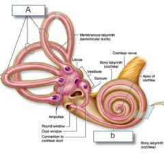
What is A?
What is B? |

A-Bony labyrinth
B-Membranous labyrinth |