![]()
![]()
![]()
Use LEFT and RIGHT arrow keys to navigate between flashcards;
Use UP and DOWN arrow keys to flip the card;
H to show hint;
A reads text to speech;
27 Cards in this Set
- Front
- Back
|
Leukemia - definition |
-Cancer (Malignancy) of blood and blood forming tissues -Abnormal function: cells don’t work |
|
|
Leukemia - outcomes |
-Immunosuppression: severe, life threatening infections -Imbalanced hemostasis: hemorrhage -Decreased oxygen carrying capacity: anemia, tissue death, organ failure |
|
|
Time of Onset (Classifications) |
-Acute: rapid, progressively worsening clinical course -Chronic: indolent clinical course |
|
|
Cell maturation (Classifications) |
-Acute: proliferation of immature cells: “arrested maturation” -Chronic: proliferation of mature looking cells: “normal maturation” |
|
|
Leukemia - symptoms |
-Anemia
-Thrombocytopenia: bleeding -Neutropenia: infections -Bone pain: expansion of marrow -Weightloss -Hepatosplenomegaly -Lymphadenopathy |
|
|
Acute leukemia - lab results |
-Decr to mod incr WBC
-N to decr PLTs -Abnormal morphology: Large, hypogranular, micromegakaryocytes |
|
|
Chronic leukemia - lab results |
-Incr to markedly incr WBC -N to incr PLTs -Anemia |
|
|
Peroxidase - Myeloperoxidase (MPO) - principle |
-Peroxidase in granules in myelocytic and monocytic cell lines
-Most strongly in myelocytic cells -Peroxidase + H2O2 oxidizes a dye substrate creating a black/blue/brown ppt -Differentiates AML vs ALL |
|
|
Myeloperoxidase - interpretation |
-Evaluate blasts only -Stains Primary Granules -neutrophils-eosinophils -monocytes -stain = Acute Myeloid Leukemias -no stain = Acute Lymphoid Leukemias |
|
|
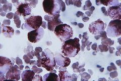
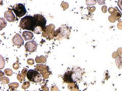
MPO stain smear |
|
|
MPO stain smear |
|
|
Sudan Black B (SBB) - principle |
-Stains lipids such as sterols, neutral fats & phospholipids -found in granules of neutrophils & monocytes -Differentiates AML vs ALL |
|
|
Sudan Black B (SBB) - interpretation |
-Consider blasts only -AML will see incr staining of myelocytic and monocytic blasts |
|
|
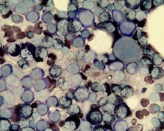
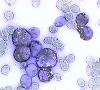
Sudan Black B smear |
|
|
Sudan Black B smear |
|
|
Esterases - principle |
-hydrolyze an ester substrate -A napthol compound is released and combines with a diazonium salt that ppts |
|
|
Specific Esterase - general |
-napthol AS-D chloroacetate
-granulocyte esterase -myelocytic blasts will stain ---primary granules ---auer rods |
|
|
Nonspecific Esterase (NSE) - general |
-Alpha-napthyl acetate or alpha-napthyl butyrate
-Monocytic cells are strongly pos, & inhibited w/ NaF
|
|
|
Combined Esterase - general |
-Combines specific and nonspecific in one dye (Not commonly used) |
|
|
Periodic Acid Schiff (PAS) - principle |
-Acid oxidizes glycogen, mucoproteins & other high molecular wt. carbs to aldehydes -Aldehydes react w/ Schiff rgt staining bright pink -Positive rxn in malignant lymphocytic & erythrocytic cells (chunky or blocky) -Negative in benign cells -Megakaryocytes will also stain, but diffuse |
|
|
Leukocyte Alkaline Phosphatase (LAP) - principle |
-In secondary granules of neutrophils -Substrate: napthol AS-BI phosphate is hydrolyzed by LAP -Hydrolyzed substrate combines w/ a dye & precipitates (color depends on substrate used) |
|
|
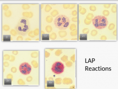
LAP - interpretation |
-Score 100 PMNs and Bands on a scale of 0 - 4+ based on the amount of ppt in the cell -Used to Investigate a shift to the left -Malignant granulocytes (CML): Decr LAP vs. leukomoid rxn (incr LAP) -Other causes of decreased LAP: PNH, Sideroblastic anemia, Myeloproliferative disorders -CML will have REDUCED score |
|
|
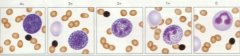
LAP scale |
|
|
LAP reactions |
|
|
Tartrate Resistant Acid Phosphatase (TRAP) - principle |
-present in almost all nonerythroid cells -hydrolyzes substrate napthol AS-BI phosphoric acid -Hydrolyzed substrate couples w/ a dye to form a red ppt -All normal isoenzymes of the enzyme are inactivated by tartaric acid and will not stain. -In Hairy Cell Leukemia isoenzyme #5 is resistant to tartaric acid and will stain |
|
|
Terminal deoxyribonucleotidal Transferase (TdT) - principle |
-A specific cell marker (enzyme) that catalyzes the polymerization of deoxynucleotides found only in lymphocytic precursors. Absent in lymphocytes -Not a cytochemical stain: is an immunofluorescent stain (flow cytometry) or immunoperoxidase technique |
|
|
TdT - interpretation |
-Positive in acute lymphocytic leukemias ( L1 & L2 ) -Must interpret carefully: TdT has been observed in AML |