![]()
![]()
![]()
Use LEFT and RIGHT arrow keys to navigate between flashcards;
Use UP and DOWN arrow keys to flip the card;
H to show hint;
A reads text to speech;
105 Cards in this Set
- Front
- Back
|
how many bones are in the vertebral column? |
33 bones |
|
|
What are the normal physiological curves of the spine? |
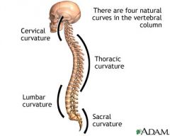
sacral convex posteriorly lumbar concave posteriorly thoracic convex posteriorly cervical concave posteriorly |
|
|
what is kyphosis? |
excessive posterior curvature of thoracic spine |
|
|
what is scoliosis and most common area for it to occur? |
lateral curvature of the spine -most often in thoracic spine |
|
|
What is lordosis? |
increased anterior curvature most often in lumbar spine |
|
|
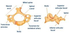
What parts of the vertebra make up the vertebral arch? |
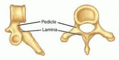
pedicles and laminae |
|
|
where are the lamina in comparison to the pedicles? |
lamina are more posterior than pedicles |
|
|
what do the laminae form? |
laminae on each vertebra meet to form the spinous process
(mnemonic: Lamina Like each other so they combine to form SP) |
|
|
What do the pedicles join? |
pedicles join the vertebral body to the transverse process |
|
|
what are the other parts of each vertebra (other than pedicles and laminae) |
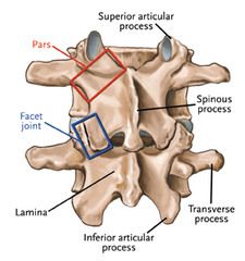
2 transverse processes 4 articular processes |
|
|
what forms the vertebral canal? and what is in the canal? |
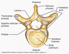
the arches of successive vertebrae form the vertebral canal -contains spinal cord
|
|
|
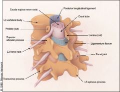
what is the intervertebral foramen |
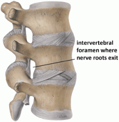
lateral opening formed by the stacking of vertebra through which spinal nerve roots exit from the SC |
|
|
what is a regional characteristic unique to cervical vertebrae? |
(7) cervical -foramina of transverse process contain vertebral arteries -C1 has no spinous process |
|
|
what is a regional characteristic unique to thoracic vertebra? |
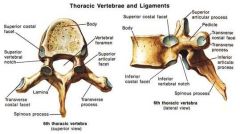
(12) thoracic facets on vertebral bodies for articulation with the ribs |
|
|
what is a regional characteristic unique to lumbar vertbrae? |
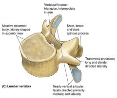
(5) Lumbar massive bodies and sturdy laminae to support weight of the body |
|
|
what is a regional characteristic unique to sacral vertebrae? |
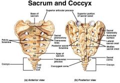
5 sacral -fused vertebrae with 4 foramina on each side |
|
|
what is a regional characteristic unique to coccyx? |
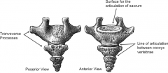
tiny incomplete (no vertebral arch) and 4 fused vertebrae |
|
|
which cervical vertebra are easily palpated? |
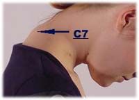
C3-C5 are short and deep to the surface = difficult to palpate C7: vertebra prominens and easy to palpate |
|
|
where can transverse processes be palpated? |
in thoracic and lumbar region |
|
|
what type of lever do the intervertebral joint form? |
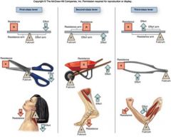
each segment forms a first class lever system facets = fulcrum paraspinals = force component weight of upper body and head = resistance component |
|
|
what is a spinal/vertebral segment? |
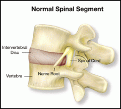
two adjacent vertabrae with the disc in between |
|
|
How does the size of the vertebral bodies change as you move inferiorly down column? |
from T4 inferiorly vertebral bodies become progressively larger to bear incr weight |
|
|
what types of fractures can occur in the vertebral bodies? |
wedge fracture burst fracture tear drop fracture |
|
|
what is a vertebral wedge fracture and MOI? |
compression of anterior aspect of vertebral body from forced flexion of the thoracic or lumbar spine |
|
|
what is the MOI of a burst vertebral body fracture? |
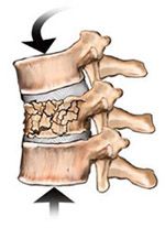
excessive compression of the vertebral body |
|
|
what is a teardrop vertebral body fracture? |
compression of anterior vertebral body causing a triangular fragment to split from body anteriorly |
|
|
what is the structure of the intervertebral disk? |
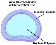
2 portions inner portion= nucleus pulposus -mostly water and is maintained in central position by the annular rings that are firmly attached superiorly and inferiorly to adjacent vertebral bodies outer structure = annulus fibrosis = layers of concentric fibers |
|
|
what is the function of the IV disc? |
it is flexible -shock absorption to dissipate compressive load |
|
|
when is the IV disc at risk of injury? |
during trunk ROTATION, IV pressure increases and annular fibers oriented in direction of the rotatory movement become taut -ROTATION with COMPRESSIVE FORCES (e.g. while under a load from lifting) increases risk of injury |
|
|
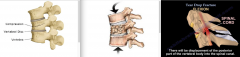
What injury can occur to the IV disc? |
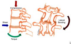
rotation with compressive force can cause herniated or protruding nucleus pulposus -usually occurs posterolaterally and may compress an adjacent spinal nerve root |
|
|
What type of joint are the zygapophyseal joints? |
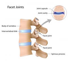
aka facet joints plane synovial joints between inferior and superior articular processes of adjacent vertebra |
|
|
What covers the joint surfaces of the facets and what surrounds each facet joint? |
hyaline cartilage covers the flat surface of each facet -each facet joint is surrounded by a thin, loose articular capsule |
|
|
what is the function of the facet joints? |
-permit GLIDING between the vertebrae -in cervical and lumbar regions, facet joints bear some weight -help to control flexion, ext, and rotation of adjacent vertebrae |
|
|
Where does the majority of the vertebral column movement take place? |
in cervical and lumbar regions |
|
|
when can the facet joints become injured and what can result? |
-quick FLEXION and ROTATION movement can cause impingement of the articular capsule -disease such as OA -with injury to facet joints, the related spinal nerves are often affected -this can cause pain and mm spasm along related dermatomes &/or myotomes. |
|
|
what ligaments lie between the spinous processes of adjacent vertebrae? |
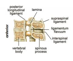
supraspinous ligament and interspinous ligament |
|
|
where does the ligamenta flava lie? |
connects laminae of adjacent vertebral arches |
|
|
where is the posterior longitudinal ligament? |

along posterior aspect of the vertebral bodies and lies in the vertebral canal |
|
|
where is the anterior longitudinal ligament? |
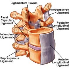
connects anterior aspects of the vertebral bodies and intervertebral discs |
|
|
what does the ALL and PLL prevent? |
ALL (broader than PLL) prevents hyperextension and supports the intervertebral discs -PLL (narrower and weaker than ALL) prevents hyperflexion and posterior protrusion of the nucleus pulposus |
|
|
what do the iliolumbar ligaments connect? |
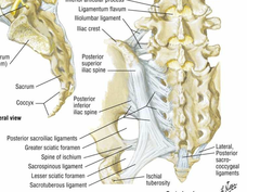
2 bands superior band runs from L4 TP to iliac crest inferior band runs from L5 TP to iliac crest, anterior surface of SI joint and lateral sacral ala |
|
|
what is the function of the iliolumbar ligaments? |
provide structural support to the lower lumbar spine -connect sacrum and ilium to the lumbar spine |
|
|
what is the implication of the iliolumbar ligaments to dysfunction and LBP? |
dysfunction of the SI joint may influence structure and function of the L4 and L5 vertebra |
|
|
what are the 3 groups of back muscles? |
SID: 1. Superificial 2. Intermediate 3. Deep
superficial and intermediate are considered the EXTRINSIC muscles deep = INTRINSIC muscles |
|
|
what is the function of the superficial back muscles? |
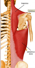
connect the upper limb to the trunk and provide movement of the limbs (e.g. trapezius and latissimus dorsi) |
|
|
what is the function of the intermediate back muscles? |
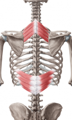
=respiratory muscles (e.g. serratus posterior) |
|
|
what is the overall function of the intrinsic back muscles? |
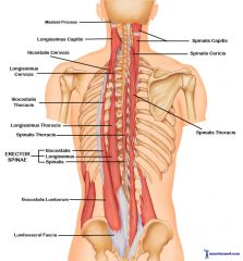
the deep muscles (e.g. erector spinae) maintain posture and move the vertebral column and head |
|
|
What are the 3 layers of the intrinsic back muscles? |
superficial layer, intermediate layer, deep layer |
|
|
what muscles are in the superficial layer of the intrinsic back muscles? |
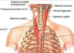
splenius muscle (capitis and cervicis) -acting alone = laterally flex and rotate head and neck to same side -together = extend head and neck |
|
|
what muscles are in the intermediate layer of the intrinsic back muscles? |

erector spinae in 3 vertical columns: Think "I Like Spaghetti" 1. iliocostalis (lateral) 2. longissimus 3. spinalis (medial) Action: bilat: extend head and vertebral column unilaterally: lateral flexion of head and vertebral column -concentrically straighten the flexed spine and eccentrically allow vertebral column to bend forward |
|
|
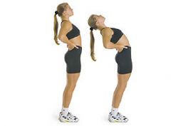
what is a back strain? |
results from extreme movements usually flexion and rotation of the vertebral column -some degree of stretching or microscopic tearing of muscle and/or ligament fibers -usually erector spinae are involved |
|
|
what muscles are in the deep layer of the intrinsic back muscles? |
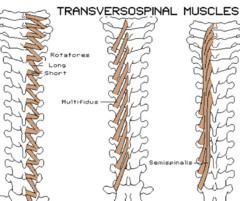
1. SEMISPINALIS 2. MULTIFIDUS 3. ROTATORES collectively these = transversospinal muscles bc fibers attach between tranverse processes and spinous processes of the vertebrae Action: bilaterally: extend and stabilize spine unilat: lateral flexion to same side and rotate trunk to opposite side |
|
|
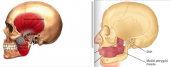
what is the function of the quadratus lumborum? |
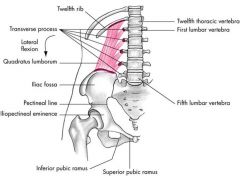
lumbar spine stabilizer unilateral: elevates ilium bilat: assists in forced exhalation and extends back |
|
|
what is the iliopsoas action and when is it active? |
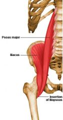
iliacus + psoas muscles that share common insertion and action Action: hip flexion and ER active during walking, sitting, and standing |
|
|
what other action besides hip flexion and ER can the psoas assist in? |
lumbar extension plays significant role in maintaining upright stance |
|
|
What are the actions/functions of the piriformis? |
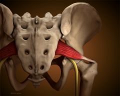
when hip is extended and in nonweightbearing: hip ER -when hip flexed at 90 deg: hip abd -in weightbearing, piriformis resists hip IR |
|
|
What can muscle spasm of the piriformis cause? |
-mm spasm can put pressure on the sciatic nn and cause piriformis syndrome -diskogenic radicular symptoms are common |
|
|
what level is the thyroid cartilage located at? |

C4-C5 |
|
|
what vertebral level are the vocal cords located at? |
at the level with the midpoint of the anterior border of the thyroid cartilage (C4-C5) |
|
|
where does the hyoid bone lie? |
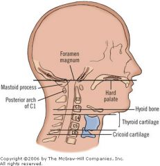
superior to the thyroid cartilage at C3 level |
|
|
what are the superificial neck muscles? |
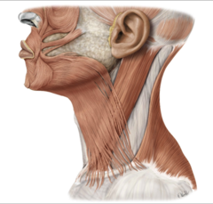
platysma, SCM and Trapezius |
|
|
what is the function/action of the platysma? |
-tenses the skin of the neck -assists in depressing the mandible -draws the mouth inferiorly -one of the facial expression muscles |
|
|
what is the function/action of the SCM? |
unilaterally: tilts the head to the same side and rotates the face in opposite direction bilat: flexes the neck (e.g. when raising the head off a pillow) |
|
|
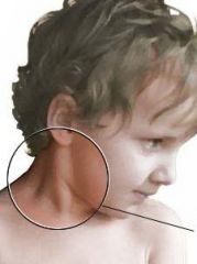
when is one injury of the SCM? |
at birth the muscle can be injured causing congenital torticollis or wryneck |
|
|
what action does the trapezius have? |
unilat: lateral flexion of the neck to the same side bilat: shrugs the shoulders when the neck is stabilized |
|
|
what is an injury of the trapezius? |
forced forward flexion of the cervical spine (e.g. when in a head-on car collision) may cause strain and associated muscle spasms of the mm fibers in the trapezius |
|
|
where does the external carotid artery lie? |
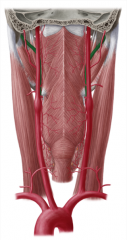
in a groove created by the trachea and the strap muscles |
|
|
what does absence of a cartoid pulse indicate? |
cardiac arrest and is a medical emergency |
|
|
where do the vertebral arteries originate? and what is their pathway? |
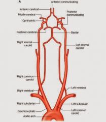
branch off the subclavian artery and ascends vertically through transverse foramina of the cervical vertebrae into the brain at the level of the foramen magnum |
|
|
what can occlusion of the vertebral arteries do? |
cause Vertebral artery syndrome |
|
|
what is Vertebral Artery Syndrome? |
occlusion or stenosis of the vertebral arteries can result in dizziness, tinnitus, fainting, nystagmus, and transient diplopia |
|
|
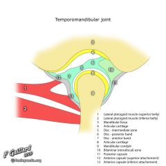
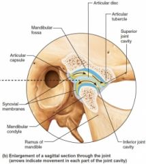
what are the bony landmarks of the TMJ? |
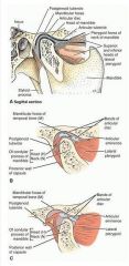
temporal bone forms the roof of the TMJ from posterior to anterior landmarks are mandibular fossa, articular eminence, articular crest, articular tubercle |
|
|
What are the articulating surfaces of the TMJ? |
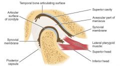
articular eminence-crest-tubercle area is the primary articulating surface of the temporal bone that articulates with the mandibular condyle of the mandible |
|
|
what lies in the mandibular fossa at rest and with teeth in occlusion (closed)? |

posterior band of the disk occupies mandibular fossa |
|
|
Describe the anatomical bony landmarks of the mandible? |
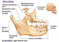
-mandibular condyle is convex (varies in shape and size from person to person) -landmarks on either side = medial and lateral poles -neck = inferior to condylar head -coronoid process = anterior to the neck and anterior to mandibular notch -ramus = inferior to neck; starts the body of the mandible -mandibular body = houses the lower arch of the teeth |
|
|
what are the articulating surfaces of the TMJ covered by? |
dense fibrous connective tissue that is avascular and aneural -has greater potential to remodel and less likely to breakdown over time than hyaline cartilage. |
|
|
how is the TMJ unlike any joint in the body? |
end range is unique because rigid end point of closure created by lower arch of the teeth contacting the upper arch |
|
|
what detemines the final end point of the TMJ articulation? |
the occlusion determines the final end point of the condyle to disc to temporal bone relationship when the posterior teeth are together |
|
|
When can TMJ be injured? |
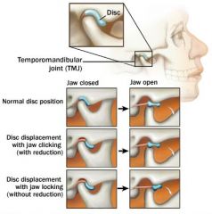
-improper closure of mandible may cause MICROTRAUMA and STRAIN joint structures -disc may DISPLACE ANTERIORLY and MEDIALLY -variation in bony and muscular anatomy may affect loading of the articular surfaces |
|
|
where can TMJ be palpated? |
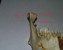
lateral pole of the mandibular condyle can be palpated anterior to external auditory meatus of the ear esp when opening and closing the mouth |
|
|
what is the composition of the articular disc of the TMJ? and the blood supply and neural supply? |
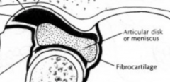
dense fibrous connective tissue avascular aneural |
|
|
Which bone is the TMJ disc more firmly attached to? |

more firmly attached to mandible than to temporal bone |
|
|
how does the disc move with the moving TMJ? |
when head of mandible slides anteriorly on the articular tubercle as the mouth opens, the articular disc slides anterior agains the posterior surface of the tubercle |
|
|
what medial/lateral attachments does the TMJ disc have? |
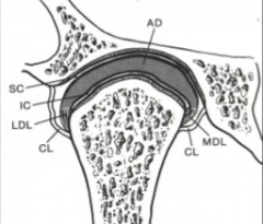
attaches to the medial and lateral poles of the condyle by the medial and lateral collateral ligaments |
|
|
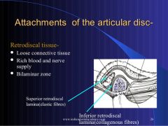
What is the TMJ disc attachments posteriorly? |
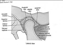
-disc is continuous with posterior attachment which consists of bilaminar zone and retrodiscal pad -bilaminar zone = superior and inferior lamina with the retrodiscal pad in between -superior lamina: fibroelastic tissue with high elastin content -attaches posteriorly to the tympanic plate -passive tension applied when band is stretched to assist in returning disk to normal postion when disk is forward (during closing of the mouth) -inferior lamina: mainly collagneous fibers with little elastic tissue -attaches to neck of condyle -limits forward motion of disk -retrodiscal pad: loose connective tissue rich in arterial and neural supply |
|
|
what are the anterior attachments of the TMJ disc? |

joint capsule and superior lateral pterygoid muscle -joint capsule limits posterior movement of disk -superior 1/3 of superior lateral pterygoid attach to anterior and medial disc -remaining fibers of superior lat. pterygoid and all of fibers of inferior lat. pterygoid attach to medial 1/3 of the neck of the condyle
|
|
|
what is the function of the anterior attachment of the TMJ disc? |
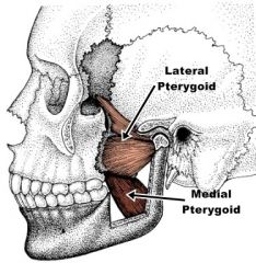
Lat. pterygoid muscle actively assists in controlling posterior movement of disk during mouth closing through eccentric contraction |
|
|
what normally prevents anterior displacement of the disc in relation to the mandibular condyle? |
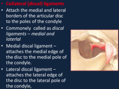
-disc has a biconcave shape creating "self-seating" effect -tight medial and lateral collateral ligaments -together these prevent anterior displacement |
|
|
what does the TMJ joint capsule attach to? |
attaches to the margins of the articular area on the temporal bone and around the neck of the mandible |
|
|
what is located in the TMJ joint capsule? |
-HIGHLY VASCULAR, SYNOVIAL producing membrane that supplies nutrients to nonvascularized tissues within the capsule -articular MECHANORECEPTORS for kinesthetic and perceptional awareness of the mandible are located inside capsule |
|
|
What other ligaments besides the medial and lateral collateral ligaments are found around the TMJ? |
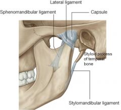
Temporomandibular ligament (TM) stylomandibular ligament sphenomandibular ligament |
|
|
what is the function of the temporomandibular ligament? |
-allows 20-25 mm of mandibular opening before it becomes tight -after 25 mm, the condyle translates anteriorly to allow further opening -i.e. max of 20-25 mm of opening occurs with rotation and no translation of the condyle |
|
|
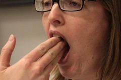
Describe a possible injury of the TMJ? |
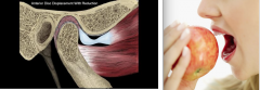
TMJ dislocation anteriorly during yawning or large bites -contraction of lateral pterygoid may cause head of mandible to dislocate or pass anterior to articular tubercle-->mandible remains open and person cannot close it |
|
|
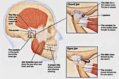
what type of joint is the TMJ and what movements occur at the joint? |
modified hinge synovival joint with 2 arthrokinematic movements 1). anterior gliding 2). hinge-like rotation Osteokinematic movements: 1). elevation/depression 2). protrusion/retrusion 3). lateral deviation |
|
|
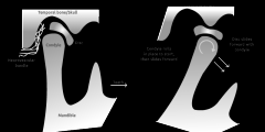
What are the arthrokinematics that occur during mandibular depression? |
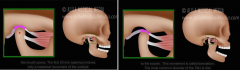
2 phases 1). rotation of condyle around long axis of the condylar heads during 1st 10-15 mm 2). head of mandible and articular disc glide anteriorly until head lies inferior to articular tubercle |
|
|
what motions does functional TMJ/mouth opening combine? and how much is this? |
combines rotation and translation and is ~40 mm (3 knuckles) |
|
|
what is TMJ protrusion? |
anterior translation in the horizontal plane and involves bilateral anterior condylar translation |
|
|
what is TMJ lateral excursion? |
![mandible moves laterally in the horizontal plane
-involves anterior translation on the contralateral side and spin on the ipsilateral side.
(e.g. right lateral deviation/excursion [jaw moves toward the right side] = right condyle rotates ...](https://images.cram.com/images/upload-flashcards/27/57/54/8275754_m.jpg)
mandible moves laterally in the horizontal plane -involves anterior translation on the contralateral side and spin on the ipsilateral side.
(e.g. right lateral deviation/excursion [jaw moves toward the right side] = right condyle rotates and left translates anteriorly) |
|
|
what muscles are involved in elevating the mandible? |
masseter temporalis medial pterygoid |
|
|
what muscles protrude the mandible? |
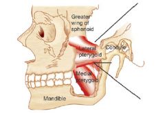
lateral and medial pterygoid |
|
|
what muscles retrude the mandible? |
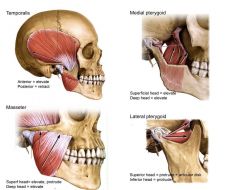
posterior fibers of temporalis |
|
|
what muscles depress the mandible? |
primarily occurs by gravity |
|
|
what muscles act to lateral deviate the TMJ/mandible? |
ipsilateral to side of movement = temporalis and masseter contralateral to side of movement = medial and lateral pterygoid |
|
|
What does the TMJ disc divide? What are the compartments in this divide? |
the joint cavity; superior and inferior compartments |
|
|
Visual of the posterior aspect of the TMJ disc |
|