Reading...
![]()
Play button
![]()
Play button
![]()
Use LEFT and RIGHT arrow keys to navigate between flashcards;
Use UP and DOWN arrow keys to flip the card;
H to show hint;
A reads text to speech;
214 Cards in this Set
- Front
- Back
|
FIV
family / genus |
feline immunodeficiency virus
retroviridae / lentivirus |
|
|
Distemper
family / genus |
canine distemper virus
paramyxoviridae / morbiliivirus |
|
|
What are the reservoirs of rabies in the united states?
how are they infected by the virus? |
skunk, fox, racooon, bat, coyote
rabies virus infects these mammals and that it will eventually kill them – however, they take longer to develop the clinical signs They transmit the virus amongst the species and maintain the virus |
|
|
Pathogenesis of rabies replication
|
virus replicates locally at bite wound specifically in muscle fibers in the vicinity of the neuromuscular junction. enters nerve endings and migrates up nerve by axoplasm flow. reaches the cns and infects neurons. virus then spreads centripetally to salivary glands and to other organs via nerve cells. in the salivary glands, rabies virus replicates in the acinar cells and is excreted in the saliva. there is no viremia. virus may be present in the saliva before clinical signs of rabies are seen
|
|
|
EIA / swamp fever
family / genus |
retroviridae / lentivirus
|
|
|
presenting clinical signs of rabies in the dog
|
most common form is the furious form: loose fear of humans, abnormal aggressive behavior, attack moving objects without provocation, change in voice-laryngeal paralysis. peculiar howling, salivation due to paralysis of the pharyngeal muscles. abnormal sexual behavior
|
|
|
what other viruses can cause CNS disease in dogs?
|
distemper, pseudorabies, infectious canine hepatitis (CAV-1), non resp parainfluenza virus infection, EEE/VEE, powassan, st louis encephalitis, la crosse virus
|
|
|
Equine viral arteritis
family / genus |
equine arteritis virus
arteriviridae / arterivirus |
|
|
what vaccines are available to prevent rabies infection in dogs?
|
killed, attenuated
|
|
|
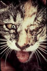
presenting clinical signs of rabies in cats
|
most common form is the furious form: loose fear of humans, abnormal aggressive behavior, attack moving objects without provocation, change in voice-laryngeal paralysis. peculiar howling, salivation due to paralysis of the pharyngeal muscles. abnormal sexual behavior. Cats are often anxious, staring, with a blank look on their eyes, spooky
|
|
|
viruses that cause CNS dz in cats
|
pseudorabies, panleuk, FIP, FeLV, FIV, FSE
|
|
|
BVDV
family / genus |
bovine viral diarrhea virus
flaviviridae / pestivirus |
|
|
presenting clinical signs of rabies in cattle
|
aggression, bellowing, abnormal sexual behavior, tenesmus with paralysis of the tail and anal sphincter muscles, etc
|
|
|
viruses that cause CNS dz in cattle
|
pseudorabies, BSE, BHV-5,, MCF, EEE/VEE, BTV, BVDV, Akabane
|
|
|
blue tongue
family / genus |
bluetongue virus
reoviridae / orbivirus |
|
|
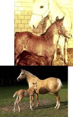
presenting clinical signs of rabies in horses
|
lameness!
|
|
|
differentials for CNS dz in horses
|
EEE/WEE/VEE, West Nile, pseudorabies, EHV-1, powassan, main drain virus, st louis encephalitis, la crosse virus
|
|
|
when are reservoirs most likely to transmit rabies?
|
when they become clinically ill
|
|
|
most telling sign of rabies infection in wild mammals?
|
behavioral changes
|
|
|
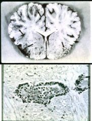
pathognomonic sign for rabies
|
negri bodies (in all but 15-20% of cases)
|
|
|
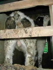
scrapie
|
a non febrile chronic and fatal dz of sheep and goats, characterized clinically by pruritis, which is manifested by rubbing affected parts against objects and biting the flank
|
|
|
Which animals has CWD been diagnosed in?
|
mule deer, elk, moose
|
|
|
clinical signs of CWD
|
teeth grinding, abnormal behavior, excressive drinking, marked weight loss
|
|
|
If thoracocentesis is performed on a cat with a thymic lymphosarcoma, what cells would you expcet to see in this fluid?
|
large lymphocytes/lymphoblasts
|
|
|
what affect has vaccination had on thymic lymphosarcomas?
|
decreased incidence
|
|
|
which cats are more prone to mediastinal or thymic lymphomas?
|
cats less than 3 years of age. 80-90% of all cases are FELV associated
|
|
|
signs of thymic lymphoma
|
pleural effusion, dyspnea, occasional regurgitation (from esophageal pressure)
|
|
|
which cats are more prone to alimentary lymphosarcoma?
|
older cats, often test negative by the SNAP test
|
|
|
if you wanted to detect the presence of felv in the cells on a blood smear, what test would you use?
|
IFA test
|
|
|
IFA procedure for Felv testing
|
1st ab is mouse ab specific for p27, and second ab is a rabbit antimouse ab tagged with fluorescent molecule
|
|
|
what kind of leukemia does felv cause?
|
acute lymphoblastic leukemia
|
|
|
felv vaccination?
|
recombinant poxvirus, given ID
|
|
|
are gingivitis and stomatitis more often associated with FeLV or FIV?
|
FIV
|
|
|
primary cuase of gingivitis and stomatitis in FIV
|
FCV that starts to replicate in the mucosal epithelium and causes the lesions
|
|
|
how will a high percentage of cats with FIP present?
|
anorexic and icteric
|
|
|
what organ is frequently involved with FIP
|
liver
|
|
|
what type of fluid is typically seen with the wet form of FIP?
|
clear, straw colored, clots on standing, slimy to the touch. cellularity is low (predominantly macrophages, some neutrophils). AG ratio <.45; total protein >3.5
|
|
|
What affect does FIP have on the eyes?
|
fibrin deposition/precipitates
retinal hemorrhate, cuffing, color change of iris, keratic precipitates |
|
|
What affect does FIP have on the eyes?
|
fibrin deposition/precipitates
retinal hemorrhate, cuffing, color change of iris, iritis/keratic precipitates anterior uveitis |
|
|
What clinical signs would you expect to observe in cats with the dry form of FIP?
|
pyogranulomas in liver, spleen, lungs, kidneys, lymph nodes, and eyes.
|
|
|
histologic lesions of FIP (dry form)
|
perivascular granulomas or pyogranulomas with systemic vasculitis or thrombovasculitis
|
|
|
Ovine progressive pneumonia/Visna
family / genus |
OPPV
retroviridae / lentivirus |
|
|
composition of FIP pyogranuloma?
|
macrophages predominate and are accompanied by some neutrophils, lymphocytes and plasma cells
|
|
|
what is the typical posture of cats with feline panleukopenia virus?
|
head over water bowl, hunched posture
|
|
|
what type of dz in FPV?
|
acute dz characterized by vomiting and dehydration. NOT DIARRHEA IF ACUTE
|
|
|
blood results of FPV?
|
severe leukopenia, primarily neutropenia
|
|
|
what cells does FPV infect?
|
crypt cells leading to secondary bacterial infection
|
|
|
clinical signs of panleuk
|
seen primarily in <6mo old kittens. peracute dz: overhwelming infection with no clinical signs and death within 24hrs
acute- fever, depression, anorexia, and vomiting. extreme dehydration, hunched posture, tender abdomens. cerebellar hypoplasia |
|
|
clinical signs of CPV-2
|
frequent, bloody diarrhea, anorexia, dehydration
|
|
|
what does the CPV SNAP test detect and how?
|
solid phase ELISA that uses a CPV2 MAb to capture the virus on a membrane. performed on fecal or intestinal material
|
|
|
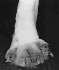
Clinical signs of CDV infection?
|
copious mucopurulent nasal discharge, sunken eyes, dehydration,
cough GI form- vomiting, diarrhea CNS signs- seizures, ataxia, myoclonus, chorea, ascending paralysis hyperkeratosis of pads |
|
|
what wild animals does CDV frequently infect?
|
raccoons
|
|
|
what symptom can CDV cause in young, growing, animals?
|
enamel hypoplasia
|
|
|
when are CNS signs of CDV seen?
|
1-3 weeks after infection or 2-4 weeks after dog seemingly recovers from a mild respiratory dz
|
|
|
CDV is aka
|
hard pad disease
|
|
|
CDV inclusions
|
histopath- both IN and IC inclusions can be seen.
generally only see IC inclusions except in brain and bladder wall where both are seen |
|
|
signs of ICH
|
echymotic hemorrhages in the mucosa, pale mucosa from anemia. enlarged tonsils
|
|
|
what does ICH/CAV1 cause in the fox?
|
CNS disease
|
|
|
most common form of CAV-1 in the dog?
|
hepatitis
|
|
|
ICH/CAV1 is aka
|
blue eye
|
|
|
what is blue eye?
|
a keretatisis caused by Ab binding the cells of the cornea, on the descemet membrane. Transient lesion that does not cause blindness
|
|
|
caprine arthritis encephalitis
family / genus |
retroviridae / lentivirus
|
|
|
Why do we vaccinate with CAV2 not CAV1?
|
CAV1 vaccine can cause blue eye lesions
|
|
|
typical lesion of canine herpesvirus that are seen at necropsy?
|
mottled kidneys with focal disseminated echymotic hemorrhages
|
|
|
how can you prevent CHV1 infections in a litter?
|
hyperimmune serum, increase body temp
|
|
|
EIA family / genus
|
retroviridae, lentivirus
|
|
|
signs of acute EIA infection
|
After an incubation period of 7-21 days, infected horses develop a high fever, severe anemia, anorexia, ataxia, profound weakness, and thrombocytopenia with resultant petechial hemorrhages on the mucosal surface particularly the conjunctiva and under the tongue
Rapid weight loss and dependent edema of the abdomen and legs are also notedduring this acute episode Signs will last for about a week during which time the horse may die or recover from the infection and become a healthy carrier capable of transmitting the virus – a persistent EIA virus carrier |
|
|
signs of recurrent EIA infection
|
Seen in infected horses weeks to months after the initial acute attack
The recurrent attacks of fever is what characterizes this disease The recurrent attacks are characterized by fever, anemia, weakness (wobbly, tired), emaciation, ventral edema, thrombocytopenia, and hypergammaglobulinemia The horse may die during the recurrent attack, recover to undergo more recurrent attacks, or may remain healthy for the rest of its life without undergoing any more recurrent attacks |
|
|
necropsy of an EIA horse
|
Depends upon the stage of infection. If death occurs during acute disease, see widespread hemorrhage, splenomegaly, emaciation, anemia, and enlarged reddish‑brown liver caused by liver necrosis and hemosiderin deposition. During the chronic stages of disease, see evidence of hematopoiesis - yellow marrow of long bone being replaced by red marrow.
|
|
|
what do the c-ELISA and AGID detect?
|
circulating serum ab to the core p26 protein
|
|
|
what is the anemia of EIA due to?
|
hemolysis and erythrophagocytosis of complement coated RBC
|
|
|
Hypergammaglobulinemia seen during each recurrent episode of EIA is due to
|
Ab production during clonal expansion of memory/plasma cells brought on by the high degree of virus replication and viremia
|
|
|
Understand the pathogenesis of EIA ventral edema
|
p26ag/ab complexes in vasculature
|
|
|
pathogenesis of EIA anemia
|
erythrophagocytosis
|
|
|
when are abortions due to EHV1 commonly seen?
|
after the 6th month of pregnancy
|
|
|
symptoms of EHV1
|
myeloencephalopathy
ataxia posterior paresis eventually quadiplegia |
|
|
what animals are susceptible to the respiratory form of EHV1
|
weaned foals, a few weeks preceding the outbreak of abortion storms
|
|
|
respiratory dz due to EHV-1
|
EHV-1 spreads naturally from older infected horses to susceptible younger horses by aerosol droplets and by close contact (mucosa-to-mucosa)
A mild respiratory tract disease may spread amongst young horses at weaning, or a few months later; this is characterized by fever, and nasal catarrhal that becomes mucopurulent. This respiratory infection coincides with period when mares are 6-11 months pregnant However, most EHV-1 respiratory infections are subclinical and go undetected – seroconversion is the only evidence of infection More severe respiratory disease associated with EHV-1 has been observed in foals from birth to a few months old, sometimes coinciding with myeloencephalopathy in mares |
|
|
EHV-1 myeloencephalopathy is observed
|
adult horses usually where horses congregate
It is observed in adult mares (1) during and following abortion storms, (2) 1-3 months after foaling, and (3) a few weeks after a respiratory disease outbreak in either mares and in foals |
|
|
signs of EHV1 myeloencephalopathy
|
Signs include incoordination, ataxia, posterior paresis (weakness in the hind quarters), paraplegia leading to quadriplegia, recumbency, and sometimes death
|
|
|
paralytic or myeloencephalitic form of EHV-1 infection is caused by the
|
neuropathogenic EHV-1 which can be differentiate from the respiratory virus by a mutation in the DNA polymerase gene; however, the respiratory virus can cause CNS disease and the neuropathogenic virus is transmitted via the respiratory route and can cause severe pneumonia in young foals
|
|
|
what is responsible for the ataxia, paresis and paralysis seen with EHV1
|
hemorrhage and necrosis in various parts o the spinal cord
|
|
|
two most important viruses associated with abortion in mares
|
EHV-1 is most important
also EVA |
|
|
histopath to diagnoses EHV1 abortion
|
lesions in aborted fetal liver and spinal cord. multiple focal areas of necrosis in the liver with IN inclusions in infected cells
|
|
|
Vaccination protocols for EHV1
|
Many vaccines available, but none are completely satisfactory
MLV vaccines given IM are not very immunogenic; 2 doses induce immunity in mares and foals, but does not protect fully against abortions. Inactivated vaccines provide good immunity, but must be given frequently. Vaccinate at weaning, a month later, 6 months later and then annually. Pregnant mares should be vaccinated with the inactivated virus vaccine at 5th, 7th and 9th months of pregnancy |
|
|
equine viral arteritis:
system affected AKA clinical signs viral pathogenesis |
A respiratory disease primarily
Common name is pinkeye because it causes severe conjunctivitis Depressed horse with ventral edema, nasal and occular discharges Virus replicates in endothelial cells of blood vessels and causes arteritis, hence the fluid effusion (edema) and the name of the disease An important virus that causes abortions in mares |
|
|
EVA family / genus
|
arteriviridae / arterivirus
|
|
|
EVA transmission
role of the stallion? types of horses most susceptible? |
Transmission is by (1) aerosol transmission during outbreaks of respiratory disease and (2) venereally through infected semen
Transmission can also take place via mucosal contact with aborted foals/placenta Of all the various means of transmission, the most important means of transmission on breeding farms is through infected semen venereally. Stallions can shed virus in semen for up to 2 years post-infection. Frozen semen used in AI is a very important source of EAV and an important way to introduce the virus onto a breeding farm STB's most susceptible |
|
|
two viral diseases associated with ventral edema in horses
|
EVA and EIA
|
|
|
most common way EVA is introduced onto a farm
|
via infected semen from stallions who are carriers (natural service or AI)
|
|
|
another name for infectious bovine rhinotracheitis
virus that causes IBR What does IBR cause? |
red nose
BHV-1 necrosis of nasal epithelium and turbinates |
|
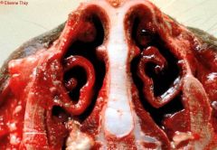
An intense inflammation is observed in the nasal cavity:
hyperaemia, edema, pseudomembranes and ulcers. |
BHV-1
|
|
|
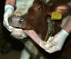
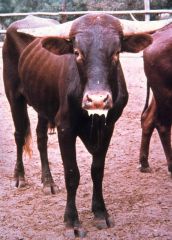
two viruses that cause open mouth breathing in cattle
|
IBR-BHV1 and BRSV
|
|
bovine dz that causes frequent urination, tail switching and slight vaginal discharge in cows. small pustules and small white necrtotic areas and ulcers can be seen on the vulva and vaginal mucosa
|
Infectious Pustular vulvovaginitis
BHV-1 genital form- |
|
|
When are BHV1 abortions commonly seen?
What lesions are seen at necropsy? Diagnose by? |
Commonly seen in last trimester of pregnancy in animals that have not been vaccinated
Typically see multifocal disseminated necrosis in liver and lungs with intranuclear inculsions Dx by IPX and PCR |
|
Family/ genus?
Transmission? |
Bovine leukemia virus
family Retroviridae and is a deltaretrovirus BLV Transmission PRIMARILY via blood inoculation Insect vectors may play a role in transmission (Tabanids and Stomoxys) Blood transfusion, dirty needles, vaccination, traumatic injuries, tattooing, all can transmit the disease if blood infected leukocytes are present on the instruments. Cattle are the only animal that are naturally infected… sheep are highly susceptible to lymphoma when experimentally infected |
|
|
what cells are infected and transformed with BLV?
Is there viremia? |
B-cells are infected and transformed
No free virus in blood |
|
|
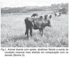
clinical signs associated with BLV lesions
|
All cases of LSA are characterized by decreased appetite, decreased milk production, WEIGHT LOSS, and anemia. Additional signs are seen with LSA in certain specific organs:
Diarrhea and melena are observed with abomasal LSA Hydropericard, hydrothorax, and edema of brisket with cardiac LSA |
|
|
BVD infection of pregnant cattle and fetal infection
|
Exposure of cattle to virus at estrus may lead to failure of conception. Insemination of seronegative cattle with BVD infected semen can lead to poor conception initially. However, after the animal has seroconverted, conception is normal and calf is born healthy. Infection during 0-45 days leads to decreased conception rate and return to estrus.
Infection of fetus 45-125 days can lead to death of the fetus (mummification or abortion), congenital abnormalities, or persistent infection. Persistent infection occurs when the fetus is infected during 45-125 days with a NCP strain. The fetus recognizes the virus as self. It is born normal except that it secretes the virus consistently. PI calves will develop mucosal disease when the (1) NCP virus mutates into a CP virus, or (2) infected with a CP virus that is homologous (antigenically similar) to the NCP virus. Mucosal disease can occur within the first 2 years of life. PI calves will develop a normal immune response to other strains of BVDV that are antigenically different to the one they are persistently infected with. They can even respond well to vaccines!!! Infection of the fetus during the period 125-175 days of gestation will result in congenital defects. Infection of the fetus after 180 days of gestation results in a fully competent immune response by the fetus, with elimination of the virus. Calf is born with antibodies to the virus and is virus free. |
|
|
BVD family / genus
how do you diagnose BVD? |
family Flaviviridae and genus pestivirus
Lekopenia in acute mucosal disease (< 50% of normal) Erosive lesions (NOT VESICULAR) Virus Isolation. Virus can easily be grown on TC. Virus in TC is identified as BVDV by FA test or IPX. Approximately 5-7 days required for identification. NCP BVD does not cause CPE!! A whole blood sample is the best sample for virus isolation (virus in lymphocytes). Nasal swabs can be submitted. Feces is not a good sample. Direct virus identification in tissues. This can be by FA, IHC , or PCR. Ear notch sample is a sample that is easily obtained and on which IHC or PCR is performed. Serology can be performed on paired samples taken 3-4 weeks apart. The antibody titer is determined by the SNT. Screening of herds for BVD can be done using single serum samples. |
|
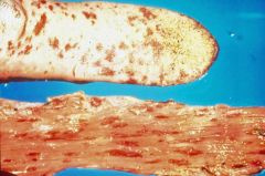
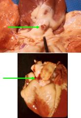
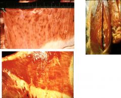
Note the erosions on the tongue (upper), and the erosive/ulcerative lesions in the esophagus (lower)
What diseases can cause these lesions? |
MCF and BVD mucosal dz
BHV-1 in very young calves Rinderpest |
|
|
transmission of BVDV
|
is primarily through PI animals excreting the NCP BVDV in all secretions
|
|
|
when can a bovine fetus be infected with BVDV to cause cerebellar hypoplasia?
|
125-175d
|
|
|
what can occur with BVDV fetal infections?
|
Cerebellar hypoplasia and hydranencephaly is common and calves are unable to stand and walk normally after birth. Retinal atrophy and dysplasia, optic neuritis, microphthalmia, can lead to various degrees of blindness.
|
|
|
An animal that is transiently viremic with BVDV (undergoing acute infection) will be ICH _____ if ear notch sample is tested
|
negative
|
|
|
how cna BVDV PI animals be diagnosed?
|
IHC on ear notch samples
|
|
|
MCF virus reservoirs / specific viruses
2 forms: |
MCF Reserviors
The virus that causes MCF belongs to the virus family Herpesviridae and is a gamma-herpesvirus. Alcelaphine herpesvirus type 1 (AHV-1) is a virus that infects wildebeest naturally Ovine herpesvirus type 2 (OHV-2) is a virus that infects sheep naturally 2 Forms of MCF are recognized. (1) The African form of MCF is associated with AHV-1 in wildebeest. The disease is endemic in Africa but also occurs in the US in zoos and on exotic animal farms that show and breed wilderbeest. (2) The Sheep-associated or US‑European form of MCF is caused by OHV-2 of sheep and is endemic in the US. OHV-2 is transmitted from sheep to cattle, bison, and deer in the US. |
|
|
describe the eye lesions seen with MCF
|
Corneal opacity starts at the limbus and progresses towards the center
Interstitial keratitis |
|
|
most common form of MCF seen
clinical signs |
The head and eye from is the form most commonly seen:
It is characterized by sudden onset of high fever (106-107 F), extreme depression, anorexia, photophobia with blepharospasm, profuse mucopurulent nasal and ocular discharges, bilateral corneal opacity, and diarrhea. There is a generalized lymphadenopathy and lymph nodes are unusually large. Lamness is often noted due to coronitis Corneal opacity, described as chronic bilateral stromal keratitis, begins at limbus and progresses centrally, and is accompanied by blepharospasm and congestion of scleral vessels Examination of the mouth will reveal erosions on the soft palate, tongue and gums. The nasal mucosa and nasal passages are deep red, necrotic, and covered with fibrinopurulent exudates CNS signs may appear and is characterized by incoordination, muscle tremor, and head pressing Death occurs in 7-10 days after onset of signs |
|
|
two other forms of MCF and their clinical signs
|
The peracute and alimentary tract form is common in deer and characterized by high fever, dyspnea and acute gastroenteritis. The typical head and eye from may not be present. Death occurs in 1-3 days
The mild from is characterized by transient fever and mild erosion. |
|
|
pathognomonic lesion in cattle with MCF
where does virus replicate? what does MCF cause? |
Necrotic vasculitis is the pathognomonic lesion in cattle with MCF
Virus replicates in vascular endothelial cells and CD8+ cells kill these viral infected cells Lymphoproliferation in various tissues, with widespread perivascular lymphoplasmacytic infiltration |
|
|
what species does MCF cause high mortaliity in?
|
deer
|
|
|
what vector transmits Bluetongue Virus?
seasonality of BTV? |
cullicoides
late summer/fall |
|
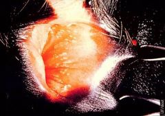
pathognomic lesion for BTV
|
BTV
Hemorrhage at the base of the pulmonary artery This is a pathognomonic lesion of BT in sheep |
|
|
Two viruses that cause hydranencephaly in sheep
|
Bluetongue virus
akabane virus |
|
|
define hydranencephaly
define hydrocephalus |
Hydranencephaly is accumulation of fluid within the white matter as a result of virus induced necrosis
Hydrocephalus is fluid accumulation in ventricles |
|
|
Congenital infections with BTV
|
Dumb calf
BTV, particularly the attenuated vaccine virus, will cross the placenta and infect calves and lambs Brain lesions include hydranencephaly and arthrogryposis |
|
|
BTV in cattle
cattle act as _____ of BTV clinical signs |
BT infection in cattle is usually sub-clinical or inapparent
Cattle act as amplifiers of BTV Clinically, it is characterized by lameness, erosions in the nostril, and by peeling of the skin of the nasal septum |
|
how transmitted?
mortality? Seasonality? lesions at necropsy |
Epizootic hemorrhagic Dz
seasonal disease – transmitted by Cullicoides Mortality may be high in deer and it is observed in late summer/fall Carcass of dead deer will show extensive hemorrhages |
|
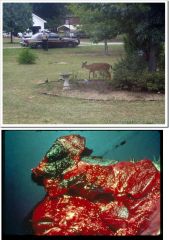
One form of this dz is ____ and deer die as a result of ___________. __________________ is commonly observed with this form
|
epizootic hemorrhagic dz
One of the forms of EHD in deer is the pulmonary form Deer die as a result of acute lung edema Frothing at the mouth/nostril is commonly observed in this form Observe the prominent lung edema - interlobular fluid accumulation is evident in this lung |
|
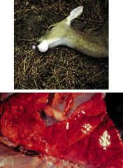
how does this dz get its name?
|
EPIZOOTIC HEMORRHAGIC DISEASE GETS ITS NAME FROM THE FACT THAT THERE ARE HEMORRHAGES EVERYWHERE PARTICULARLY IN THE MUSCLES
|
|
|
three diseases that cause high mortality in wild and farmed deer
|
EHD, BT, MCF
MCF- presumably acquired from contact with sheep. sporadic and highly fatal in deer! |
|
|
four vesicular dzs
|
vesicular stomatitis
vesicular exanthema of swine foot and mouth dz swine vesicular dz |
|
|
Vesicular stomatitis
incubation period clinical signs recovery, morbiidty, mortality |
Incubation period is average – about a week.
Fever, anorexia, and excess salivation are the first signs usually – mild fever with profuse "ropy" saliva. In horses and cattle see vesicles on dorsum of tongue, dental pads, buccal mucosa, teats, coronary bands. When vesicle rupture, it leaves a small shallow erosion/ulcer. Lamness is often noted. In swine vesicle on the snout and feet causing lameness. In horses, tongue lesions are most pronounced. Confusion arises when there are no vesicles present. Vesicle may dehydrate by fluid seepage resulting in an eroding rough area or dry necrotic lesion. Difficult to diagnose in this case. Recovery in 3 - 4 days Morbidity = 10-80% Mortality = 0% |
|
|
Vesicular exanthema of swine
incubation period clinical signs recovery, morbiidty, mortality what type of virus causes? |
An acute, febrile, contagious disease of swine characterized by the formation of vesicles on the snout, in the mouth, and on the feet. Incubation period: 18 ‑ 72 hours.
Morbidity is high, mortality low . Importance is that it is clinically indistinguishable from F&M, swine vesicular disease, and vesicular stomatitis. Caused by a calicivirus. Apart from outbreaks in Iceland and Hawaii, VESV has only been diagnosed in the US. |
|
|
Foot and Mouth Dz
incubation period clinical signs |
Incubation period is short: 2‑4 days
In Cattle: starts with lameness, smacking of lips and salivation. Vesicles may appear in mouth (gums, tongue, lips, dental pad), on muzzle, on feet (interdigital space, dew claw), on teats and udder. Vesicles starts as blanched areas, rupture, with complete healing after 2 weeks. In calves, death may occur from myocarditis. In Pigs: lameness and vesicles on snout are prominent signs. |
|
|
swine vesicular dz
clinical signs |
- It is a disease of pigs characterized by vesicular lesions in the mouth and on the feet ‑ minimal loss of condition and lesions heal rapidly.
|
|
Name the 2 viruses that cause vesicular disease in cattle
How is the virus transmitted with each of these viruses What samples would you collect in clinically affected cattle |
Two viruses that cause vesicular disease in cattle
Vesicular Stomatitis Virus Foot and Mouth Disease Virus (An aphtovirus belonging to the virus family Picornaviridae.) Virus Transmission of Vesicular Stomatitis Transmitted primarily by the sand flies (eye gnats) and blackflies. See outbreak along river drainages. Wild life species in Central America act as reservoir, where VSV is enzootic. Outbreaks in the US is believed to be windborne, initiated by infected carrier insects blown up from Central America. Many animals are infected asymptomatically and serve as amplifiers of VSV. Transmission during an outbreak is through mucosa or broken skin. Saliva and vesicular fluid from infected animals are highly infectious. Virus remains viable in envionment Virus Transmission of Foot and Mouth Transmission: In acutely ill animals, virus in saliva and most body fluids (also in semen). Spread by direct contact with infected animals and fomites, by inhalation (aerosols), by ingestion (garbage feeding). What samples would you collect in clinically infected animals Vesicular Stomatitis: Samples include vesicular fluid and paired serum samples on which RT-PCR and virus isolation are performed Foot and Mouth: Vesicular fluid** and epithelial covering |
|
|
Name the 2 vesicular viruses that can cause vesicles on the teat of cows
One other dz that can cause vesicules on teats |
Vesicular Stomatitis Virus
Foot and Mouth Disease Virus Bluetongue Virus |
|
|
Name the 4 viruses that cause vesicular disease in Pigs
How is the virus transmitted with each of these viruses What samples would you collect in clinically affected pigs |
4 vesicular disease viruses
Vesicular Stomatitis Virus Vesicular Exanthema of Swine Foot and Mouth Disease Virus Swine Vesicular Disease (an enterovirus in the virus family Picornaviridae ) Virus Transmission Transmitted primarily by the sand flies (eye gnats) and blackflies. See outbreak along river drainages. Wild life species in Central America act as reservoir of the virus where VSV is enzootic in low-lying areas receiving heavy rainfall with high vector population. Outbreaks in the US is believed to be windborne, initiated by infected carrier insects blown up from Central America. Many animals are infected asymptomatically and serve as amplifiers of VSV. Feeding of infected meat (garbage) Transmission: In acutely ill animals, virus in saliva and most body fluids (also in semen). Spread by direct contact with infected animals and fomites, by inhalation (aerosols form milk trucks, wind), by ingestion (garbage feeding). Not listed in the notes… Samples to collect Samples include vesicular fluid and paired serum samples on which RT-PCR and virus isolation are performed Virus is diagnosed in fluid from vesicles – RT-PCR and virus isolation Vesicular fluid and epithelial covering |
|
|
vesicular stomatitis
virus family / genus |
Caused by a vesiculovirus in the virus family Rhabdoviridae
(bullet shaped virion) |
|
|
what arthropod transmits vesciular stomatitis?
|
primarily by the sand flies (eye gnats) and blackflies.
|
|
|
only vesicular dz that affects horses
|
vesicular stomatitis
|
|
vesicular dz
Know the vesicular diseases and the clinical signs in each species What would you expect to observe if you examined the mouth of this animal? |
Foot and Mouth dz
Clinical Signs - In Cattle: starts with lameness, smacking of lips and salivation. Vesicles may appear in mouth (gums, tongue, lips, dental pad), on muzzle, on feet (interdigital space, dew claw), on teats and udder. Vesicles starts as blanched areas, rupture, with complete healing after 2 weeks. In calves, death may occur from myocarditis. - In Pigs: lameness and vesicles on snout are prominent signs. |
|
|
most important consequence of suid herpesvirus 1 in sows
|
SMEDI
|
|
|
pseudorabies in cattle is AKA
symptoms |
Mad itch
intense pruritis (flank/hind legs) leading to frenzy. eventual pharnygeal paralysis, salivation, mania, bellowing and convulsions. death within 48 hours |
|
|
Pseudorabies in dogs and cats
clinical signs |
intense pruritis (head region). scratching, violent rubbing. Bulbar paralysis, salivation. frantic, frenzy. generalized convulsions. NO aggression. Howling, mewling, vomting and diarrhea are common
|
|
|
how is encephalomyocarditis virus transmitted?
what does EMV cause in piglets? |
by rodents, especially rats
in piglets- convulsions and acute heart failure |
|
|
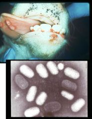
what type of virus is this?
|
coronavirus
|
|
|
TGE is caused by a ____virus
clinical signs in young piglets what cell types are affected? what is the result of this? vaccinate protocol for sows? |
coronavirus
100% mortality in piglets less than 10d old- severe diarrhea, vomiting, dehydration, death infects and kills columnar epithelial cells resulting in villous atrophy- reduces digestive and absorptive capacity vaccinate sows 3 weeks prior to farrowing with MLV |
|

this virus is also referred to as ______
what cell types are infected? mortality? |
Rotavirus diarrhea is often referred to as “white scours” or “milk scours”
Rotavirus infects and destroys the terminally differentiated enterocytes lining the tips of the intestinal villi causing intestinal malabsorption and maldigestion Mortality due to rotavirus is < 20 % amongst suckling pigs It is a major factor in post-weaning diarrhea resulting in poor weight gain |
|
|
what causes porcine postwearning multisystemic wasting syndrome?
where does the virus replicate? why does it cause substantial mortality? |
porcine circovirus-2
Replicates in lymphoid tissues causing swollen lymph nodes It can cause substantial mortality in young pigs because of its immunosuppressive properties |
|
|
what does porcine repro resp syndrome cause
|
respiratory disease in growing pigs and SMEDI in adult sows
|
|
clinical signs?
|
SWINE INFLUENZA CAUSED BY H1N1 AND H3N2
Clinically see sudden onset of fever, anorexia, followed by extreme prostration (do not want to move, muscle pain, stiffness), labored breathing, "thumps", deep cough, watery discharge from eyes and nose. Disease looks bad, but most recovery rapid after few days Mortality 1-4%. |
|
|
Five viruses that have been associated with SMEDI in sows
What does SMEDI stand for and why is it so important? Pathogenesis of SMEDI? |
Know the 5 viruses that cause SMEDI
Pseudorabies (Suid Herpesvirus-1) Encephalomyocarditis Virus Porcine Postweaning Multisystemic Wasting Syndrome (Porcine Circovirus type-2) Porcine Reproductive and Respiratory Syndrome Virus (Caused by an arterivirus – Prototype virus is called the Leylytstad virus) Porcine Parvovirus Porcine Teschovirus types 2-7 and 11-13 Classical Swine Fever (CSF virus is a Pestivirus belonging to the virus family Flaviviridae.) SMEDI- stillbirth, mummification, embryonic death and infertility SMEDI Pathogenesis - Sows infected in later part of pregnancy will abort premature piglets, stillborn piglets, and mummified fetuses |
|
|
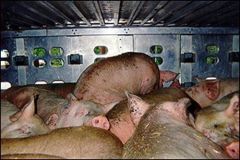
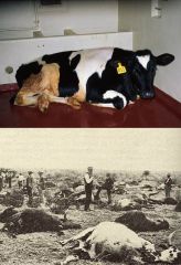
what do classical and african swine fever cause?
how can these viruses be transmitted? most important necropsy lesions? |
Hog Cholera and African Swine fever cause very high mortality in pigs
The viruses causing these diseases remain in meat products and will be transmitted to pigs fed the garbage containing the virus tainted meat During an outbreak the virus is easily spread between pigs through contact with infected secretion – feces, saliva, etc.. Most important necropsy lesion is wide spread hemorrhages throughout the body Both are exotic viruses in the US and are reportable diseases |
|
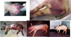
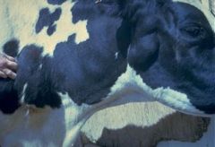
clinical signs include by high fever, depression, conjunctivitis, diffuse purplish discoloration (hyperemia) of skin,and Nervous signs (circling, convulsions)
|
hog cholera - clasical swine fever
|
|
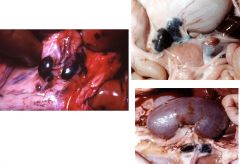
Enlarged haemorrhagic mesenteric lymph nodes
Enlarged hemorrhagic gastro-hepatic lymph node Enlarged hemorrhagic renal lymph nodes with petechiation of the renal cortex |
african swine fever
|
|
|
how is ASF transmitted, and what animals can serve as as reservoir?
|
argasid ticks
wild hogs |
|
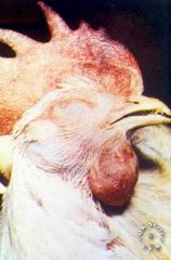
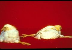
very important virus of chickens with 100% mortality.
Characterized by various clinical signs including sneezing, coughing, edema of the face and head, diarrhea, and CNS signs Widespread hemorrhages at necropsy |
Highly pathogenic avian influenza
|
|
|
what disease can cause a very similar clinical presentation to HPAI and is indistinguishable?
|
Newcastle dz- avian paramyxovirus-1
|
|
|
what clinical signs are seen with both HPAI and NCD
|
open mouth breathing.
80% mortality not uncommon virus spreads quickly other signs inlucde CNS dz (tremors, torticollis, weird postures) and diarrhea |
|
|
signs of HPAI in ducks?
|
generally no clinical signs, but may see CNS signs
|
|
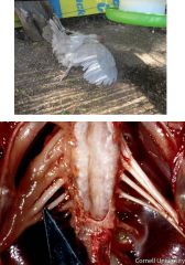
HPAI and VVND
what are these pictures showing what is pathognomic for these diseases? |
Echymotic hemorrages in the heart muscle and in the fat around the heart.
Hemorrage in the proventriculus - this is almost pathognomonic for HPAI and VVND viruses Echymotoc hemorrages are also seen in the intestinal tract and respiratory tracts |
|
|
what can the viscerotropic velogenic pathotype cause?
|
100% mortality with no accompanying clinical signs.
When clinical signs are present, it usually starts with respiratory disease characterized by fever (43 C), dullness, thirst, ruffled feathers, hemorrhagic comb, edema of the head, respiratory distress (open mouth breathing and gasping for air, coughing) and death |
|
|
in the US what maintains the ND virus
what species has NDV affected? |
domestic birds and cormorants
ND outbreaks have been diagnosed in chickens, guinea fowl, turkeys, pheasants, ducks, geese, pigeons, and wild birds |
|
|
pathogenesis of avian paramyxovirus-1
|
Like influenzavirus, the insertion of multiple basic AA, e.g. lysine and arginine, just before the cleavage site of the F protein results in increased virulence
The F protein of the lentogenic pathotypes are cleaved by trypsin-like enzymes present on the epithelial cells of the respiratory and gastro-intestinal tracts, and the virus cannot go systemic The F protein of the velogenic pathotypes, on the other hand, can be cleaved by furin-like proteases within cells. Intracellular cleavage means that virus leaves the cells as infectious particles and in that way can go viremic and infect cells in all organs of the body |
|
|
how has VVND been introduced into the country?
|
through smuggling of exotic birds, or through illegal trade of poultry and poultry products.
|
|
|
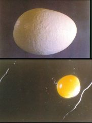
early signs of NCD?
|
typically include a dramatic drop in egg production. In layers that produce brown eggs, eggs that are laid may be discolored. The light eggs in this image are abnormal. This discoloration is caused by abnormal pigment deposition of the egg while in the oviduct of infected chickens.
|
|
|
infectious laryngotracheitis
caused by? common symptom? pathogenesis? |
Caused by Gallid herpesvirus 1
Open mouth breathing is common because of the necrosis in the tracheal epithelium and the accumulation of caseous exudate in the trachea and larynx |
|
infectious bronchitis
caused by? causes? what cells are infected? what does this result in? |
Caused by a coronavirus – it causes mild respiratory disease in adult chickens
This respiratory virus (like other respiratory viruses!) also infects the epithelial cells of the oviducts Because of the inflammation, calcium deposition in the egg shell is interfered with causing abnormal misshapen eggs with thin egg shells Albumin is also watery |
|
|
Differentials for egg deformities and decline in egg production:
|
Infectious Bronchitis (decline in #’s for layers, deformed)
Lentogenic Newcastle Disease Virus (deformed) – Low Pathogenic Avian Influenza (decline in #’s) Mesogenic NDV (deformed, decline in #’s) |
|
|
what is infectious bronchitis virus associated with in young chicks?
|
Associated with very high morbidity, but low mortality in young chicks - (explosive) respiratory infection characterized by sneezing, coughing, and gasping for air.
|
|
|
infectious bronchitis virus in layers
|
Although IBV infection in layers, is often subclinical, it is accompanied by a marked drop in egg production, with many soft‑shelled and malformed eggs with watery albumin
|
|
|
Mareks dz
caused by? where does virus replicate? transmission? pathogenesis? |
Marek’s disease is caused by Gallid herpesvirus 2
During the acute infection, the virus replicated in the cells lining the feather follicles and are transmitted when the feathers fall out The virus is inhaled by susceptible birds and this is how the infection spreads Pathogenesis Virus is inhaled and reaches the lungs where it is taken up by lung macrophages. Infects lymphocytes disseminates throughout the body via infected lymphocytes Virus reaches the follicles in the skin and infects epithelial cells lining the feather follicles Replication in the epithelial cells of the feather follicles allows the virus to be released into the environment as “dry skin aerosol” when the feather “fall off” After the infected chicken mounts an immune response, the virus goes latent in T‑cells These T-cells can eventually be transformed into cancer cells by the virus. |
|
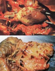
marek's dz
what is the bottom picture showing |
Note the swollen sciatic nerve on the left and how different it is from its pair on the right – the nerve is infiltrated with virus infected lymphocytes and causes nerve swelling resulting in paralysis
Marek’s disease is caused by Gallid herpesvirus 2 |
|
|
how is marek's dz transmitted and acquired?
|
MDV is transmitted via virus replication in the epithelial cells of the feather follicles, and being aerosolized when the feathers are shed
Therefore, MDV is a acquired through the respiratory system |
|
the top is a typical clinical picture of what disease?
what is the pathogenesis of this disease that leads to the lesion seen in the bottom picture? |
Mareks DZ
MDV eventually transforms the lymphocytes in some infected birds and the transformed lymphocytes infiltrate the nerves and causes paralysis of the legs (Sciatic nerve) or wings (brachial nerve) Sciatic nerve is enlarged (unilaterally) and looses whiteness and striation |
|
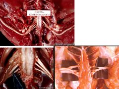
these lesions are typical of what dz?
what are the nerves infiltrated with? |
Mareks Dz
Sciatic and brachial nerves are enlarged and loose their typical striation patterns These nerves are infiltrated with herpesvirus transformed T-cells |
|
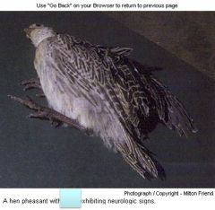
what dz is causing this pheasant to show neuro signs?
|
EEE
|
|
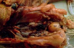
Infection causes swelling, edema, hyperemia and sub-serosal hemorrhages in the gland
what disease is this? what is the BF? |
Infectious Bursal Dz
BF= bursa fabricius (Where chicken manufactures b-cells) |
|
|
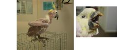
what is Pacheco's dz? what birds are most affected?
|
Pacheco's Disease is a highly contagious and highly fatal disease of all psittacine birds and affects all ages. Amazon parrots account for the majority of cases, followed by African grey parrots, macaws, and cockatoos
|
|
|
what virus causes pacheco's dz?
how is it transmitted? clinical signs? |
Psittacine herpesvirus-1
Healthy birds that have recovered from the disease are carriers and can excrete virus in feces during time of stress (breeding, loss of mate, change in environment, pet shops!) Very often, parrots are described as normal one day and dead the next! Signs include lethargy, anorexia, regurgitation, diarrhea, ruffled feathers, and neurological signs characterized by tremors of neck, wings, and legs |
|
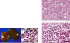
Birds die of massive liver necrosis characterized by enlarged liver, spleen, and kidneys
See multifocal hepatic and splenic necrosis with eosinophilic intranuclear inclusion bodies |
Pacheco's Dz
|
|
what virus causes this high contagious dz of parrots (electus spp, african greys and cockatoos)
|
psittacine beak and feather dz
psittacine circovirus |
|

name the viruses that can cause infectious tracheobronchitis
|
Canine Parainfluenza virus
Canine Influenza virus Canine Adenovirus type-2 (Infectious canine laryngotracheitis) Canine Adenovirus-1 Canine Distemper virus Canine Herpesvirus-1 Canine Reoviruses |
|
what bacterial pathogen is associated with ITB
|
Bordatella Bronchiseptica
mycoplasma |
|
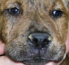
this virus causes severe eye lesions that includes keratitis and corneal ulceration
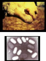
It may also cause oral erosions or ulcerations Sneezing, nasal discharge, anorexia, fever, are the other commonly seen clinical signs This virus infects young kittens usually and they remain latently infected after they recover |
feline viral rhinotracheitis
caused by feline herpesvirus-1 Adults = conjunctivits only Causes oral ulcerations when nasal passages are infected Can recrudesce |
|
|
what are the most importan diseases that cause bovine respiratory dz?
|
bhv-1
PI-3 BRSV |
|
|
what three viruses are associated with bovine interstitial pneumonai
|
PI-3
BRSV Bovine adenovirus |
|
|
what two virses are most frequently associated with bovine enzootic pneumonia of young calves and veal calves
|
PI-3
BRSV |
|
|
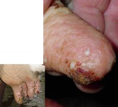
pathogenesis of Jaagseikte sheep retrovirus
|
Virus transforms the pneumocytes of the lungs which secrete excessive amounts of fluid that fill up the alveolar space
This fluid pours out of the nostril when the sheep’s head is down or when you hold up the animals like a wheelbarrow! |
|
|
Ovine progressive pneumonia pathogenesis
|
Virus transforms macrophages which inflitrate the lungs and other tissues including the udder and brain
Lungs are heavy and infiltrated with macrophages |
|
|
viruses that can cause equine respiratory dz
|
EHV-1
EHV-4 Equine Influenza EVA Equine Rhinitis A Virus Equine Adenovirus Equine Reovirus EHV-2 Hendra virus |
|
|
What is the common names for EHV-4?
What are the clinical signs associated with infection with EHV-4 and the age group of horses? What samples to submit for a diagnosis of equine herpesvirus 4? What type of vaccines are available to protect against EHV-4 infection and what is the recommended vaccination program? |
Equine viral rhinopneumonitis
Foals & yearlings – fever, mucopurulent nasal catarrh & conjunctivitis Nasal secretions for PCR & virus isolation MLV – 2 doses Inactivated – frequently Pregnant mares – 5, 7 & 9 months |
|
|
Influenza and Rhinitis A
What are the most important clinical signs associated with these 2 viral infection and what clinical signs might help differentiate between these 2 infections? Are vaccines available? If so, what type are available and the vaccination routine followed. What sample would you submit to the diagnostic laboratory for diagnosis of these infections |
Equine Influenza - fever, dry hacking coughing, slight nasal discharge, depression, inappentance
Equine Rhinitis A – copious nasal discharge that becomes mucopurulent & coughing for 2-3 weeks 2. Equine influenza – bivalent inactivated vaccine – foals at 6 months and again 3-8 weeks later; booster 6 months post 2nd dose; revaccinated biannually in April & October Equine Rhinitis A – no vaccine Equine influenza – deep nasal or pharyngeal swabs & paired serum samples |
|
common virus of horses that rarely causes clinical dz
however, in certain arab foals, it can be fatal |
Equine adenovirus is a common virus of horses that rarely causes clinical disease in normal horses
It spreads easily between horses and cause subclinical infections However, in Arab foals that are born with combined immunodeficiency, this virus will infect the foals and cause a fatal disease These foals usually start to show clinical disease after the first month or two and respiratory disease is the most common disease associated with this condition They will normally die by the time they reach 4-6 months of age |
|
extensive hemorrhage in the cortex and perivascular cuffing
only lesion seen with this dz- there are no systemic lesions |
EEE
|
|
|
Perivascular cuffs are commonly seen in the CNS of animals ________
|
infected with all types of “viral encephalitides”
|
|
two most important viruses that cause diarrhea in calves?
how are calves infected? |
The 2 most important viruses that cause diarrhea in calves is Bovine Rotavirus and Bovine Coronavirus
Adults secrete both viruses in their feces just before and at time of calving – they are asymptomatic carriers and contaminate the environment |
|
|
what cells do bovine rotavirus and coronavirus infect?
what does this result in? treatment? vaccination? |
Bovine Rotavirus and Bovine Coronavirus infect the epithelial cells of the small intestine and replicate and destroy the mature columnar cells lining the villi
Decrease ability to digest and adsorb nutrients Very voluminous watery diarrhea with severe dehydration Must cut off milk for 2 days and give lots of electrolytes by mouth, or sub cut or IV Explosive outbreaks may occur on farms that do not have good hygiene or management Vaccinate dam with inactivated vaccine before calving both IM and Intramammary |
|
this dz is caused by a type B coronavirus.
The most significant clinical sign is profuse and watery diarrhea, which is greenish to brownish with occasional blood streaks Persists for about 5 days Anorexia, depression, and drastic decrease in milk production is also noted |
Winter Dysentery
|
|
this dz is caused by a morbiliivirus
causes very high morbidity and mortalitiy Profuse watery diarrhea erosive with mucosal lesions throughout the gastro intestinal tract |
rinderpest
|
|
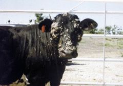
causes warts most often in young cattle on the face/neck.
Virus? how is it spread? treatment? vaccine? |
bovine papilloma
Spread via rubbing on posts and leaving the virus on the wood or metal Will eventually regress by themselves Vaccine is available – Killed vaccine given intradermally Efficacy of vaccine difficult to judge because warts go away by themselves anyway |
|
two types of papillomas
pathogenesis |
Papilloma are either epithelial papilloma or fibropapillomas
All warts have similar structural features Papilloma virus stimulates the cells of the basal layer to replicate – this is through one viral protein translated in the viral infected cells These infected cells do not contain any structural viral proteins or infectious virus Infectious virus present in dead cells overlaying the wart |
|
|
where are papillomas most often seen in cows?
transmission? control? |
Papilloma are often seen on the teat of milking cows
Easily transmitted between cattle via milking machine if not careful “Rice grain” or “frond like” warts are described Difficult to control and get rid of! |
|
proliferative lesion on the teat of a cow that progresses to a scab
caused by a parapoxvirus and is zoonotic |
pseudocowpox
|
|
what type of virus causes this dz?
what samples need to be taken to confirm? the lesions are proliferative, but not erosive or ulcerative |
Bovine papular stomatitis, caused by a parapoxvirus
samples to confirm: EM exam of lesion scraping or saliva |
|
|
Viruses that can cause teat lesions
|
Bovine Papallomavirus (BPV-1 and BPV-6)
Psuedocowpox (parapoxvirus) Cowpox/Buffalopox (orthopoxvirus) Bovine Herpesvirus-2/ Bovine Herpes Mammillitis Virus Vaccina Virus |
|
painful, ulcerative lesions on teat and udder
|
bovine herpes mammillitis virus
(BHV-2) |
|
Typical proliferative lesions on the lips of sheep – young lambs especially
Human infection with the virus – starts as a vesicular lesion, progresses to a pustule, and then forms a firm adherent scab Shearers commonly infected |
contagious ecthyma
|
|
proliferative/scabby lesions caused by a parapoxvirus
age affected?vaccination? |
contagious ecthyma
Disease of in young lambs 2-6 months old Must vaccinate the lambs at 2 months for protection Vaccinating the ewe is useless for protecting the lambs |
|
|
know the three diseases caused by a parapoxvirus
are they all zoonotic? |
pseudocowpox
bovine papular stomatitis contagious ecthyma all are zoonotic |
|
|
pseudocowpox lesions
|
Lesions start with small papule, which quickly grows into a vesicle or pustule, which ruptures after 48 hours forming a thick scab. Scab is elevated with granulation tissue underneath. After 7-10 days, scab drops off leaving behind an area of 0.5‑2.5 cm diameter. This area is encircled by small scabs giving it the appearance of a "horseshoe-shaped ring" - crusty reddish raised border with a wart like granulomatous lesion in the middle
|
|
|
bovine papular stomatitis?
importance? |
Characterized by raised and not depressed lesions - raised hyperemic areas, which rapidly become roughened plaque-like with irregular borders. Occurs on lips, dental pads, muzzle, buccal mucosa. Not economically important. Importance in that it must be differentiated from the vesicular diseases.
|
|
|
contagious ecthyma lesions
|
characterized by cutaneous vesiculo-papular eruptions followed by development of pustule and thick friable crusts. Commonly affects young lambs ‑ lesions most common on muzzle, lips, occasionally in mouth, on the feet, face. In ewes, see lesions primarily on the udder. Morbidity is high, but case fatality is low. Lambs loose weight due to suckling difficulties.
|
|
|
when are papillomas most frequently seen in dogs?
treatment? |
Most frequently seen in younger dogs
Not cause for alarm – will regress normally in 6-8 weeks May remove surgically if interferes with eating or collects bacteria and gets infected |
|
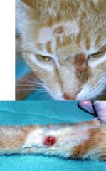
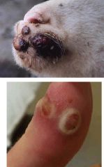
solitary lesions that are proliferative in nature
why lesions on the face and leg? zoonotic? |
Cowpox
Lesion associated with cowpox virus that cats pick up from rodents (mice, rats) – lesions on face/leg because that is what contacts the rodent! it is zoonotic |
|
when will these lesions be seen on cats?
lesions are solitary, proliferative lesions that can occur anyway. they resemble raised plaques. |
feline papilloma
occur in older cats that are immunodeficient as a result of FIV infection |
|
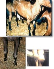
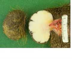
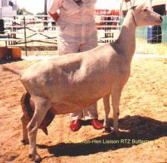
Inflammation of the synovial membrane resulting in painful joint swelling
Adults become progressively more lame and looses weight |
caprine arthritis encephalitis
aka Big knee |
|
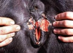
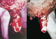
venereal dz of horses
Lesions consist of vesicular or pustular eruptions on the vulva and perineum of mares and on the penis of stallions. Lesions may occur on the teats and on muzzle of suckling foals. |
equine coital exanthema
EHV-3 |
|
dz caused by a lentivirus
what cells are transformed? what does this cause? |
caprine arthritis encephalitis
transforms monocytes which then infiltrates joints, CNS, and Udder Chronic progressive lameness in adult goats |
|
|
many goats affected with CAE develop ____ and the virus spreads through the ____
|
mastitis
milk |
|

lamb has hair, rahter than wool and presents as ataxia and trembling
|
hairy shaker / border disease
|