![]()
![]()
![]()
Use LEFT and RIGHT arrow keys to navigate between flashcards;
Use UP and DOWN arrow keys to flip the card;
H to show hint;
A reads text to speech;
19 Cards in this Set
- Front
- Back
|
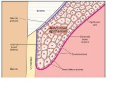
Basal Lamina |
Superficial part of basement membrane; within dentogingival junction has both an external and internal basal lamina surrounding junctional epithelium. |
|
|
External Basal Lamina |
is a structure similar to basal lamina that surrounds the sarcolemma of muscle cells. It is secreted by myocytes and consists primarily of Collagen type IV, laminin and perlecan (heparan sulfate proteoglycan). |
|
|
Internal Basal Lamina |
|
|
|
Col |
Interdental gingiva apical to contact area assumes nonvisible concave form between facial and lingual gingival surfaces. |
|
|
Dentogingival Junction |
Junction between tooth surface and gingival tissue. |
|
|
Dentogingival Junctional Tissue |
Tissue that includes sulcular epithelium and junctional epithelium. |
|
|
Epithelial Attachment (EA) |
Device that attaches junctional epithelium to tooth surface. |
|
|
Epithelium |
Basic tissue that covers and lines external and internal body surfaces. |
|
|
Junctional Epithelium (JE) |
Deeper extension of sulcular epithelium. |
|
|
Pocket Epithelium (PE) |
Epithelium linning periodontal pocket. |
|
|
Sulcular Epithelium |
Epithelium that stands away from the tooth creating gingival sulcus. |
|
|
Free Gingival Crest |
Most superficial part of marginal gingiva. |
|
|
Free Gingival Groove |
Groove that separates attached gingiva from marginal gingiva. |
|
|
Gingival Crevicular Fluid |
Fluid in gingival sulcus. |
|
|
Gingival Hyperplasia |
Overgrowth of mainly interproximal gingiva. |
|
|
Gingival Recession |
Inferiorly placed margin of free gingival crest. |
|
|
Gingivitis |
Inflammation of the gums. |
|
|
Periodontal Pocket |
Gingival and periodontal pockets are extensions of the gingival sulcus, which exists in health. |
|
|
Periodontitis |
Inflammation of the tissue around the teeth, often causing shrinkage of the gums and loosening of the teeth. |