Reading...
![]()
Play button
![]()
Play button
![]()
Use LEFT and RIGHT arrow keys to navigate between flashcards;
Use UP and DOWN arrow keys to flip the card;
H to show hint;
A reads text to speech;
23 Cards in this Set
- Front
- Back
- 3rd side (hint)
|
PICA
|
arise from Vertebral artery at level of the medulla
Supply lateral medulla and inferior occipital surface of the cerebellum |
|
|
|
AICA
|
arise from proximal basilar artery at the level of the caudal ppons
Supply the lateral caudal pons and small region of cerebellum |
|
|
|
SCA
|
arise from the top of the basilar artery at the level of the rostral pons
Supply superior cerebellum and small region of rostral laterodorsal pons |
|
|
|
supraclinoid ICA branches
|
Ophthalmic, PCOM, anterior choroidal, anterior cerebral, middle cerebral arteries
|
|
|
|
ACA
|
travel in interhemispheric fissures
2 main branches: pericallosal and callosomarginal supply anterior medial surface: frontal to anterior parietal |
|
|
|
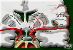
MCA
|
turns laterally and enters the Sylvian fissure
usually bifurcates to superior and inferior divisions but variable |
|
|
|
MCA superior division
|
supply lateral frontal lobe, usually including the peri-Rolandic cortex
|
|
|
|
MCA inferior division
|
supply lateral temporal lobe and variable portion of the parietal lobe
|
|
|
|
PCA
|
supply the inferior-medial temporal lobe and medial occipital lobe
wrap aroid the midbrain, supplying it |
|
|
|
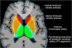
lenticulostriate arteries
|
arise from initial portion of the MCA before it enters the Sylvian fissure
supply large regions of the basal ganglia and internal capsule |
|
|
|
anterior choroidal artery
|
arise from the ICA
supply portions of the globus palidus, putamen, thalamus and posterior limb of the internal capsule |
|
|
|
recurrent artery of Heubner
|
arise from initial portion of the ACA
supply portions of the head of the caudate, anterior putamen, globus palidus, and internal capsule |
|
|
|
thalamoperforator arteries
|
arise from proximal PCA
supply the thalamus and sometimes extend to the posterior limb of the internal capsule |
|
|
|
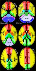
Superficial cerebral vessels
|
ACA, MCA, PCA
|
|
|
|
Deep cerebral vessels
|
lenticulostrate, anterior choroidal, recurrent artery of Heubner, and thalamoperforators
|
|
|
|
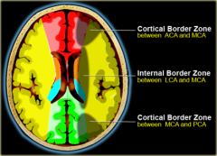
Cortical border zone infarctions
|
Infarctions of the cortex and adjacent subcortical white matter located at the border zone of ACA/MCA and MCA/PCA
|
|
|
|
Internal border zone infarctions
|
Infarctions of the deep white matter of the centrum semiovale and corona radiata at the border zone between lenticulostriate perforators and the deep penetrating cortical branches of the MCA or at the border zone of deep white matter branches of the MCA and the ACA.
|

|
|
|
Medial lenticulostriate arteries
|
Branches of the A1-segment of the anterior cerebral artery.
They supply the anterior inferior parts of the basal nuclei and the anterior limb of the internal capsule. |
|
|
|
Lateral lenticulostriate arteries
|
Branches of the horizontal M1-segment of the middle cerebral artery.
They supply the superior part of the head and the body of the caudate nucleus, most of the globus pallidus and putamen and the posterior limb of the internal capsule. |

|
|
|
Fetal Origin of the Posterior Cerebral Artery
|
Fetal origin of the posterior cerebral artery occurs when the embryonic posterior cerebral artery fails to regress. It may occur on the right side (10% of the general population), the left side (10% of the general population), or bilaterally (8% of the general population)
|
|
|
|
Fenestration of the anterior cerebral artery
|
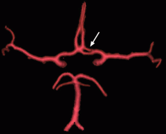
The prevalence of fenestration of the A1 segment is between 0% and 4% in anatomic imaging studies (10) and 0.058% in angiographic studies
|
|
|
|
Middle cerebral artery duplication
|
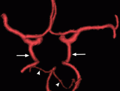
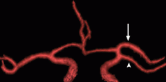
Middle cerebral artery duplication occurs when a middle cerebral artery branch arises above the bifurcation of the internal carotid artery. The duplicate vessel parallels the main middle cerebral artery and supplies the anterior temporal lobe (13) (Fig 5). The reported prevalence of middle cerebral artery duplication is 0.2%–2.9% (14).
|
|
|
|
Basilar artery fenestration
|
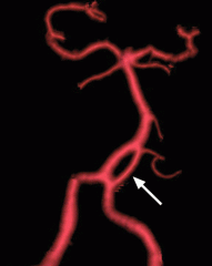
Basilar artery fenestration has been found in 0.6% of angiographic examinations (19) and approximately 5% of autopsies (20). Basilar artery fenestrations are most commonly located in the proximal basilar trunk, close to the vertebrobasilar junction (3)
|
|