![]()
![]()
![]()
Use LEFT and RIGHT arrow keys to navigate between flashcards;
Use UP and DOWN arrow keys to flip the card;
H to show hint;
A reads text to speech;
32 Cards in this Set
- Front
- Back
|
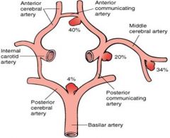
Describe the circle of willis. |
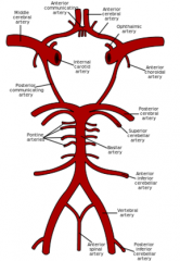
Note the opthalmic artery comes off the internal carotid. |
|
|
Give some features of the blood supply of the brain. |
Brain = 2% bw, 20% O2 and 15% CO. 20s anoxia -> unconsciousness >5m -> permanent unconsciousness Flow autoregulated. Decreased O2/increased CO2 - increases flow Arteries, thin walled, easily blocked, distorted or ruptured Veins, no valves, thin walled, no muscles or elasticity to help return |
|
|
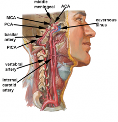
Describe the blood supply of the brain when viewed laterally. |
|
|
|
What is seen on a normal angio? |
Under normal situations anterior communicating and posterior communicating closed Classic Circle of Willis seen in 34.5% Common variations One posterior communicating small one large Anterior communicating large |
|
|
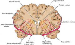
Describe the deep perforating arteries of the MCA and what they perfuse. |
|
|
|
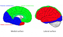
Describe the blood supply to the cerebral cortex from both a medial and a lateral view. |
|
|
|
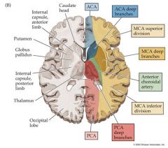
Describe the perfusion of the brain from a transverse/horizontal plane. |
|
|
|
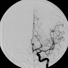
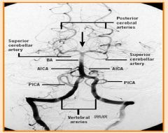
Describe the posterior circulation of the brain as viewed on an angiogram. |
|
|
|
What do the a) vertebral, b) basilar and c) PCA supply? |
Vertebral - SC + dorsal medulla of BS via PICA
Basilar - pons + cerebellum
PCA - Inferior and medial aspects of temporal occipital cortex, thalamus and posterior internal capsule, midbrain, anastomose with MCA |
|
|
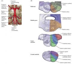
HARD/LEARN THIS Describe the posterior circulation to the brainstem. |
|
|
|
Describe the superficial venous drainage of the brain. |
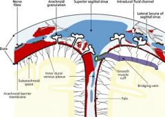
Superior cerebral veins cross the subarachnoid space - pierce dura (bridging veins) as they enter intracranial (dural) venous sinuses. Arachnoid granulations allow CSF to flow into venous blood of sinuses but prevent backflow of blood into subarachnoid space. |
|
|
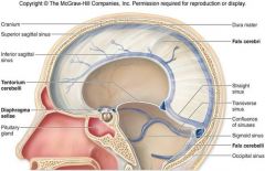
Describe the venous sinus anatomy of the brain. |
|
|
|
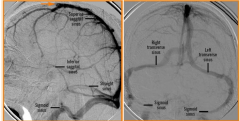
Remember this venous angiogram. |
|
|
|
What are the main causes of CVAs? |
Atherosclerosis Hypertension Aneurysm Elderly Head injury (trauma) Alcoholics Arteriovenous malformation |
|
|
What are the causes of a) ischemic stroke b) haemorrhagic? |
a) atherosclerosis, thromboembolism
b) trauma, spontaneous |
|
|
What are the effects of MCA stroke on a) the dominant hemisphere b) the nondominant? |
a) global aphasia, sensorimotor loss on contralateral face, ipsilateral upper limb and trunk
b) neglect syndrome |
|
|
What are the effects of a) ACA and b) PCA strokes? |
a) contralateral sensorimotor loss below the waist, urinary incontinence, personality defects, split brain syndrome.
b) contralateral homonymous hemianopia, reading and writing deficits, impaired memory |
|
|
What is an epidural haematoma? |
Extradural - traumatic, blood between dura and skull, rapid arterial bleeding, dura is peeled off skull. May present with lucid period immediately after trauma followed by unconsciousness. Bleed rapid arterial (or large venous sinuses) Middle meningeal A. (temperoparital area, pterion) Ant. Ethmoidal A. (frontal) |
|
|
What is a subdural haematoma? |
Dura still attached to skull, traumatic, blood between dura mater and arachnoid mater, bridging veins, acute/chronic.
Acute, subacute or chronic Acute after high speed acceleration and deceleration Associated with cerebral contusions Slower onset as venous bleed |
|
|
What is a subarachnoid haematoma? |
Spontaneous, between arachnoid and pia, ruptured aneurysm or head injury, 1-7% strokes, ARTERIAL |
|
|
What are the symptoms and diagnosis of an epidural haematoma? |
As blood collects it compresses intracranial structures Compress cranial nerve III Weakness of extremities on opposite side of lesion (crossed pyramid pathways Loss of visual field opposite to lesion (compress of PCA)
CT or MRI - convex lens, Expansion stops at the sutures because dura is more tightly attached here so then presses inwards |
|
|
What are the symptoms and diagnosis of subdural haematoma? |
Symptoms: Irritability, Seizures, Headache, Numbness, Disorientation
Diagnosis: CT Crescent shaped with concave surface They can have convex appearance so can look like extradural But they can cross the suture lines |
|
|
What are the symptoms and diagnosis of a subarachnoid haematoma? |
Symptoms: Severe headache (thunderclap) Vomiting Confusion Lowered level of consciousness
Diagnosis: CT White signal diffuse over sulci on both sides Occasionally lumbar puncture Evidence of blood in 3% of people with normal CT Also bilirubin |
|
|
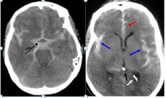
How does an epidural haematoma appear on a CT? |
|
|
|
How does a subdural haematoma appear on a CT? |
|
|
|
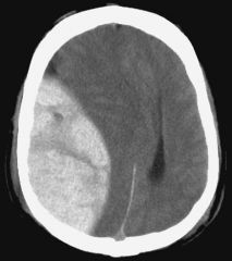
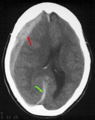
How does a subarachnoid haematoma appear on a CT? |
|
|
|
Describe some features of a cerebral aneurysm. |
3 types: Saccular Fusiform Berry
1:15 people develop a brain aneurysm Women at higher risk 3:2 Danger comes if it ruptures |
|
|
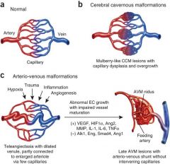
Describe the appearance and formation of a cerebral arteriovenous malformation. |
|
|
|
What are the symptoms of an AVM? |
The most frequently observed problems related to an AVM are headaches and seizures while at least 15% of the population at detection have no symptoms at all. Other common symptoms are a pulsing noise in the head, progressive weakness and numbness and vision changes as well as debilitating, excruciating pain |
|
|
What is a TIA? |
Temporary loss of brain function (<30 mins) Sudden onset but resolves within 24 hrs Diagnosis based on symptoms alone Warning sign of heart attack or stroke 4-8% in 1st month 12-13% in 1st year 24-29% in 5 years |
|
|
What are the effects of TIA on the anterior and posterior circulations? |
Anterior circulation: Motor weakness Hemi-sensory loss Dysarthria Transient monocular blindness
Posterior circulation: Vertigo Diplopia Ataxia amnesia |
|
|
Summarise what the anterior and posterior circulation do. |
Posterior circulation supplies brainstem, thalamus, temporal and occipital lobes • Anterior circulation supplies, frontal, parietal, temporal lobes • MCA is most important branch of anterior circulation |