Reading...
![]()
Play button
![]()
Play button
![]()
Use LEFT and RIGHT arrow keys to navigate between flashcards;
Use UP and DOWN arrow keys to flip the card;
H to show hint;
A reads text to speech;
27 Cards in this Set
- Front
- Back
|
Describe the three layers of blood vessels.
Describe bordering layers. |
Tunica Intima: layer of simple squamous ENDOTHELIUM
Internal Elastic Lamina Tunica Media: circularly-arranged smooth muscle (constrict or dilate), collagen fibers, ground substance, elastic fiber External Elastic Lamina Tunica Adventitia: CT and elastic fibers |
|
|
How does the tunica adventitia differ in large vessels?
|
Contains VASA VASORUM--vessels supplying vessels
|
|
|
How do arteries generally differ in their layering (when compared to veins)?
|
Thicker tunica media, more elastic fiber
|
|
|
Contrast the layers of the following vessel types:
Elastic Arteries Muscular Arteries Arterioles Provide examples of categories (if applicable), describe innervation (if applicable) |
Elastic: close to heart, under high pressure, ex: aorta, pulmonary trunk
Tunica Media: TONS of elastic (alternates with smooth muscle and collagen to form ELASTIC LAMELLAE) Tunica Intima: thicker than in other arteries bc of layer of subjacent CT Tunica Aventitia: Loose CT and BV's (VASA VASORUM) Medium Sized: ex: coronary arteries Intima: Thinner Media: mostly smooth muscle; collagen, some elastic Arterioles: Smallest Intima: endothelial nuclei may bulge into lumen Media: 1-3 layers of smooth muscle--regulate blood flow to target tissues Innervation from symp and parasymp Final branching gives rise to capillary microcirculation |
|
|
How would you differentiate between an arteriole and vein under the microscope?
|
Vein would look flimsier and wouldn't have thin layer of muscle
|
|
|
How do veins appear under the microscope?
|
Collapsed, thin tunica media
|
|
|
What is microcirculation? What structures control it? Why would you want control? How does this occur?
|
Blood flow from arterioles to capillaries
Metarterioles with precapillary sphincters close and prevent blood from entering capillary beds; instead flows directly from arteriole to venule, allows blood to be diverted to where its needed |
|
|
What is a metarteriole?
|
Links arterioles and capillaries; it's a short arterial capillary
Has individual muscle cells (not tunica media) to form precapillary sphincters to encricle capillary |
|
|
What is the function of arteriovenous anastomoses?
|
Provide circulation detours and shunts (connections between artery and vein) to allow bypass or shunting by capillary bed
|
|
|
What is portal circulation? Provide an example. What is the benefit of portal circulation?
|
Occurs when capillary bed drains into another capillary bed through veins. Ex: hepatic portal system
Allows for substance secreted by first capillary bed to be modified, received, etc. by second capillary bed |
|
|
Describe the cellular structure of capillaries. What cells associate with them? What do they do?
|
Capillary = endothelial cell tube
Pericytes associate with walls--they're mesenchymal-like; may be contractile in function (microfilaments apparent) |
|
|
When viewing a longitudinal section through a capillary, how would you identify a capillary?
Hint: There are RBC's present. |
Use capillary's width; it's about the diameter of a RBC
|
|
|
How would you identify a capillary on a cross-section?
|
There'd be only one RBC in the lumen of the vessel and it'd be spanning the width of the vessel
ALSO THERE IS NO SMOOTH MUSCLE IN WALL OF CAPILLARIES! |
|
|
Describe the three types of capillaries. Where is each type found?
|
Continuous: Muscle, CNS, thymic cortex; pinocytotic vesicles for transporting molecs (except in CNS)
Have continuous endothelial cells and continuous basal lamina. No openings, no fenestrations, plenty of tight junctions. Fenestrated: have small openings (fenestrations) which allow components of blood/interstitium to bypass endothelial cells on their way to/from tissue; continuous basal lamina (acts as filter) With diaphragm--Fenestrae closed by thin membrane (diaphragm); found in areas where rapid exchange needed, ex: kidneys, intestines, endocrine organs Without diaphragm--Fenestrae lack thin membrane covering (diaphragm), THICK basal lamina; found in areas where rapid exchange needed; only in renal glomerulus Discontinuous: found in sinusoids; fenestrated endothelial cells (no diaphragms), incomplete basal lamina; found in areas where free exchange of substance/cells needed, ex: bond marrow, liver, spleen |
|
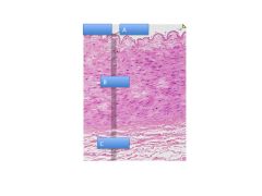
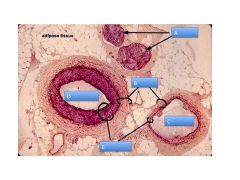
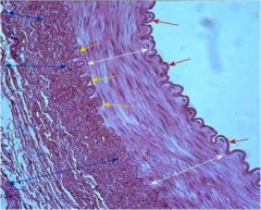
Artery or Vein?
Identify. |
Artery
A: Tunia Intima—thick; look at underlying CT B: Tunica Media C: Tunia Adventitia |
|
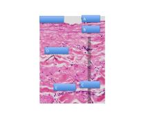
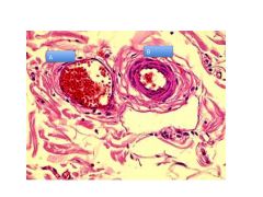
Artery or Vein?
Identify. |
Artery or vein? Vein
A: Intima B: Media C: Adventitia D: Collagen E: Smooth Muscle |
|

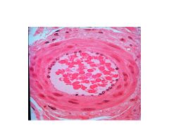
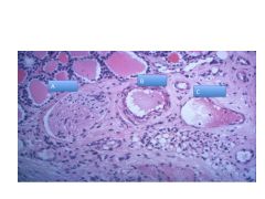
Artery or vein? Subtype?
|
Large Elastic Artery: tons of elastic fiber
|
|
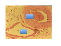
Artery or vein? Subtype?
|
Arteriole: 1-3 layers smooth muscle, bulging nuclei
|
|
|
A: Artery: much more muscular; scalloped edge
B: Vein: kind of collapsed, no muscular lining |
|
|
A: Nerve
B: Adventitia C: Vein D: Artery E: Media |
|
|
A: Venule; thinly walled
V: Arteriole: ~3 layers of smooth muscle |
|
|
A: Nerve
B: Arteriole C: Vein |
|
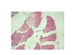
Identify.
|
Capillary: RBC in middle
|
|
|
Capillary: transverse cut; width of RBC
|
|
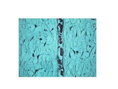
Identify.
|
Capillary: single cell lining
|
|
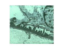
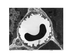
Capillary type?
|
Fenestrated without diaphragm
|
|
Artery or vein? Subtype?
|
Medium-sized muscular artery;
Lots of smooth muscle in media (not too much) |