Reading...
![]()
Play button
![]()
Play button
![]()
Use LEFT and RIGHT arrow keys to navigate between flashcards;
Use UP and DOWN arrow keys to flip the card;
H to show hint;
A reads text to speech;
63 Cards in this Set
- Front
- Back
|
Muscle Tissue
|
one of four primary tissues, divided into 3 types
|
|
|
What are the three types of muscle tissue?
|
skeletal muscle, cardiac muscle and smooth muscle
|
|
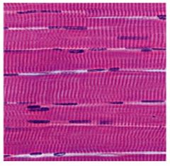
skeletal muscle
|
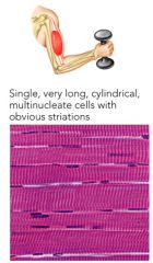
moves body by pulling on bones striated, ~ voluntary
|
|
|
cardiac muscle
|
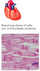
pumps blood throughout body straiated, involuntary
|
|
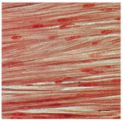
smooth muscle
|
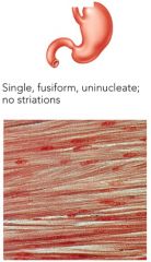
pushes fluids & solids along digestive tract & regulates diameters of small arteries non-striated, involuntary
|
|
|
Skeletal Muscle Functions
|
1. Produce skeletal muscle
2. Maintain body posture & position 3. Support soft tissues 4. Guard body openings (e.g. digestive tract - voluntary control over swallowing) 5. Maintain body temperature (muscle contractions use energy, heat released) 6. Store nutrient reserves (when diet is low in proteins & calories; muscle protein is broken down) |
|
|
Skeletal Muscle Fibers
|
skeletal muscle cells = muscle fibers Different from "typical" cells:
• Large cells (large diameter & can span between 2 tendons → up to 12 inches) • Multinucleate - 1 cell has 100s of nuclei ∙ genes in nuclei control production of enzymes/proteins (more copies = faster protein production) |
|
|
sarcolemma
|

cell membrane of muscle cell
|
|
|
sarcoplasm
|

cytoplasm of muscle fiber
|
|
|
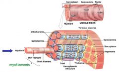
Transverse tubules
|

narrow tubes continuous with sarcolemma; extend into sarcoplasm
• filled with extracellular fluid • transmit action potential through cell • allow entire muscle fiber to contract simultaneously |
|
|
myofibrils
|

lengthwise subdivisions within muscle fiber Made up of bundles of protein filaments (myofilaments)
|
|
|
What are the two types of myofilaments?
|

thin filaments & thick filaments
|
|
|
thin filaments
|
made up of protein actin
|
|
|
thick filaments
|

made up of myosin
|
|
|
When myofibrils shorten they will cause ……
|
muscle fiber contraction
|
|
|
Skeletal Muscle Structures Hierarchy of structure
|
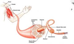
Muscle → Fascicle → Muscle fiber → Myofibril → Myofilament
|
|
|
Fascicle
|
bundles of muscle cells
|
|
|
Muscle Fibers
|
multinucleated, elongated cell
|
|
|
Myofibril
|
bundle of overlapping myofilaments
|
|
|
Myofilament
|
protein filaments (thick and thin filaments)
|
|
|
sarcoplasmic reticulum
|

membranous structure surrounding each myofibril
• helps transmit action potential to myofibril • similar in structure to smooth endoplasmic reticulum • forms expanded chambers (terminal cisternae) attached to T tubules |
|
|
cisternae
|

concentrate Ca²+ (via ion pumps)
~40,000 times more calcium in cisternae than sarcoplasm • stored Ca²+ is released into sarcomeres to begin muscle contraction |
|
|
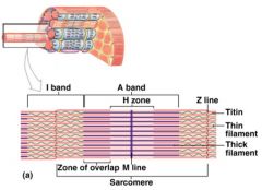
sarcomeres
|

contractile unit
• contractile units of muscle • structural units of myofibrils • form visible patterns within myofibrils |
|
|
Muscle striations
|
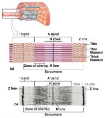
striped or striated pattern within myofibrils: alternating dark, thick filaments (A Bands) and light, thin filaments (I Bands)
|
|
|
What kinds of bands do sarcomeres have? Identify which is thick and thin filaments
|

A Bands - dark, thick filaments I Bands - light, thin filaments
|
|
|
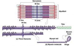
M line
|
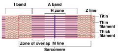
center of A band; at midline os sarcomere - connects thick filaments
|
|
|
Z lines
|

centers of I bands; at 2 ends of sarcomere
|
|
|
Zones of overlap
|

where thick and thin filaments overlap
|
|
|
H zone
|

area around M line; has thick filaments but no thin filaments
|
|
|
Titin
|

strands of elastic protein - extend from tips of thick filaments to Z line Function: stabalize the filaments → recoils after stretching during muscle contraction
|
|
|
What is the function of Titin?
|
stabilize the filaments →recoils after stretching during muscle contraction
|
|
|
muscle contraction
|
Transverse tubules encircle the sarcomere near zones of overlap Ca²+ released by sarcoplasmic reticulum causes thin and thick filaments to interact
|
|
|
Thick filaments
|
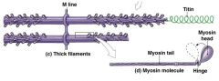
Myosin subunits twisted around filament
• tail - binds to other myosin molecules • head - made of 2 globular protein subunits; projects toward nearest thin filament |
|
|
During muscle contraction, myosin heads:
|
• interact with actin filaments, forming cross-bridges
• pivot, producing motion |
|
|
Thin filaments
|
contain 3 major thin filament proteins
|
|
|
What are the three major thin filament proteins?
|
actin, tropomyosin and troponin
|
|
|
actin
|

2 twisted strands of globular polypeptide subunits (G actin) form long actin filaments (F actin)
• "actin sites" on G actin strands bind to myosin |
|
|
tropomyosin
|
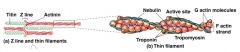
double stranded; covers active sites on actin to prevent actin-myosin interaction
|
|
|
troponin
|

globular protein; binds tropomyosin to G actin; controlled by Ca²+ via a Ca²+ binding side
• resting conditions = low Ca²+ concentration |
|
|
Sliding Filament Model
|
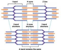
during muscle contraction → thin filaments of sarcomere slide toward M line between thick filaments
• width of A band stays the same • I band and H zone shorten • Z lines move closer together |
|
|
Thick and Thin filaments ____________ in length; ……
|
Thick and Thin filaments do not change in length; they slide past one another to shorten muscle fiber
|
|
|
What are the 5 Steps of the Crossbridge Cycle?
|
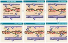
1. Exposure of active sites
2. Formation of cross-bridges 3. Pivoting of myosin heads 4. Detachment of cross-bridges 5. Reactivation of myosin |
|
|
What occurs during Step 1 of the Crossbridge Cycle?
|
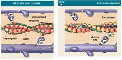
a) resting sarcomere - tropomyosin covers active sites on thin filaments (preventing myosin binding)
b) active site exposure = Ca²+ binds to troponin; rotates & swings tropomyosin away from active site • resting sarcomere - tropomyosin covers active sites on thin filaments (preventing myosin binding) • Ca²+ released → binds to troponin • weakens bond between troponin-tropomyosine complex • troponin rotates & swings tropomyosin away from active site |
|
|
What occurs during Step 2 of the Crossbridge Cycle?
|
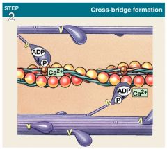
• active sites are exposed
• "energized" myosin heads bind to active sites forming cross bridges |
|
|
What occurs during Step 3 of the Crossbridge Cycle?
|
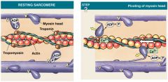
resting position - myosin points away from M line
• myosin goes back to "ready" position (~like a spring in a mousetrap - ready to spring forward) • myosin becomes energized by breaking down ATP into ADP & Phosphate • energy is "stored" in energized head as potential energy • after cross bridge formation - stored energy is released • myosin head pivots toward M line = power stroke • ADP and Pi are released • myosin head - pulls on actin filament sliding it towards M line |
|
|
What occurs during Step 4 of the Crossbridge Cycle?
|
• link between myosin & actin is broken when ATP binds to myosin heads
• active site is now exposed and ready to form another cross bridge |
|
|
What occurs during Step 5 of the Crossbridge Cycle?
|
• ATPase in free myosin head hydrolyzes ATP into ADP + P
• energy released in process = moves myosin head back to "energized" position • energy is stored in myosin head as potential energy |
|
|
The Entire Contraction Cycle can be repeated as long as:
|
• duration of neural stimulus
• calcium ion concentration is high • ATP is available |
|
|
What do you think would happen if the body suddenly ran out of ATP?
|
1. ATP energizes the myosin heads and without ATP you can't energize the myosin heads
2. ATP breaks the crossbridge |
|
|
Regulation of Muscle Contraction: Structures
Neuromuscular junction |
connection between motor neuron & muscle cell
|
|
|
Regulation of Muscle Contraction: Structures
presynaptic cell (motor neuron) |
delivers AP to axon terminal
|
|
|
Regulation of Muscle Contraction: Structures
ACh secretes across _________ |
synaptic cleft
|
|
|
Regulation of Muscle Contraction: Structures
ACh binds to ACh receptors on ______ |
postsynaptic cell (muscle cell)
|
|
|
Regulation of Muscle Contraction: Structures
Motor end plate |
region of sarcolemma (highly folded; many ACh receptors)
|
|
|
Regulation of Muscle Contraction: Structures
T Tubules |
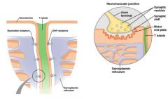
conduct APs deep into muscle fiber
|
|
|
Excitation-Contraction Coupling: Structures
DHP receptors |
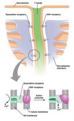
functions as "voltage sensors"
|
|
|
Excitation-Contraction Coupling: Structures
Sarcoplasmic Reticulum |

stores & releases Ca²+ to myofilaments
closely associated with T tubules |
|
|
Excitation-Contraction Coupling: Structures
Ryanodine receptors |

acts as Ca²+ channels
|
|
|
Excitation-Contraction Coupling: Structures
Ca²+ pumps |
actively transport Ca²+ back into SR into cytosol
|
|
|
Relaxed state
|

tropomyosin covers actin's myosin binding site
• prevents crossbridge formation |
|
|
Contraction state
|
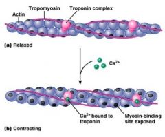
Ca²+ binds to troponin → causes conformational change in troponin
• troponin shifts tropomyosin's position • myosin binding site is exposed |
|
|
What are the steps of Excitation in Excitation-Contraction Coupling?
|
1. Action potentials travels down motor neuron to axon terminal through voltage-gated Ca²+ channels open causing Ca²+ to enter cell
2. Vesicles in axon terminal fuse with membrane → exocytosis of Acetylcholine into synaptic cleft 3. Acetylcholine diffuses across the synaptic cleft → binds to receptors on motor end plate → Na+ rushes in muscle fiber 4. Action potential travels across sarcolemma → and down the T tubules 5. DHP receptors "sense" voltage change → shape change occurs → open ryanodine/Ca²+ channels 6. Ca²+ exits SR → enters sarcoplasm |
|
|
What are the steps of Contraction in Excitation-Contraction Coupling?
|
1. Ca²+ binds to troponin → shape change → moves tropomyosin away from active site (on actin)
2. Myosin binds to active site = crossbridge formation 3. Myosin head pivots → pull thin filaments toward M line = power stroke - sarcomere shortens 4. ATP binds to ATPase on myosin head = breaks crossbridge 5. ATP →(ATPase)→ ADP + Pi → causes myosin to return to "energized" position 6. Ca²+ is actively transported back into SR → troponin/tropomyosin cover active site |