Reading...
![]()
Play button
![]()
Play button
![]()
Use LEFT and RIGHT arrow keys to navigate between flashcards;
Use UP and DOWN arrow keys to flip the card;
H to show hint;
A reads text to speech;
122 Cards in this Set
- Front
- Back
|
Olfactory
|
CN I
Type: sensory (smell) |
|
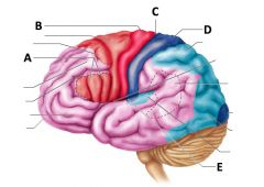
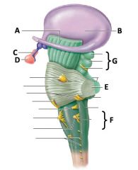
what is C?
|
central sulcus
|
|
|
Optic
|
CN II
Type: sensory (visual) |
|

what is A?
|
frontal lobe
|
|
|
Oculomotor
|
CN III
Type: motor (contracts eye muscles to control eye movements; constricts pupil; elevates eyelid) |
|

what is B?
|
parietal lobe
|
|
|
Trochlear
|
CN IV
Type: motor (superior oblique eye muscle) |
|

what is C?
|
occipital lobe
|
|
|
Trigeminal
|
CN V
Type: sensory (skin of face, oral/nasal/sinus mucosa, teeth) & motor (muscles of mastication) |
|
what is D?
|
temporal lobe
|
|
|
Abducens
|
CN VI
Type: motor (lateral rectus eye muscle) |
|

what is A?
|
cerebral peduncle
|
|
|
Facial
|
CN VII
Type: sensory (taste/anterior 2/3 of tongue) & motor (facial expressions, salivary glands) |
|

what is B?
|
thalamus
|
|
|
Acoustic/Vestibulocochlear
|
CN VIII
Type: sensory (hearing and balance) |
|

what is D?
|
pituitary gland
|
|
|
Glossopharyngeal
|
CN IX
Type: sensory (poster 1/3 of tongue, pharynx, gag reflex) & Motor (parotid gland) |
|

what is E?
|
cerebellar peduncles
|
|
|
Vagus
|
CN X
Type: sensory (pharynx, larynx, lungs heart, GI tract) & motor (palate, pharynx, larynx, trachea, bronchial tree, heart, GI tract) |
|
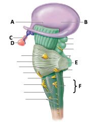
what is F?
|
medulla oblongota
|
|
|
Spinal Accessory
|
CN XI
Type: motor (SCM,trapezius) |
|

what is C?
|
infundibulum
|
|
|
Hypoglossal
|
CN XII
Type: motor (innervates tongue muscle that promotes movement of food & talking) |
|
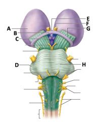
what is A?
|
thalamus
|
|

what is B?
|
hypothalamus
|
|

what is C?
|
mamillary bodies
|
|

what is D?
|
cerebellar peduncles
|
|

what is E?
|
optic chiasm
|
|

what is F?
|
optic nerve
|
|

what is G?
|
cerebral peduncle
|
|

what is H?
|
pons
|
|
what is G?
|
corpora quadrigemina
|
|

what is J?
|
optic tract
|
|

what is A?
|
dura mater
|
|

what is B?
|
periosteal dura
|
|
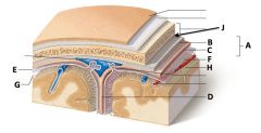
what is C?
|
meningeal layer of dura
|
|

what is D?
|
falx cerebri (there is also falx cerebelli - not pictured)
|
|

what is E?
|
subdural space
|
|

what is E?
|
subdural space
|
|

what is F?
|
arachnoid mater
|
|

what is G?
|
subarachnoid space
|
|

what is H?
|
pia mater
|
|

what is J?
|
epidural space
|
|

What is A and the function of it?
|
lacrimal sac; tears drain into it from the lacrimal canaliculi
|
|

what is B and its function?
|
lacrimal gland; tear producing gland
|
|
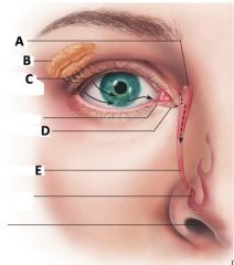
what is C and its function?
|
lacrimal duct; ducts that transport tears from the gland to the surface of the eye
|
|

what is D and its function?
|
lacrimal canaliculi; drains tears from the surface of the eye to the lacrimal sac
|
|

what is E and its function?
|
nasolacrimal duct; transports tears from the lacrimal sac into the nasal cavity
|
|
|
what is the fibrous tunic?
|
the outermost layer of the eye consisting mostly of collagenous connective tissue
|
|
|
what is the vascular tunic?
|
aka the uvea, it is the middle layer of the eye
|
|
|
what is the nervous tunic?
|
aka the retina, it is the innermost layer of the eye consisting of three layers of neurons; the layer closest to the uvea contains specialized neuronal cells that are sensitive to visible light
|
|

what is A?
|
the choroid
|
|

what is B?
|
ciliary body
|
|

what is C?
|
the iris
|
|

what is D?
|
the pupil
|
|

what is E?
|
the optic disc
|
|

what is F and it's structure?
|
the macula lutea; high concentration of cone cells0 critical for visual acuity
|
|

what is G and its structure?
|
fovea centralis - central region of the macula lutea; highest concentration of cones and the source of sharpest vision
|
|
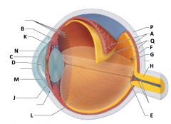
what is H?
|
optic nerve
|
|

what is J?
|
lens
|
|

what is K?
|
suspensory ligaments (aka ciliary zonule fibers)
|
|

what is L?
|
vitreous chamber (contains vitreous humor)
|
|

what is M?
|
anterior chamber (contains aqueous humor)
|
|

what is N?
|
cornea
|
|

what is P?
|
the sclera
|
|

what is Q?
|
the retina
|
|
|
what is the function of cones?
|
photoreceptors responsible for sensing bright light and color
|
|
|
what is the function of rods?
|
photoreceptors responsible for sensing light in a dim environment
|
|
|
what are constrictor pupillae?
|
smooth muscle cells arranged in a circle around the pupil - controlled by PNS
|
|
|
what are the dilator pupillae?
|
radially oriented myoepithelial cells which contract to dilate the pupil
|
|
|
what is the posterior chamber?
|
it is located between the lens and the iris containing aqueous humor
|
|
|
what are ciliary processes?
|
projections from the ciliary body covered in epithelium that produce aqueous humor; suspensory ligaments connect the ciliary processes to the lens
|
|
|
what is the innervation and movement of the superior rectus muscle?
|
CN III - elevate and abduct
|
|
|
what is the innervation and movement of the inferior rectus muscle?
|
CN III - depress and adduct
|
|
|
what is the innervation and movement of the medial rectus muscle?
|
CN III - adduct
|
|
|
what is the innervation and movement of the inferior oblique muscle?
|
CN III - elevate and adduct
|
|
|
what is the innervation and movement of the lateral rectus muscle?
|
CN VI - abduct
|
|
|
what is the innervation and movement of the superior oblique muscle?
|
CN IV - depress and medial
|
|

what is A?
|
malleus bone
|
|

what is B?
|
incus bone
|
|

what is C?
|
stapes bone
|
|

what is D?
|
semicircular canals
|
|

what is E?
|
vestibular nerve
|
|

what is E?
|
vestibular nerve
|
|
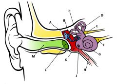
what is F?
|
cochlear nerve
|
|

what is G?
|
cochlea
|
|

what is H?
|
eustachian tube
|
|

what is L?
|
tympanic membrane
|
|

what is M?
|
external auditory canal
|
|
|
parietal peritoneum
|
lines the body walls the enclose the abdominal cavity
|
|
|
visceral peritoneum
|
wraps around various organs within the abdominal cavity
|
|
|
peritoneal cavity
|
potential space between the parietal and visceral peritoneum filled with serous fluid
|
|
|
intraperitoneal
|
organs that are suspended by mesenteries
|
|
|
retroperitoneal
|
organs that are not surrounded/suspended by mesenteries
|
|
|
greater omentum
|
mesentery that descends from the greater curvature of the stomach and drapes over central surface of most digestive organs - attaches to the transverse colon
|
|
|
lesser omentum
|
continuation of the visceral peritoneum that connects to inferior side of the liver with the lesser curvature of the stomach
|
|
|
falciform ligament
|
suspends liver from the diaphragm and anterior body wall
|
|
|
three types of salivary glands
|
parotid, submandibular, sublingual
|
|
|
cardia
|
small region of stomach that joins with distal esophagus
|
|
|
fundus
|
dome-shaped region just inferior to diaphragm
|
|
|
body
|
largest portion of the stomach
|
|
|
pyloric region
|
distal end of stomach (pyloric antrum narrows to form canal)
|
|
|
caudate lobe
|
of liver - superior
|
|
|
quadrate lobe
|
of liver - inferior
|
|
|
round ligament
|
inferior continuation of falciform ligament
|
|
|
common hepatic duct
|
combination of left/right hepatic ducts exiting from liver; joins the cystic duct
|
|
|
porta hepatis
|
exit point of common hepatic duct from liver
|
|
|
cystic duct
|
bile duct from gallbladder - joins with the common hepatic duct
|
|
|
common bile duct
|
combination of common hepatic and cystic duct
|
|
|
hepatopancreatic ampulla
|
joining of common bile and pancreatic ducts which enters the duodenum
|
|
|
hepatopancreatic sphincter
|
entry point of hepatopancreatic ampulla into duodenum
|
|
|
three portions of the small intenstine (proximal to distal)
|
duodenum --> jejunum --> ileum (DJI)
|
|
|
plicae circulares
|
"circular folds" - visible circular ridges along the wall of the SI
|
|
|
cecum
|
first part of large intestine
|
|
|
segments and bends of the large intestine (proximal to distal)
|
cecum --> ascending --> hepatic flexure --> transverse --> splenic flexure --> descending --> sigmoid colon --> rectum --> anal canal --> anus
|
|
|
teniae coli
|
longitudinal muscle running the lnegth of the large intestine
|
|
|
haustra
|
pouches formed along the length of the large intestine
|
|
|
four major layers of the GI tract (innermost to outermost)
|
mucose --> submucosa --> muscularis externa --> serosa or adventitia
|
|
|
parietal cell
|
pink staining cytoplasm with central nucleus; pump H+ into lumen
|
|
|
chief cell
|
purplish-blue cytoplasm; produce pepsinogen
|
|
|
paneth cell
|
cells lining the base of intestinal glands that produce antibacterial secretions; many bright pink secretory vesicles
|
|
|
enteroendocrine cells
|
intestinal cells that produce hormones secreted into underlying lamina propria
|