![]()
![]()
![]()
Use LEFT and RIGHT arrow keys to navigate between flashcards;
Use UP and DOWN arrow keys to flip the card;
H to show hint;
A reads text to speech;
75 Cards in this Set
- Front
- Back
|
Joints of lower limb |
Hip Knee Knee |
|
|
Hip joint composed of |
femar and acetabulum |
|
|
type of joint Hip |
ball and socket |
|
|
Knee joints composed of |
femur and patella fembur and tibia |
|
|
type of joint femur and patella |
plane |
|
|
type of joint femur and tibia |
hinge |
|
|
anatomy of tibia |
lateral/medial condyle tibial tuberosity medial malleolus |
|
|
anatomy of fibula |
fibular head lateral malleolus |
|
|
compartments of lower leg |
anterior deep and superficial posterior lateral |
|
|
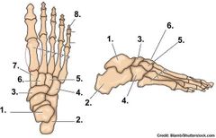
1. talus 2. calcaneus 3. navicular 4. cuboid 5. lateral cuneiform 6. intermediate cuneiform 7. medial cuneiform 8. distal phalange 9. middle phalange 10. proximal phalange Red circle: metatarsals Also: sesamoid bones on inferior side |
|
|
Tarsal bones |
1. Calcaneus 2. Talus 3. Navicular 4. Cuboid 5. 3 cuneiforms |
|
|
muscles of anterior compartment |
1. tibialis anterior 2. extensor hallucis longus 3. extensor digitorum longus 4. fibularis tertius |
|
|
Tibialis Anterior Origin |
1. lateral condyle of tibia 2. upper half/lateral surface of tibia 3. interosseous membrane |
|
|
Tibialis anterior insertion |
1. medial cuneiform 2. based of 1st metatarsal |
|
|
action tibalis anterior |
1. dorsiflexion 2. inversion |
|
|
innervation of tibialis anterior |
deep fibular |
|
|
Origin of extensor digitorum longus (EDL) |
1. lateral condyle of tibia 2. ant surface of fibula 3. IO membrane |
|
|
insertion of extensor digitorum longus (EDL) |
middle and distal phalanges of the 2-5 toes |
|
|
action of extensor digitorum longus (EDL) |
1. extension of toes 2. dorsiflexion of ankle |
|
|
innervation of extensor digitorum longus |
deep fibular |
|
|
origin of extensor hallucis longus (EHL) |
1. middle 1/2 of ant fibula 2. IM (intraosseous membrane) |
|
|
insertion of extensor hallucis longus (EHL) |
distal phalanx of great toe |
|
|
action of extensor hallucis longus (EHL) |
1. extend great toe 2. dorsiflex |
|
|
innervation of extensor hallucis longus (EHL) |
deep fibular |
|
|
which muscles inverts ankle? |
tibialis anterior |
|
|
what nerve innervates anterior compartment? |
deep fibular |
|
|
origin of fibularis tertius |
1. distal 1/3 of ant fibula 2. IM (intraosseous membrane) |
|
|
insertion fibularis tertius |
dorsum shaft of 5th metatarsal |
|
|
action of fibularious tertius |
1. forefoot eversion 2. weak ankle dorsiflexion |
|
|
innervation of fibularis tertius |
deep fibular |
|
|
which muscle in anterior compartment everts foot? |
fibularis tertius |
|
|
Actions of lateral compartment muscles |
foot eversion |
|
|
innervation of lateral compartment muscles |
superficial fibularis (peroneal) nerve |
|
|
muscles of lateral compartment |
1. fibularis (peroneus) longus 2. fibularis (peronenus) brevis |
|
|
origin of fibularis longus |
head and upper 2/3 of lateral fibula |
|
|
insertion of fibularis longus |
inferolateral surface of medial cuneiform (tendon of insertion runs underneath foot from lateral side to medial) |
|
|
action of fibularis (peroneus) longus (FL) |
1. eversion 2. plantar flexion |
|
|
innervation of fibularis (peroneus) longus (FL) |
superficial fibular (peroneus) |
|
|
origin of fibularis (peroneus) brevis (FB) |
lower 2/3 of lateral fibula |
|
|
insert of fibularis (peroneus) brevis |
tuberosity on base of 5th metatarsal (tendon of insertion going behind and under ankle) |
|
|
action of fibularis (peroneus) brevis |
1. planter flexion 2. eversion |
|
|
innervation of fibularis (peroneus) brevis |
superficial fibular (peroneal) |
|
|
actions of superficial posterior compartment muscles |
plantar flexion |
|
|
muscles of superficial posterior compartment |
1. gastrocnemius 2. plantaris 3. soleus |
|
|
origin of gastrocnemius |
2 heads 1. posterior surface of medial condyle 2. posterior surface of lateral condyle |
|
|
insertion of gastrocnemius |
achilles tendon to calcaneum |
|
|
innervation of gastrocnemius |
tibial nerve |
|
|
action of gastrocnemius |
1. plantar flexion 2. knee flexion |
|
|
innervation of superficial posterior compartment |
tibial nerve |
|
|
origin plantaris |
lateral supracondylar ridge of femur |
|
|
insertion of plantaris |
calcaneum |
|
|
action of plantaris |
1. planter flexion 2. knee flexion |
|
|
innervation of plantaris |
tibial nerve |
|
|
origin of soleus |
shafts of tibia and fibula |
|
|
insertion of soleus |
achilles tendon of calcaneum |
|
|
action of soleus |
plantar flexion |
|
|
innervation of soleus |
tibial nerve |
|
|
muscles of deep posterior compartment |
1. popliteus 2. flexor digitorum longus 3. flexor hallucis longus 4. tibialis posterior |
|
|
origin of popliteus |
lateral condyle of femur lateral meniscus |
|
|
insertion of popliteus |
proximal shaft of tibia |
|
|
action of popliteus |
1. flex knee 2. medially rotate knee |
|
|
origin flexor digitorum longus |
posterior shaft of tibia |
|
|
insertion of flexor digitorum longus |
inferior side of distal phalanges 2-5 |
|
|
action of flexor digitorum longus |
1. plantar flexion 2. inversion 3. toe flexion |
|
|
innervation of flexor digitorum longus |
tibial nerve |
|
|
origin of flexor hallucis longus |
posterior shaft of fibula |
|
|
insertion of flexor hallucis longus |
base of distal phalanx of big toe |
|
|
innervation of flexor hallucis longus |
tibial nerve |
|
|
action of flexor hallucis longus |
1. plantar flexion 2. inversion 3. big toe flexion |
|
|
innervation of deep posterior compartment |
tibial nerve |
|
|
origin of tibialis posterior |
1. posterior shafts of tibula and fibula 2. IM (intraosseous membrane) |
|
|
insertion of tibialis posterior |
navicular tuberosity |
|
|
innervation of tibialis posterior |
tibial nerve |
|
|
action of tibialis posterior |
1. plantar flexion 2. inversion |
|
|
toe joints |
DIP -distal interphalange PIP -proximal interphalange MP -metatarsal phalange |